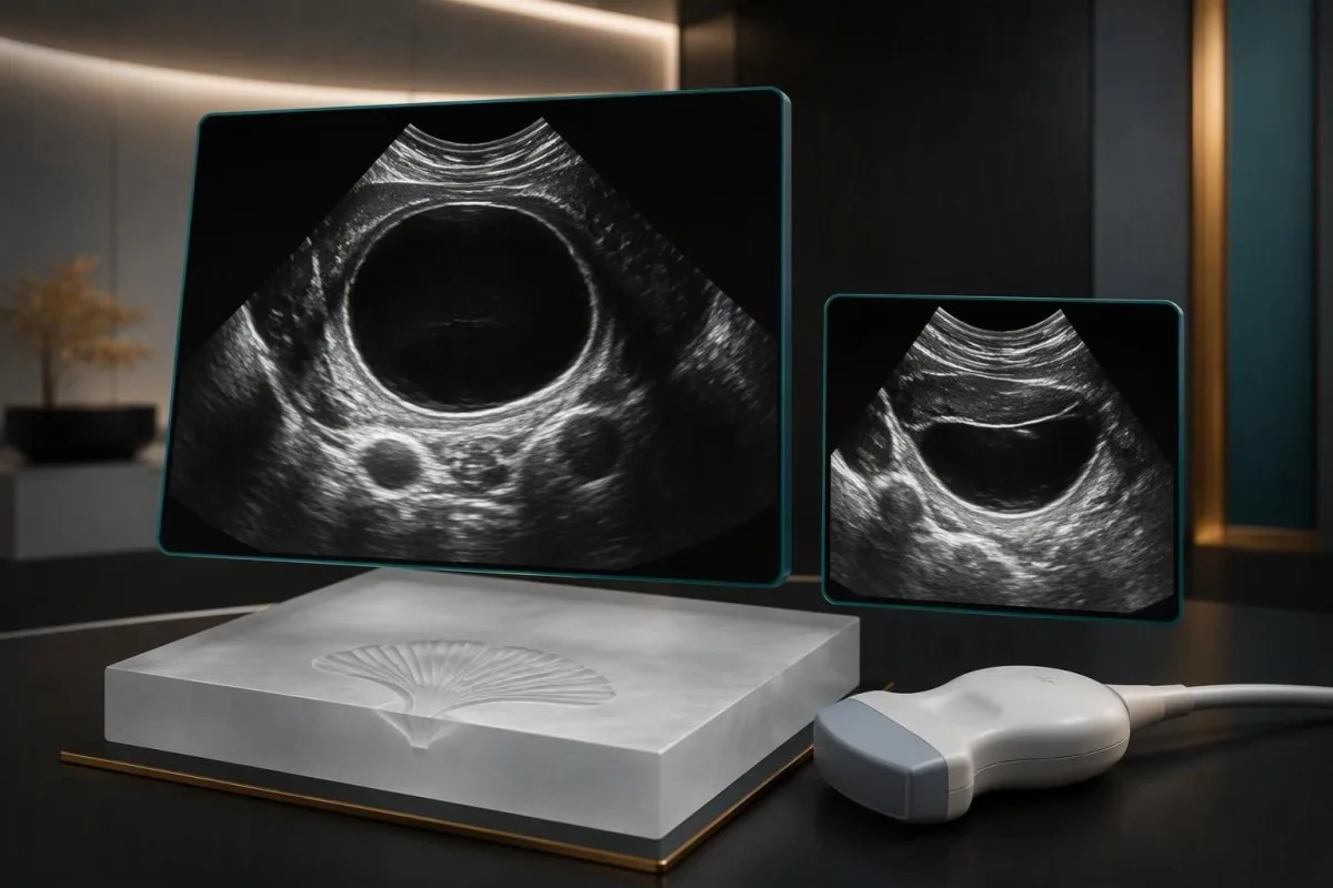
ගෙඩිය පුපුරා යාමෙන් දියර පිටවීමට හෝ අභ්යන්තර රුධිර වහනයට හේතු විය හැක; ඩිම්බකෝෂය ඇඹරීමෙන් එහි රුධිර සැපයුම අනතුරට ලක් වේ. දෙකම හදිසි ශ්රෝණි වේදනාව ඇති කළ හැකි බැවින්, රෝග ලක්ෂණ පමණක් මත ඒවා ආරක්ෂිතව වෙන්කර හඳුනාගත නොහැක.


- ඩිම්බකෝෂ ගෙඩියක රෝග ලක්ෂණ එක් පැත්තක ශ්රෝණි ප්රදේශයේ වේදනාව, පීඩනය, උදරය පිම්බීම හෝ ලිංගික සංසර්ගයේදී වේදනාව ඇතුළත් විය හැකි නමුත්, බොහෝ ගෙඩි කිසිදු රෝග ලක්ෂණයක් ඇති නොකරයි.
- පුපුරා ගිය ඩිම්බකෝෂ ගෙඩියක රෝග ලක්ෂණ යෝනි මාර්ගයෙන් සුළු රුධිර වහනයක් සමඟ හදිසි වේදනාවක් ඇතුළත් විය හැක; සැලකිය යුතු රුධිර වහනය මුළුමනින්ම උදරය ඇතුළත සිදු විය හැක.
- ඩිම්බකෝෂය ඇඹරීමේ රෝග ලක්ෂණ බොහෝ විට එක් පැත්තක හදිසි වේදනාව සහ ඔක්කාරය හෝ වමනය ඇතුළත් වේ, නමුත් ඇඹරීම කඩින් කඩ සිදුවන විට වේදනාව අඩු වැඩි විය හැක.
- හදිසි ඇගයීම හදිසි දැඩි ශ්රෝණි වේදනාවක් සඳහා අවශ්ය වේ, විශේෂයෙන් වමනය, ක්ලාන්ත ගතිය, උරහිස් අගෙහි වේදනාව, උණ හෝ ගැබ් ගැනීමේ හැකියාව ඇති විට.
- ගෙඩියේ ප්රමාණය සෙ.මී. 5 පමණට වඩා වැඩි වීමෙන් ඩිම්බකෝෂය ඇඹරීමේ අවදානම වැඩි වේ, නමුත් කුඩා ගෙඩියක් තිබීම හෝ ගෙඩියක් නොපෙනීම ඩිම්බකෝෂය ඇඹරීම බැහැර නොකරයි.
- ගැබ් පරීක්ෂණය ගැබ් ගැනීම ජීව විද්යාත්මකව සිදුවිය හැකි සෑම අවස්ථාවකම, උපත් පාලන ක්රම භාවිත කළත්, හදිසි ශ්රෝණි වේදනාව තක්සේරු කිරීමේ කොටසකි.
- ඩොප්ලර් සමඟ අල්ට්රාසවුන්ඩ් පරීක්ෂණය ගෙඩියක්, ශ්රෝණි තරලය සහ රුධිර සංසරණය තක්සේරු කිරීමට උපකාරී වේ, නමුත් ඩොප්ලර් රුධිර ප්රවාහය පවතින බව ඩිම්බකෝෂය ඇඹරීමක් නොමැති බව තහවුරු නොකරයි.
- අනුක්රමික පරීක්ෂණ (Serial testing) සැක සහිත රුධිර වහනයකදී නැවත හිමොග්ලොබින් පරීක්ෂා කිරීම සහ සායනිකව ස්ථාවර, ස්ථානය නොදන්නා ගර්භණීභාවයකදී පැය 48ක පමණ පරතරයකින් නැවත සෙරුම් hCG පරීක්ෂා කිරීම ඊට ඇතුළත් විය හැක.
ඩිම්බකෝෂ ගෙඩියක් පුපුරා යාම සහ ඩිම්බකෝෂය ඇඹරීම අතර වෙනස කුමක්ද?
ගෙඩියක් පුපුරා යාම ගෙඩියේ අන්තර්ගතය සහ ඇතැම්විට රුධිරය ශ්රෝණිය තුළට මුදාහරියි; ඩිම්බකෝෂය ඇඹරීම ඩිම්බකෝෂයට ආධාරක වන පටක ඇඹරී, එහි රුධිර සැපයුම අඩාල විය හැක. මේ දෙකම හදිසි, එක් පැත්තක ශ්රෝණි වේදනාවක් ඇති කළ හැකි අතර, වේදනාවේ ස්වභාවය හෝ යෝනි මාර්ගයෙන් රුධිර වහනය නොමැති වීම ඒවා විශ්වාසදායක ලෙස වෙන්කර හඳුනා නොගනී.
හදිසි, දැඩි ශ්රෝණි වේදනාවක් ඇති විට හේතුව නිවසේදී සොයා බැලීමට උත්සාහ කරනවාට වඩා හදිසි වෛද්ය තක්සේරුවක් අවශ්ය වේ. වෛද්යවරු සාමාන්යයෙන් ජීව ලක්ෂණ, පරීක්ෂාව, ගර්භණී පරීක්ෂණ සහ ශ්රෝණි අල්ට්රාසවුන්ඩ් පරීක්ෂණය ඒකාබද්ධ කරයි; මුල් ස්කෑන් පරීක්ෂණයෙන් නිශ්චිත නිගමනයක් ලබාගත නොහැකි වුවද, සැක සහිත ඩිම්බකෝෂ ඇඹරීමකට හදිසි ශල්යකර්මයක් අවශ්ය විය හැක.
ඩිම්බකෝෂ ගෙඩියක් යනු තරලය අඩංගු ව්යුහයක් වන අතර, එය තිබීමෙන්ම ප්රතිකාර අවශ්ය රෝගයක් බව අදහස් නොවේ. බොහෝ ක්රියාකාරී ගෙඩි මාසික චක්ර 2–3ක් තුළ සුව වී යන අතර, එන්ඩොමෙට්රියෝමා, ඩර්මොයිඩ් ගෙඩි සහ වෙනත් දිගටම පවතින ගැටිති වෙනස් ලෙස හැසිරේ; අපගේ කාන්තා සෞඛ්ය මාර්ගෝපදේශය චක්රයට අදාළ පසුබිම පැහැදිලි කරයි.
වෛද්ය තෝමස් ක්ලයින් ලෙස ලියන මගේ සායනික ප්රමුඛතාව වන්නේ, කාලය සංවේදී විය හැකි හදිසි තත්ත්වයක් සහ පසු විපරමට සුදුසු ගෙඩියක් වෙන්කර හඳුනා ගැනීමයි—වේදනාව විස්තර කරන ආකාරය පමණක් මත එයට නමක් දීම නොවේ. Kantesti හි සංවිධානමය පසුබිම අපගේ කාර්යභාරය පැහැදිලි කරයි: Kantesti යනු රුධිර පරීක්ෂණවල ප්රතිඵල පැහැදිලි කළ හැකි AI රුධිර පරීක්ෂණ විශ්ලේෂකයකි, නමුත් එයට ගෙඩියක් පුපුරා යාම හඳුනා ගැනීමට හෝ ඩිම්බකෝෂ ඇඹරීමක් බැහැර කිරීමට නොහැක.
සංකූලතා නොමැති ඩිම්බකෝෂ ගෙඩියක් නිසා ඇති විය හැකි රෝග ලක්ෂණ මොනවාද?
ඩිම්බකෝෂ ගෙඩියක රෝග ලක්ෂණ අඳුරු ශ්රෝණි වේදනාවක්, එක් පැත්තක පීඩනයක්, බඩ පිපීමක්, ලිංගික එක්වීමේදී අපහසුතාවක් හෝ අසල ව්යුහයන් මත පීඩනය හේතුවෙන් නිතර මුත්රා කිරීමක් ඇති විය හැක. බොහෝ ගෙඩි රෝග ලක්ෂණ ඇති නොකරන අතර, අල්ට්රාසවුන්ඩ් පරීක්ෂණයකදී ගෙඩියක් සොයාගැනීමෙන් එය සෑම රෝග ලක්ෂණයක්ම පැහැදිලි කරන බව තහවුරු නොවේ.
වේදනාව පිහිටන ස්ථානය යටින් පවතින ගැටලුව හඳුනාගැනීමට පරිපූර්ණ සිතියමක් නොවේ. දකුණු පැත්තේ ගෙඩියක් මධ්යයේ අපහසුතාවක් ඇති කළ හැකි අතර, බඩවැල් පිම්බීම, ශ්රෝණි පතුලේ මාංශපේශි වේදනාව හෝ මුත්රා පද්ධතියේ තත්ත්වයක්ද සමාන සංවේදන ඇති කළ හැක; වැදගත් වන්නේ රෝග ලක්ෂණ පරීක්ෂාවට සහ රූපගත කිරීමට ගැළපේද යන්නයි, වේදනාව ඇති පැත්ත පමණක් නොවේ.
මාසික චක්රයේ වේලාව පසුබිම් තොරතුරු සපයන නමුත් තහවුරු කිරීමක් නොවේ. චක්රයේ මැද කෙටි වේදනාවක් ඩිම්බ මෝචනය සමඟ ඇති විය හැකි අතර, මාසික රුධිර වහනය සමඟ නැවත නැවත ඇතිවන වේදනාව එන්ඩොමෙට්රියෝසිස් ගැන සැක කිරීමට හේතු විය හැක; හදිසි හේතු බැහැර කළ පසු චක්ර 2–3ක් ආවරණය වන රෝග ලක්ෂණ දිනපොතක් රටා හඳුනාගැනීමට උපකාරී වේ.
බහුසිස්ටික් ඩිම්බකෝෂ සින්ඩ්රෝමය යනු එක් විශාල ඩිම්බකෝෂ ගෙඩියක් තිබීම හා සමාන දෙයක් නොවේ. PCOS සමඟ සම්බන්ධ කුඩා කූප, හදිසි වේදනාකාරී ගෙඩියකට වඩා වෙනස් වන අතර, ශ්රෝණි වේදනාව පමණක් තිබීමෙන් PCOS තහවුරු නොවේ; අපගේ PCOS රෝගනිර්ණ නිර්ණායක හෝමෝන රටා, මාසික වාර ඉතිහාසය සහ වෙනත් සොයාගැනීම් වැදගත් වන්නේ මන්දැයි පැහැදිලි කරයි.
පුපුරා ගිය ඩිම්බකෝෂ ගෙඩියක රෝග ලක්ෂණ දැනෙන්නේ කෙසේද?
පුපුරා ගිය ඩිම්බකෝෂ ගෙඩියක රෝග ලක්ෂණ බොහෝවිට හදිසි ශ්රෝණි වේදනාවක් ලෙස ආරම්භ වන අතර, ඇතැම්විට ව්යායාමයේදී හෝ ලිංගික එක්වීමේදීද, ඇතැම්විට හේතුකාරකයක් නොමැතිවද ඇති වේ. තරලය හෝ රුධිරය උදර කුහරයේ ඇතුළු පටලය උත්තේජනය කරන විට වේදනාව එක් ස්ථානයක පැවතිය හැකි හෝ වඩාත් පැතිර යා හැක; යෝනි මාර්ගයෙන් රුධිර වහනය නොතිබිය හැක.
මුදාහැරෙන තරල සුළු ප්රමාණයකින් වුවද, අනතුරුදායක රුධිර අහිමිවීමක් නොමැතිව දැඩි වේදනාවක් ඇති කළ හැක. එහි ප්රතිවිරුද්ධ තත්ත්වයද වැදගත්ය: අභ්යන්තර රුධිර වහනය දිගටම පැවතියද මුලදී මධ්යස්ථ අපහසුතාවක් පමණක් ඇති විය හැකි බැවින්, වේදනාවේ තීව්රතාව රුධිර වහනයේ බරපතළකම මැනීමට විශ්වාසදායක මිනුමක් නොවේ.
උරහිස් අග වේදනාව, කරකැවිල්ල, සිහිසුන් වීම හෝ අසාමාන්ය දුර්වලතාව සැලකිය යුතු උදර අභ්යන්තර රුධිර වහනයක් සමඟ ඇති විය හැක. විශේෂයෙන් ගර්භණීභාවයක් තිබිය හැකි විට, මෙම රෝග ලක්ෂණ සඳහා හදිසි වෛද්ය ප්රතිකාර අවශ්ය වේ; අපගේ එක්කෝ, පෑඩ් ගණනින් අභ්යන්තර රුධිර වහනය මැනිය නොහැක. heavy menstrual bleeding guidance can help distinguish separate concerns about vaginal blood loss.
After a clinician confirms an uncomplicated rupture and establishes stability, pain often begins improving over the next 24–48 hours, although tenderness can last longer. That timeframe is not permission to wait out an unassessed first episode: a presumed rupture can actually be torsion, ectopic pregnancy, or another abdominal emergency.
ඩිම්බකෝෂය ඇඹරීමේ කුමන රෝග ලක්ෂණ සැලකිලිමත් වීමට හේතු විය යුතුද?
ඩිම්බකෝෂය ඇඹරීමේ රෝග ලක්ෂණ commonly include sudden one-sided pelvic or lower abdominal pain with nausea or vomiting. Pain can come and go when the adnexa twist and partially untwist, so temporary improvement does not make torsion safe to dismiss.
An enlarged ovary or adnexal mass increases mobility and creates a mechanical opportunity for twisting. Masses around 5 cm or larger are a recognized risk factor, but size is not a diagnostic cutoff; torsion also occurs with smaller masses and without a mass.
ACOG's Committee Opinion No. 783 reports that torsion occurs without an associated mass or cyst in as many as 46% of pediatric and adolescent cases (ACOG, 2019). That adolescent finding should not be applied as an adult prevalence estimate, but it explains why a normal-looking or cyst-free scan cannot end the assessment automatically.
Fertility treatment can enlarge the ovaries and increase torsion risk, including during early pregnancy. Anyone receiving ovarian stimulation should mention the treatment and the date of their last monitoring scan during emergency assessment; our IVF monitoring explanation describes why laboratory results and ultrasound measurements are interpreted together.
රෝග ලක්ෂණ පමණක් මත ගෙඩියක් පුපුරා යාම සහ ඩිම්බකෝෂය ඇඹරීම වෙන්කර හඳුනාගත නොහැක්කේ ඇයි?
Rupture and torsion share too many symptoms for a reliable home diagnosis: abrupt pain, tenderness, nausea, and sometimes fluctuating discomfort. Appendicitis, ectopic pregnancy, kidney stones, and pelvic infection can overlap with both, making examination and testing necessary.
Consider a hypothetical 29-year-old with 30 minutes of right-sided pelvic pain and vomiting. A known cyst makes torsion or rupture plausible, but it does not eliminate appendicitis or ectopic pregnancy; anchoring on an old ultrasound report can delay recognition of the actual emergency.
Nausea makes torsion more concerning, but rupture can also provoke nausea through pain and abdominal irritation. Fever is similarly unhelpful as a gatekeeper: early torsion may occur without fever, and fever with pelvic pain broadens the differential rather than identifying one cause.
Normal inflammatory markers do not reliably exclude an early abdominal emergency. Our appendicitis testing limits explain the same principle: a reassuring white cell count is one piece of evidence, not a substitute for a repeat examination when pain persists or changes.
හදිසි ශ්රෝණි වේදනාවක් සඳහා හදිසි ප්රතිකාර අවශ්ය වන්නේ කවදාද?
Sudden severe pelvic pain requires emergency assessment, even without fever or vaginal bleeding. Seek urgent help immediately for pain with fainting, marked dizziness, repeated vomiting, shoulder-tip pain, a rigid or increasingly swollen abdomen, or possible pregnancy.
Call your local emergency number if you collapse, feel close to fainting, or cannot travel safely; do not drive yourself while dizzy or impaired by pain. There is no dependable pain-score threshold or minimum duration that makes waiting safe when torsion or internal bleeding is possible.
Anticoagulants and bleeding disorders lower the threshold for urgent assessment because a ruptured cyst may bleed more extensively. Bring the medication name and last dose, and do not stop prescribed anticoagulation without clinician advice; our coagulation test limitations explain why apparently normal clotting results may not capture every medication effect.
New persistent pelvic pain that is not severe still deserves prompt clinical advice, particularly with a known cyst, pregnancy possibility, or recent fertility treatment. Tell the service if pain has improved after a sudden episode: intermittent torsion can produce a misleadingly comfortable interval, and pain relief does not establish the diagnosis.
ඩිම්බකෝෂ ගෙඩියක වේදනාව සඳහා ගර්භණී පරීක්ෂණයක් අවශ්ය වන්නේ ඇයි?
Pregnancy testing helps identify ectopic pregnancy, a potentially life-threatening cause of pelvic pain that can resemble cyst rupture or torsion. Testing is appropriate whenever pregnancy is biologically possible, regardless of contraception use or whether the pain feels familiar.
A negative home urine test can be misleading very early in pregnancy or when urine is dilute. Many urine assays detect hCG at approximately 20–25 IU/L, but sensitivity varies; our urine pregnancy test guidance explains why the test date and menstrual history matter.
NICE guideline NG126 recommends considering pregnancy testing in reproductive-age patients even when symptoms are nonspecific and emphasizes that ectopic pregnancy can present atypically (NICE, 2019; updated 2023). Pelvic pain with a positive pregnancy test needs prompt assessment, and severe pain or circulatory symptoms require emergency care.
A pregnancy test does not determine where a pregnancy is located. A corpus luteum cyst may be a normal early-pregnancy finding, while an ectopic pregnancy can coexist with an ovarian cyst; clinicians therefore interpret the test alongside ultrasound rather than treating either finding as a complete explanation.
සෙරුම් hCG ප්රතිඵල මඟින් ඊළඟ පියවර තීරණය කිරීමට මඟ පෙන්වන්නේ කෙසේද?
Serum hCG helps establish pregnancy status and guide follow-up, but a single concentration cannot diagnose ectopic pregnancy or distinguish cyst rupture from torsion. If pregnancy is confirmed but its location is not visible, clinicians may arrange repeat hCG measurements and ultrasound.
Many laboratories report serum hCG below 5 IU/L as negative and 5–25 IU/L as indeterminate, although the assay's own interpretation takes precedence. An indeterminate result may reflect very early pregnancy, a resolving pregnancy, or another explanation; severe symptoms require assessment rather than waiting for the number to cross a threshold.
For a clinically stable pregnancy of unknown location, NICE recommends 2 serum hCG measurements as close as possible to 48 hours apart, but not earlier. Symptoms take priority over the trend, and even a falling concentration does not immediately remove ectopic risk; our hCG decline explanation discusses why follow-up continues until the clinical situation is resolved.
Kantesti is an AI blood test interpretation platform that can explain hCG units and serial laboratory changes, but an algorithm cannot locate a pregnancy. The practical limitation is substantial: the same hCG concentration can accompany different ultrasound findings, so an apparently reassuring result must never overrule new pain, faintness, or a clinician's concern.
අල්ට්රාසවුන්ඩ් පරීක්ෂණයෙන් දැකිය හැක්කේ කුමක්ද—එයින් මඟහැරිය හැක්කේ කුමක්ද?
Pelvic ultrasound can characterize a cyst, assess ovarian size, identify free fluid, and evaluate circulation with Doppler. Transvaginal imaging often provides more detail when appropriate and consented to; transabdominal imaging is an alternative or a complementary examination.
Free pelvic fluid supports several possibilities rather than one diagnosis. A small amount can be physiological, while complex or extensive fluid can raise concern for bleeding; clinicians assess its appearance and amount alongside pregnancy status, vital signs, and hemoglobin rather than calling every fluid collection a rupture.
Preserved Doppler flow does not rule out torsion because the ovary has more than one source of arterial supply and twisting can be intermittent. ACOG states that Doppler findings alone should not guide decision-making and that no clinical or imaging criteria reliably confirm torsion before surgery (ACOG, 2019).
Cyst appearance also helps distinguish a simple functional cyst from a hemorrhagic cyst, endometrioma, or another mass. Our endometriosis diagnostic explanation discusses why imaging carries more diagnostic weight than an isolated biomarker; persistent severe pain after an inconclusive scan still warrants gynecologic reassessment.
ගෙඩියක් පුපුරා යාම හෝ ඩිම්බකෝෂය ඇඹරීම තක්සේරු කිරීමට උපකාරී වන රුධිර පරීක්ෂණ මොනවාද?
Blood tests assess consequences and alternative diagnoses, not torsion itself. A complete blood count can help monitor suspected bleeding, pregnancy testing addresses ectopic risk, and additional tests depend on hydration, medications, infection concerns, and whether an operation may be needed.
Hemoglobin can initially remain normal during acute blood loss because plasma and red cells are lost together. A later fall may reflect ongoing bleeding, fluid redistribution, intravenous fluids, or several factors; our hemoglobin and hematocrit comparison explains why the trend needs clinical context.
An illustrative fall from 12.8 to 10.4 g/dL over 4 hours would prompt careful reassessment, but it is not a universal operation threshold. Clinicians also consider pulse, blood pressure, increasing abdominal tenderness, fluid on imaging, and ongoing symptoms; a stable number cannot make an unstable patient safe.
White cell count and CRP may be normal early or elevated for nonspecific reasons, including physiological stress. Our WBC and CRP interpretation explains these disagreements; the examination still matters, particularly guarding, focal tenderness, and a change between the first assessment and a repeat assessment.
පුපුරා ගිය ඩිම්බකෝෂ ගෙඩියක් තහවුරු වූ විට එයට ප්රතිකාර කරන්නේ කෙසේද?
A stable patient with a confirmed uncomplicated cyst rupture may be treated with observation and pain relief, while ongoing bleeding or clinical instability may require hospital care or surgery. The decision depends on the whole assessment, not simply whether ultrasound shows free fluid.
Kim and colleagues reported conservative management in 80.8% of 78 healthy reproductive-age women with ruptured cysts and hemoperitoneum; 19.2% underwent surgery (Kim et al., 2014). This retrospective cohort excluded important higher-risk situations and is not a guarantee that any individual patient can safely be managed at home.
Observation may involve repeat vital signs, abdominal examinations, hemoglobin measurements, and sometimes repeat imaging. Increasing pain, worsening circulatory signs, or evidence of continuing blood loss can change the plan quickly; receiving fluids or pain medicine is supportive care, not proof that the underlying problem has resolved.
After discharge, instructions should specify whom to contact, which symptoms require immediate return, and whether another scan is needed. Iron testing is useful later if blood loss or heavy periods have depleted stores—not as the first test for sudden internal bleeding; our යකඩ පරීක්ෂණ ප්රතිඵල කියවන්නේ කෙසේද explains that follow-up distinction.
ඩිම්බකෝෂය ඇඹරී ඇති බවට සැකයක් ඇති විට කඩිනම් නාරිවේද සමාලෝචනයක් අවශ්ය වන්නේ ඇයි?
Suspected ovarian torsion needs urgent gynecologic assessment because delayed treatment can compromise ovarian function. Definitive diagnosis usually occurs during laparoscopy, and treatment generally aims to untwist the adnexa and preserve the ovary whenever clinically appropriate.
There is no validated 6-hour or 12-hour deadline that guarantees whether an ovary will recover. Earlier treatment is preferred, but a longer symptom history is not a reason to abandon assessment or assume preservation is impossible; ACOG's adolescent guidance favors detorsion and preservation regardless of the ovary's appearance when feasible.
Fertility concerns should inform follow-up without delaying emergency treatment. AMH does not diagnose torsion and cannot predict the outcome of an acute episode; our AMH after ovarian surgery discussion explains why age, procedure type, and test timing affect later interpretation.
Persistent or complex cysts, particularly after menopause, require a different risk assessment from a transient functional cyst. CA-125 is often interpreted around a laboratory cutoff of 35 U/mL but is not a rupture or torsion test; our CA-125 limits and warnings explains benign elevations, while our HE4 interpretation guidance explains why kidney function can complicate another tumor-marker result.
ඩිම්බකෝෂ ගෙඩියක වේදනාවෙන් පසු පසු විපරමට ඇතුළත් විය යුත්තේ කුමක්ද?
Follow-up should confirm symptom improvement, clarify the cyst type, and provide a specific return plan. A repeat ultrasound is sometimes arranged in approximately 6–12 weeks for selected presumed functional or hemorrhagic cysts, but timing depends on the scan, age, symptoms, and local guidance.
A planned scan weeks away does not cover a new emergency today. Return for reassessment if pain becomes sudden or severe, vomiting develops, or dizziness and weakness appear; improving discomfort is reassuring only within the context of an established diagnosis and an appropriate discharge plan.
Kantesti is an AI-powered blood test analysis tool that can help explain a CBC or iron panel after clinician-directed follow-up, not provide real-time pelvic triage. Before sharing laboratory reports, review our privacy and data handling; ultrasound access, an examination, and symptom reassessment remain outside a blood-report interpretation workflow.
My advice as Dr. Thomas Klein is to keep 3 items together: the ultrasound report, the relevant laboratory results, and the discharge instructions. Kantesti's AI තාක්ෂණික මාර්ගෝපදේශය describes our interpretation approach, but neither an AI explanation nor a normal-looking panel should persuade someone to ignore recurrent symptoms suggestive of torsion or internal bleeding.
පර්යේෂණ ප්රකාශන සහ සායනික සාක්ෂි: අදාළ වන්නේ මොනවාද?
Clinical guidance on torsion and ectopic pregnancy—not AI performance reports—supports the emergency recommendations in this article. As of October 13, 2026, the cited evidence includes ACOG's 2019 torsion opinion, NICE guideline NG126, and Kim and colleagues' 2014 observational rupture-management study.
The 3 external references have different scopes: ACOG addresses adolescents, NICE addresses early-pregnancy complications, and Kim et al. studied selected healthy patients with confirmed rupture. Applying them responsibly means preserving those boundaries; an adolescent recommendation or a small retrospective cohort should not be presented as a universal diagnostic rule.
The 2 Kantesti publications listed below describe a clinical validation framework and a global blood-test report, respectively. They are platform publications hosted on Zenodo, not trials validating AI diagnosis of ovarian torsion, cyst rupture, or ectopic pregnancy; the ResearchGate and Academia.edu links are discovery searches rather than verified copies of those records.
Readers assessing our methodology can consult Kantesti's සායනික සත්යාපන තොරතුරු alongside the independently published medical references. This article provides education rather than an individual diagnosis: sudden severe pelvic pain warrants emergency assessment, and an online explanation should never delay ultrasound, pregnancy testing, or urgent gynecologic review.
නිතර අසන ප්රශ්න
ඩිම්බකෝෂ ගෙඩිවල වඩාත් සුලභ රෝග ලක්ෂණ මොනවාද?
ඩිම්බකෝෂ ගෙඩියක රෝග ලක්ෂණවලට ශ්රෝණි ප්රදේශයේ එක් පැත්තක වේදනාව, පීඩනය, බඩ පිපීම, ලිංගික සංසර්ගයේදී වේදනාව හෝ නිතර මුත්රා කිරීම ඇතුළත් විය හැකි නමුත්, බොහෝ ගෙඩිවලින් රෝග ලක්ෂණ ඇති නොවේ. ක්රියාකාරී ගෙඩි බොහෝවිට ඔසප් චක්ර 2–3ක් තුළ සුව වී යන නමුත්, වෙනත් වර්ගවල ගෙඩි දිගටම පැවතිය හැකිය. කලින් කළ ස්කෑන් පරීක්ෂණයකින් ගෙඩියක් හඳුනාගෙන තිබුණද, රෝග ලක්ෂණ තිබීමෙන් එයට හේතුව ගෙඩියක් බව තහවුරු නොවේ. හදිසියේ ඇතිවන දැඩි වේදනාවකට සාමාන්ය ගෙඩි පසු විපරමකට වඩා හදිසි ඇගයීමක් අවශ්ය වේ.
ඩිම්බකෝෂ ගෙඩියක් පුපුරා යාම සහ ඩිම්බකෝෂය ඇඹරීම අතර වෙනස හඳුනාගන්නේ කෙසේද?
ඩිම්බකෝෂ ගෙඩියක් පුපුරා යාම සහ ඩිම්බකෝෂය ඇඹරීම රෝග ලක්ෂණ පමණක් මත විශ්වාසදායක ලෙස වෙන්කර හඳුනාගත නොහැක. මේ දෙකෙන්ම එක් පැත්තක හදිසි වේදනාව සහ ඔක්කාරය ඇති විය හැකි අතර, ඇඹරීම අර්ධ වශයෙන් ආපසු හැරුණහොත් කඩින් කඩ ඇතිවන වාර ලෙස රෝග ලක්ෂණ මතුවිය හැක. සෙ.මී. 5ක් පමණ හෝ ඊට විශාල ගැටිත්තක් ඇඹරීමේ අවදානම වැඩි කරයි, නමුත් කුඩා ගැටිති සහ ගෙඩි නොමැති ඩිම්බකෝෂද ඇඹරිය හැක. ගර්භණී පරීක්ෂාව, පරීක්ෂාව සහ අල්ට්රාසවුන්ඩ් පරීක්ෂණය මගින් තත්ත්වය තක්සේරු කිරීමට මඟ පෙන්වන අතර, රූපගත කිරීමේ ප්රතිඵල නිශ්චිත නොවූවත් ඇඹරීමක් සැක කෙරේ නම් ශල්යකර්මයක් අවශ්ය විය හැක.
යෝනි මාර්ගයෙන් රුධිර වහනයක් නොවී ඩිම්බකෝෂ ගෙඩියක් පුපුරා යා හැකිද?
ඩිම්බකෝෂ ගෙඩියක් පුපුරා ගියත් යෝනි මාර්ගයෙන් රුධිර වහනයක් සිදු නොවිය හැකියි, මන්ද පිටතට නිකුත් වූ දියර හෝ රුධිරය උදර කුහරය තුළම රැඳී පැවතිය හැකි බැවිනි. එබැවින් යෝනි මාර්ගයෙන් සිදුවන රුධිර වහනයේ ප්රමාණයෙන් ගෙඩිය පුපුරා යාමේ බරපතළකම මැනිය නොහැකියි. සංකූලතා රහිත පිපිරීමක් බව වෛද්යවරයකු තහවුරු කළ පසු, වේදනාව බොහෝ විට පැය 24–48ක් ඇතුළත අඩුවීමට පටන් ගනී; එහෙත් පරීක්ෂා නොකළ සිදුවීමක් සම්බන්ධයෙන් බලා සිටීමට එය ආරක්ෂිත කාලයක් නොවේ. සිහිසුන් වීම, දැඩි කරකැවිල්ල, උරහිසේ අග ප්රදේශයේ වේදනාව හෝ වැඩිවන වේදනාව ඇති වුවහොත් හදිසි ප්රතිකාර ලබාගන්න.
සාමාන්ය ඩොප්ලර් රුධිර ප්රවාහයක් ඩිම්බකෝෂ ඇඹරීම බැහැර කරයිද?
සාමාන්ය ඩොප්ලර් රුධිර ප්රවාහයක් තිබීම ඩිම්බකෝෂය ඇඹරීමක් නොමැති බව තහවුරු නොකරයි. ඩිම්බකෝෂයට ධමනි රුධිර සැපයුමේ ප්රධාන මූලාශ්ර 2ක් ඇති අතර, අතරින් පතර හෝ අසම්පූර්ණ ඇඹරීමක් නිසා හඳුනාගත හැකි රුධිර ප්රවාහය පවතින්නට පුළුවන. ACOG හි 2019 මාර්ගෝපදේශය ඩොප්ලර් සොයාගැනීම් පමණක් පදනම් කරගෙන තීරණ ගැනීමෙන් වළකින ලෙස උපදෙස් දෙයි. අල්ට්රාසවුන්ඩ් වාර්තාව සහනදායී ලෙස පෙනුණද, සායනික සැකය දිගටම පවතී නම් හදිසි නාරිවේද ඇගයීමක් අවශ්ය වේ.
ඩිම්බකෝෂ ගෙඩියක් ඇතැයි සැක කරන විට වෛද්යවරු ගර්භණීභාවය පරීක්ෂා කරන්නේ ඇයි?
ගර්භාෂයෙන් පිටත ගැබ් ගැනීමක් ගෙඩියක් පිපිරීමකට හෝ ඩිම්බකෝෂය ඇඹරීමට සමාන විය හැකි අතර, අභ්යන්තරයේ භයානක රුධිර වහනයක් ඇති කළ හැකි බැවින් වෛද්යවරු ගැබ් ගැනීමක් තිබේදැයි පරීක්ෂා කරති. බොහෝ මුත්රා ගර්භණී පරීක්ෂණ මඟින් hCG දළ වශයෙන් 20–25 IU/L මට්ටමේදී හඳුනාගන්නා බැවින්, ගැබ් ගැනීමේ මුල් අවධියේ ලබාගත් හෝ තනුක වූ මුත්රා සාම්පලයක් ව්යාජ-ඍණාත්මක ප්රතිඵලයක් ලබා දිය හැකිය. ධන පරීක්ෂණ ප්රතිඵලයකින් ගැබ් ගැනීම පිහිටි ස්ථානය හඳුනාගත නොහැක; අල්ට්රාසවුන්ඩ් පරීක්ෂණයක් සහ ඇතැම් විට අනුක්රමික සෙරුම් hCG පරීක්ෂණ අවශ්ය වේ. ගැබ් ගැනීමේ හැකියාවක් සමඟ ඇති දැඩි ශ්රෝණි වේදනාව, නිවසේදී කළ පරීක්ෂණයක් ඍණාත්මක වූ පසුවද, හදිසි වෛද්ය ඇගයීමක් අවශ්ය කරයි.
සාමාන්ය රුධිර පරීක්ෂණවලින් පුපුරා ගිය ගෙඩියක් හෝ ඩිම්බකෝෂය ඇඹරීමක් බැහැර කළ හැකිද?
සාමාන්ය රුධිර පරීක්ෂණවලින් ඩිම්බකෝෂය ඇඹරීම හෝ මුල් අවධියේදී සිදුවන රුධිර වහනය සහිත ගෙඩියක් පුපුරා යාම බැහැර කළ නොහැක. උග්ර රුධිර අහිමිවීමකදී මුලදී හිමොග්ලොබින් මට්ටම සාමාන්යව පැවතිය හැකි අතර, සුදු රුධිරාණු ගණන හෝ CRP මුල් අවධියේදී සාමාන්යව පැවතිය හැකිය, නැතහොත් විශේෂිත නොවන හේතු නිසා ඉහළ යා හැකිය. සායනිකව ස්ථාවර, ස්ථානය නොදන්නා ගර්භණීභාවයකදී, සාමාන්යයෙන් සෙරුම් hCG පරීක්ෂාව පැය 48ක පමණ පරතරයකින් නැවත සිදු කරනු ලැබේ; එහෙත් රෝග ලක්ෂණ උග්ර වුවහොත් ඊට පෙර නැවත ඇගයීමක් අවශ්ය වේ. පරීක්ෂණය, රූපගත කිරීම, ජීව ලක්ෂණ සහ රසායනාගාර ප්රතිඵලවල ප්රවණතා එකට සලකා අර්ථ දැක්විය යුතුය.
මට ඩිම්බකෝෂය ඇඹරී ඇති බව සිතන්නේ නම්, කොපමණ වේලාවක් බලා සිටිය හැකිද?
ඩිම්බකෝෂය ඇඹරීමක් (ovarian torsion) ඇතැයි සැක කරන්නේ නම්, නිවසේ නිරීක්ෂණය කරමින් සිටීම වෙනුවට හදිසි ඇගයීමක් අවශ්ය වේ. ඩිම්බකෝෂය යථා තත්ත්වයට පත්වීම සහතික කරන හෝ බලා සිටීම ආරක්ෂිත බව තහවුරු කරන, වලංගු බව තහවුරු කළ පැය 6ක හෝ පැය 12ක කාල සීමාවක් නැත. විශේෂයෙන් ඔක්කාරය හෝ වමනය සමඟ ඇතිවන, එක් පැත්තක හදිසියේ ඇතිවන දැඩි වේදනාව, පසුව අඩු වුවද, සැලකිලිමත් විය යුතු ලක්ෂණයකි. හැකි විට ඩිම්බකෝෂයේ ක්රියාකාරිත්වය රැක ගනිමින් ඇඹරීම හඳුනා ගැනීමට හොඳම අවස්ථාව සලසන්නේ කඩිනම් නාරිවේද ඇගයීමයි.
අදම AI බලයෙන් රුධිර පරීක්ෂණ විශ්ලේෂණය ලබාගන්න
තත්පර කිහිපයකින් ක්ෂණික හා නිවැරදි රසායනාගාර පරීක්ෂණ විශ්ලේෂණය සඳහා Kantesti විශ්වාස කරන ලොව පුරා මිලියන 2කට වැඩි පරිශීලකයන්ට එක්වන්න. ඔබගේ රුධිර පරීක්ෂණ ප්රතිඵල උඩුගත කර, තත්පර කිහිපයකින් 15,000+ ජෛව සලකුණු පිළිබඳ සවිස්තර අර්ථකථනය ලබාගන්න.
📚 යොමු කර ඇති පර්යේෂණ ප්රකාශන
Kantesti (2026). Clinical Validation Framework v2.0 (Medical Validation Page). Kantesti AI වෛද්ය පර්යේෂණ.
Kantesti (2026). AI රුධිර පරීක්ෂණ විශ්ලේෂකය: විශ්ලේෂණය කළ පරීක්ෂණ 2.5M | ගෝලීය සෞඛ්ය වාර්තාව 2026. Kantesti AI වෛද්ය පර්යේෂණ.
📖 බාහිර වෛද්ය යොමු
ඇමරිකානු ප්රසව හා නාරිවේද වෛද්යවරුන්ගේ සංගමය (2019). Adnexal Torsion in Adolescents: ACOG Committee Opinion No. 783. Obstetrics & Gynecology, 134(2), e56–e63.
National Institute for Health and Care Excellence (2019). ගැබ්ගෙඩිය (ectopic pregnancy) සහ ගබ්සා වීම: රෝග නිර්ණය සහ ආරම්භක කළමනාකරණය. NICE guideline NG126; updated 2023.
Kim JH et al. (2014). Successful Conservative Management of Ruptured Ovarian Cysts with Hemoperitoneum in Healthy Women. PLOS ONE, 9(3), e91171.
⚕️ වෛද්ය වියාචනය
මෙම ලිපිය අධ්යාපනික අරමුණු සඳහා පමණක් වන අතර වෛද්ය උපදෙස් ලෙස නොසැලකේ. රෝග නිદાન සහ ප්රතිකාර තීරණ සඳහා සෑම විටම සුදුසුකම් ලත් සෞඛ්ය සේවා සපයන්නෙකුගෙන් උපදෙස් ලබාගන්න.
E-E-A-T විශ්වාස සංඥා
අත්දැකීම්
වෛද්යවරයා විසින් මෙහෙයවන ලද රසායනාගාර අර්ථකථන ක්රියාවලි පිළිබඳ සමාලෝචනය.
ප්රවීණතාව
සායනික සන්දර්භය තුළ ජෛව සලකුණු (biomarkers) හැසිරෙන ආකාරය පිළිබඳ රසායනාගාර වෛද්ය විද්යා අවධානය.
අධිකාරීත්වය
ආචාර්ය තෝමස් ක්ලයින් විසින් ලියන ලද අතර ආචාර්ය සාරා මිචෙල් සහ මහාචාර්ය ආචාර්ය හෑන්ස් වෙබර් විසින් සමාලෝචනය කරන ලදී.
විශ්වසනීයත්වය
අනතුරු ඇඟවීම් අඩු කිරීමට පැහැදිලි පසුකැඳවීම් මාර්ග සහිත සාක්ෂි-පාදක අර්ථකථනය.