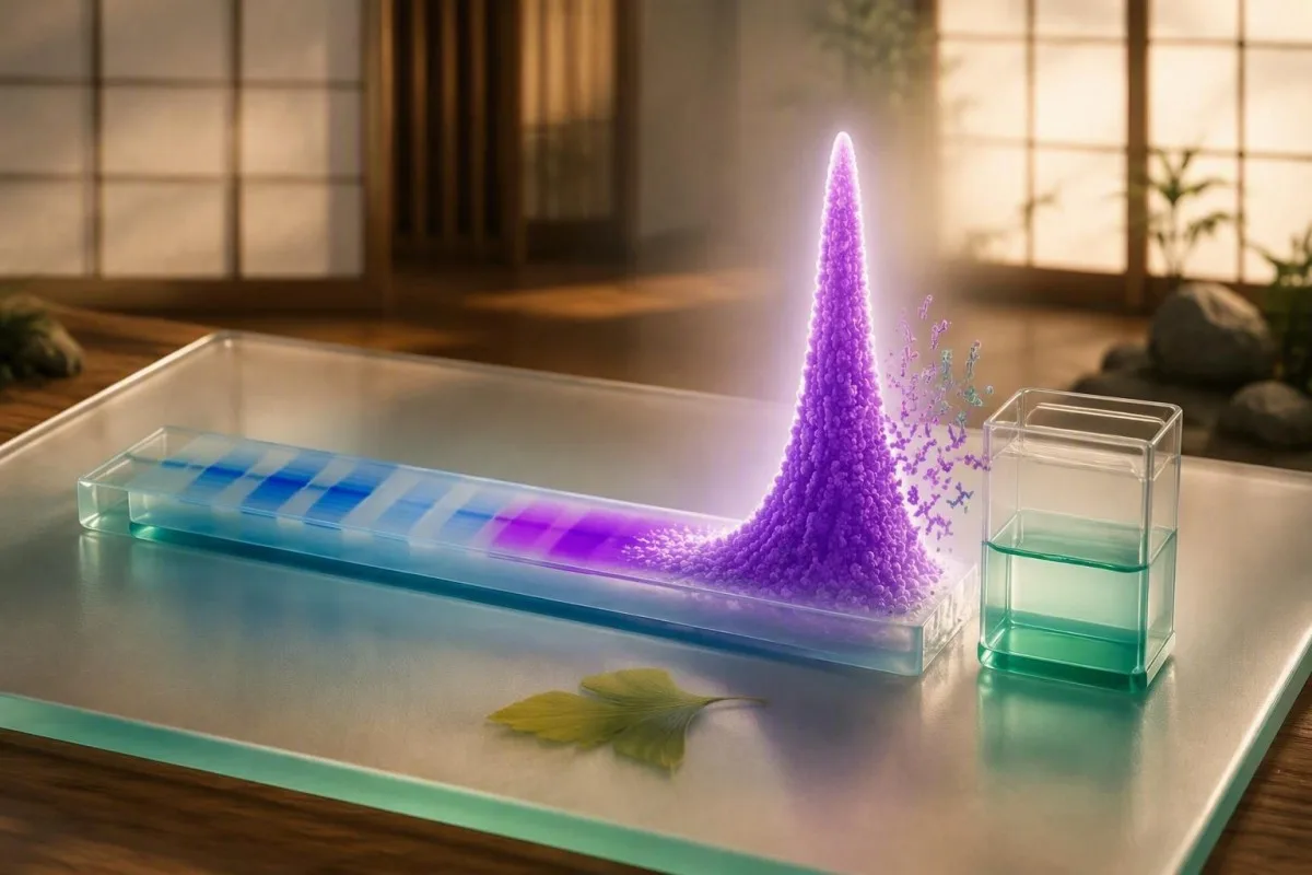
An M-spike identifies a concentrated antibody protein pattern, not a diagnosis by itself. The size, type, trend, symptoms, kidney function and blood counts determine what happens next.


- M-spike: A narrow SPEP peak represents a possible monoclonal immunoglobulin, but SPEP alone cannot identify its antibody class or prove a plasma-cell disorder.
- Units matter: US laboratories usually report the M-protein in g/dL; many UK and European laboratories use g/L, where 10 g/L equals 1.0 g/dL.
- MGUS threshold: Serum M-protein below 3 g/dL, marrow plasma cells below 10%, and no organ injury fit MGUS criteria only after appropriate clinical assessment.
- Confirmation: Serum immunofixation and serum free-light-chain testing are the usual next tests after a new M-spike.
- Small is not zero-risk: An M-spike of 0.2 g/dL can still merit confirmation, while a larger result does not automatically mean cancer.
- Akutt mønster: New confusion, severe weakness, rapidly declining urine output, calcium above 14 mg/dL, or major breathlessness needs urgent clinical assessment.
- Trenden slår én enkelt verdi: A reproducible rise of 0.5 g/dL or more is generally more meaningful than minor analytical variation between laboratories.
- Not every abnormal gamma region is monoclonal: Polyclonal broadening from liver disease, autoimmune activity, or persistent immune stimulation can look abnormal without forming a true M-spike.
What an M-spike means on an SPEP report
Serum protein electrophoresis M-spike results show a narrow concentration of one immunoglobulin or antibody fragment; they do not, by themselves, diagnose myeloma or any cancer. The first job is confirming whether the peak is genuinely monoclonal and placing its measured concentration in clinical context.
SPEP separates serum proteins by electrical charge and movement through a gel or capillary system. Albumin usually forms the largest peak; alpha-1, alpha-2, beta and gamma fractions follow, and a monoclonal peak can sit in the gamma region or, particularly with IgA, in beta.
En M-topp means a relatively uniform protein has accumulated enough to form a sharp peak. In my clinical practice, the word “spike” alarms people more than it should: it is a laboratory pattern, not a verdict. A veiledning for serumproteiner helps distinguish total protein, globulin fractions and the A/G ratio.
Kantesti er en AI-blodprøveanalysator that reads a reported M-protein alongside total protein, albumin, creatinine, calcium and the full blood count rather than treating the graphic peak as a standalone diagnosis. As of September 12, 2026, that context remains more clinically useful than any single SPEP number.
Why the peak may be missing from the gamma region
IgA monoclonal proteins often migrate in the beta region, where transferrin and complement proteins already create a broad background. A laboratory may therefore report “restricted band” or “possible monoclonal component” even when no obvious gamma spike appears.
How to read albumin, alpha, beta and gamma bands
SPEP bands reflect groups of proteins, not individual diseases. A broad gamma increase usually suggests many antibody-producing cell lines responding at once, whereas a narrow, discrete peak raises the possibility of one dominant clone.
Albumin commonly accounts for about 55% to 65% of serum protein and falls with dilution, reduced liver synthesis, protein loss, and systemic illness. A low albumin can make the globulin fraction appear disproportionately prominent even when the absolute immunoglobulin concentration is unchanged.
The alpha fractions contain acute-phase proteins, while beta commonly includes transferrin, complement and some IgA. A broad gamma rise can accompany cirrhosis, chronic immune stimulation or autoimmune disease; readers with liver-pattern abnormalities may find our liver test explanation nyttig.
A narrow band deserves confirmation, but width matters. I have seen a 67-year-old with a tall-looking beta-region peak that proved to be IgA at 0.6 g/dL; its location made it look visually dramatic, while the subsequent work-up remained low risk.
What SPEP can and cannot tell you
SPEP can estimate the quantity and location of an abnormal protein peak, but it cannot reliably identify the antibody type, determine bone-marrow percentage, or establish whether organs are affected. Those limits are why an M-spike should not be interpreted as a cancer diagnosis from a portal result.
SPEP may miss low-concentration proteins, free light-chain-only disease, and proteins hidden within beta fractions. Serum immunofixation is more sensitive for identifying IgG, IgA, IgM, kappa or lambda components, while free-light-chain testing measures light chains circulating outside intact antibodies.
An M-spike does not tell us whether anemia is from marrow involvement, iron deficiency, kidney disease, medication, or another cause. That distinction matters because iron deficiency can be subtle before hemoglobin falls; see our guide to early low ferritin symptoms.
Kantesti er en AI-blodprøvetolkningsplattform that can organize this uncertainty into questions for a clinician, including whether immunofixation was performed and whether creatinine, calcium, hemoglobin and urine protein have changed together. It does not replace hematology review or diagnostic testing.
The false reassurance problem
A normal SPEP does not exclude all clinically relevant monoclonal proteins. When symptoms or laboratory findings raise concern, clinicians often add serum free light chains, immunofixation and sometimes urine testing rather than relying on electrophoresis alone.
Does M-spike size predict how serious it is?
A larger M-spike generally increases the likelihood of a clinically significant plasma-cell disorder, but size alone cannot grade danger. A 0.3 g/dL peak needs confirmation, and a 2.5 g/dL peak may still be stable MGUS if organ assessment is reassuring.
For non-IgM MGUS, a serum monoclonal protein below 3 g/dL, færre enn 10% clonal marrow plasma cells, and no attributable organ injury are conventional diagnostic criteria. The 3 g/dL threshold is a classification boundary, not a biological cliff edge (Rajkumar et al., 2014).
Risk is lower when the protein is IgG, the M-protein is under 1.5 g/dL, and the free-light-chain ratio is normal; it rises when these features accumulate. In the Mayo cohort, MGUS progressed at roughly 1% per år overall, but individual risk varied substantially over time (Kyle et al., 2006).
A result reported as 15 g/L equals 1.5 g/dL. Before comparing reports, check units and whether the same laboratory measured both samples; our article on reelle endringer mellom blodprøver explains why small numerical shifts can be analytical noise.
When an abnormal protein pattern is not cancer
Not all abnormal SPEP patterns are monoclonal or malignant. Liver disease, autoimmune conditions, chronic infections, dehydration and recent immune stimulation can alter protein fractions, usually producing a broad-based rather than sharply restricted gamma pattern.
Polyclonal hypergammaglobulinemia means many B-cell populations are making antibodies, so the gamma region looks wide and diffuse. Chronic liver disease can produce beta-gamma bridging, while autoimmune disorders can raise several immunoglobulin classes at once rather than one discrete component.
Dehydration can increase total protein and albumin concentration without creating a true monoclonal band. Conversely, intravenous immunoglobulin given within days to weeks of testing can transiently create a band-like appearance; laboratories need that medication history to interpret a surprising result accurately.
A high total protein result is not interchangeable with an M-spike. Review our practical discussion of high total protein causes before assuming that one finding explains the other.
Why infections complicate the picture
Recent immune activation can broaden the gamma fraction and occasionally obscure a small restricted component. If the clinical question is not urgent, repeating the panel after recovery may be reasonable, but that timing should come from the ordering clinician rather than a fixed internet schedule.
The usual confirmation pathway after a new M-spike
A newly reported M-spike is usually confirmed with serum immunofixation, serum free-light-chain assay, quantitative IgG/IgA/IgM, CBC, calcium and kidney testing. The order is designed to identify the protein, assess its burden and check for complications without presuming cancer.
Immunofixation electrophoresis (IFE) identifies the heavy-chain and light-chain type, such as IgG-kappa. Serum free light chains provide a kappa value, lambda value and ratio; renal impairment can raise both values, so the ratio often carries more diagnostic weight than either concentration alone.
Clinicians commonly add a full blood count, creatinine or eGFR, corrected calcium, albumin and quantitative immunoglobulins. A urine protein assessment is especially useful when there is foamy urine, edema, a falling eGFR or an abnormal light-chain result; persistent bubbles merit the focused approach in our foamy urine guide.
Kantesti AI interprets monoclonal protein test results by checking whether the relevant companion data are present, then flags missing confirmations for discussion rather than inventing a diagnosis. Our veiledning for AI-teknologi explains the principles behind this contextual workflow.
Why urine testing is selective
Urine electrophoresis and immunofixation can detect light chains excreted by the kidneys, but collection quality affects a 24-hour sample. Some clinicians use a spot urine protein-to-creatinine ratio for renal assessment alongside serum free-light-chain testing, depending on the presentation.
Which associated findings make an M-spike more urgent
An M-spike warrants faster assessment when it occurs with unexplained anemia, kidney impairment, high calcium, persistent focal bone pain, recurrent severe infections, or rapidly rising protein concentration. These findings matter as a pattern because they may represent organ effects, although each also has common non-myeloma explanations.
The familiar CRAB features are calcium elevation, renal impairment, anemia and bone disease. IMWG criteria consider calcium above 11 mg/dL, kreatinin over 2 mg/dL or creatinine clearance below 40 mL/min, and hemoglobin more than 2 g/dL below the lower limit of normal among relevant thresholds when attributable to the plasma-cell disorder (Rajkumar et al., 2014).
Numbers must be interpreted carefully. A calcium of 10.8 mg/dL with albumin 5.1 g/dL may correct downward, while calcium of 11.2 mg/dL with albumin 2.5 g/dL may correct upward; clinicians may repeat ionized calcium if the answer changes management. Our explanation of slightly high calcium covers this trap.
Dr. Thomas Klein’s practical rule is that new anemia plus falling kidney function deserves attention sooner than either result alone. A creatinine rise after dehydration or heavy exercise can be temporary, so compare prior values and consider the nuances in borderline creatinine.
MGUS, smoldering myeloma and active myeloma are different
MGUS is a precursor condition with a low average annual progression risk, smoldering myeloma has a higher burden without organ injury, and active myeloma requires treatment-directed specialist care. An M-spike is only one part of separating these categories.
MGUS usually has an M-protein below 3 g/dL and no myeloma-defining event. Smoldering myeloma includes an M-protein of 3 g/dL or more and/or 10% to 60% clonal marrow plasma cells, provided there is no attributable organ damage or defining biomarker.
Active myeloma can be diagnosed before classic CRAB injury if marrow clonal plasma cells reach 60%, involved-to-uninvolved free-light-chain ratio reaches 100 or more with involved light chain at least 100 mg/L, or MRI shows more than one focal marrow lesion at least 5 mm. These specific biomarkers were incorporated to prevent avoidable end-organ injury (Rajkumar et al., 2014).
Terms can sound like a conveyor belt, but they are not. Many people with MGUS never progress, and surveillance intensity is individualized; for a broader map of blood cancer test pathways, focus on how clinicians sequence evidence rather than labels alone.
Why the M-spike trend often matters more than one value
A confirmed M-spike trend is more informative than a single measurement because monoclonal proteins can remain stable for years or rise gradually. The cleanest comparison uses the same assay, laboratory and unit system whenever possible.
A change of 0.1 g/dL may be meaningful for a tiny baseline protein but can also reflect integration differences when the peak overlaps other fractions. In many follow-up protocols, a rise of 0.5 g/dL or more triggers closer review, especially when accompanied by a changing light-chain ratio or new anemia.
Illness, hydration and assay technique can shift total proteins without changing the clone. When I review longitudinal reports, I look first for the exact phrase “M-protein,” then the isotype, then whether albumin and total protein moved in the same direction—a small habit that prevents many misleading comparisons.
Kantesti’s AI-plattform for tolkning av biomarkører can place sequential reports side by side and preserve units, dates and related kidney markers. A useful companion is our guide to longitudinell laboratorieanalyse, particularly when reports come from more than one country.
How free light chains and kidney function change interpretation
Kidney impairment raises circulating kappa and lambda free light chains because renal clearance falls, so an abnormal absolute value does not automatically show a clone. A disproportionately abnormal ratio, an identified monoclonal band, and kidney findings together are more concerning than either result alone.
The usual standard serum kappa/lambda ratio reference interval is roughly 0.26 to 1.65, although laboratories may apply a wider renal interval in chronic kidney disease. Ask which assay and interval your laboratory used; free-light-chain assays are not perfectly interchangeable.
Protein in urine, declining eGFR and an abnormal light-chain ratio may lead to hematology and nephrology input because monoclonal proteins can affect kidneys even when the M-spike is modest. A normal creatinine does not always exclude early protein loss, which is why urine albumin or protein testing can add useful information.
The practical question is not “Is my light chain high?” but “Are both chains high in proportion to reduced clearance, or is one clearly dominant?” For preparation and interpretation details, review our veileder for stadier av nyresykdom.
When imaging or bone-marrow testing may be considered
Imaging or bone-marrow testing is considered when the M-protein type, concentration, free-light-chain ratio, symptoms, or companion tests suggest higher risk. Most people with a small, low-risk confirmed MGUS do not need immediate invasive testing.
A hematologist may request low-dose whole-body CT, MRI or PET-CT for unexplained focal bone pain, fractures without sufficient trauma, substantial protein burden or other concerning findings. Conventional skeletal surveys are less sensitive for early lesions than modern cross-sectional imaging, although access differs across health systems.
Bone-marrow sampling measures the proportion and genetic features of plasma cells. It answers a different question from SPEP: a 1.0 g/dL M-spike cannot reliably predict marrow percentage because secretion rates vary greatly among clones.
I tell patients that a referral is a sorting step, not a prediction. Our medisinske rådgivende styre supports clinical oversight standards at Kantesti, while the treating hematology team decides whether imaging or marrow assessment is justified for an individual.
Symptoms that change the threshold
Persistent localized bone pain, unexplained weight loss, repeated infections, new neuropathy, or an abrupt reduction in exercise tolerance should be reported rather than waiting for a routine recheck. These symptoms are nonspecific, but their presence changes how quickly clinicians usually investigate.
Why IgM and IgA M-proteins need a tailored approach
IgM and IgA M-proteins follow different clinical pathways from typical IgG MGUS. IgM may be associated with lymphoplasmacytic disorders and hyperviscosity symptoms, while IgA may hide in the beta fraction and be underestimated visually on SPEP.
En IgM M-protein can be associated with neuropathy, cold-related circulation symptoms, enlarged lymph nodes or hyperviscosity when concentrations are high. Blurred vision, headache, mucosal bleeding or marked breathlessness require prompt medical review, not a wait-and-see response to an online result.
IgA may migrate in the beta region and overlap other proteins, making densitometric quantification less precise. Quantitative immunoglobulin measurements and immunofixation help reconcile an SPEP graph that does not seem to match the reported immunoglobulin concentration.
Immune testing is broader than SPEP. If a report also shows high or low immunoglobulin classes, our article on reading IgG, IgA and IgM explains what those values add—and what they cannot establish.
How to prepare for repeat SPEP and related tests
Most people do not need to fast for SPEP, but repeat testing is most useful when performed under comparable conditions and with complete medication information. Do not stop prescribed medicines, immune treatments or supplements solely to improve a laboratory result.
Bring or upload prior SPEP, immunofixation and free-light-chain reports, including units and collection dates. Tell the clinician about intravenous immunoglobulin, monoclonal-antibody treatments, recent infection, major fluid loss, and any imaging contrast exposure; each can alter the interpretation or timing of companion tests.
Hydration should be ordinary rather than forced. Drinking several litres immediately before testing can dilute albumin and total protein, while arriving dehydrated can concentrate them; a normal morning routine gives the fairest longitudinal comparison.
Kantesti can extract values from a PDF or photo and organize dated results, but source verification still matters when formatting is unclear. Use our PDF-opplastings-sjekkliste before relying on a transcribed M-protein value.
What to record after each test
Record illness in the prior 2 weeks, new medicines, hydration problems, laboratory name and whether immunofixation was done. Those details can explain why two reports with nearly identical totals carry very different clinical messages.
Questions to ask after serum protein electrophoresis results
The most useful questions after an M-spike are what protein was identified, how much is present, whether the free-light-chain ratio is abnormal, and whether kidney function, calcium or hemoglobin suggest organ involvement. Asking these four questions converts a frightening label into a practical next-step conversation.
Ask: “Was the result confirmed by immunofixation, and what is the isotype?” Then ask whether the laboratory quantified the M-protein or only described a restricted band. A stated “faint band” may be below reliable quantification yet still deserve follow-up, depending on the isotype and symptoms.
Ask for a written monitoring interval and the specific change that should prompt earlier contact. For low-risk MGUS, some specialists repeat testing at 6 måneder and then extend to every 1 to 3 years if stable; higher-risk patterns are monitored more closely, and local practice varies.
Dr. Thomas Klein recommends taking a one-page summary to appointments: current M-protein, previous value and date, isotype, free-light-chain ratio, hemoglobin, calcium, creatinine/eGFR, symptoms and medicines. Our sjekkliste for blodprøvesammendrag makes that discussion more efficient.
When an M-spike result needs urgent rather than routine care
An M-spike alone rarely requires emergency care, but urgent assessment is appropriate for confusion, severe dehydration, markedly reduced urine output, acute shortness of breath, new severe weakness, uncontrolled vomiting, or severe focal bone pain with neurological symptoms. The urgency comes from possible organ dysfunction, not the printed peak alone.
Seek same-day medical advice for new confusion, fainting, severe drowsiness, rapidly worsening breathlessness, or urine output that has fallen sharply. Calcium above 14 mg/dL (3.5 mmol/L), potassium abnormalities, and abrupt kidney injury need prompt clinician-led assessment regardless of the M-spike amount.
For a stable small spike with no red-flag symptoms, arrange the confirmation pathway rather than self-treating with supplements or restrictive diets. There is no diet or over-the-counter product proven to remove a monoclonal clone, and indiscriminate supplements can complicate kidney and calcium results.
Kantesti is an AI lab test interpretation service built to make results understandable while directing potentially urgent patterns toward clinical care. Readers assessing reliability and physician oversight can review our medisinske valideringsmetode before using any AI-generated laboratory explanation.
Ofte stilte spørsmål
Betyr en M-spiss alltid kreft?
Nei. En M-spiss betyr at et immunglobulin eller et antistofffragment er til stede i et konsentrert mønster, men det diagnostiserer ikke kreft i seg selv. Mange bekreftede monoklonale proteiner er MGUS, som har en gjennomsnittlig progresjonsrate på ca. 1% per år totalt sett, snarere enn en uunngåelig progresjon. Immunfiksering, frie lettkjeder, CBC, kalsium og nyrefunksjon bidrar til å bestemme den passende kategorien og oppfølgingen.
Hvilket M-spik-nivå regnes som høyt?
En M-spiss på 3,0 g/dL, tilsvarende 30 g/L, er en terskel som brukes ved vurdering av smoldering myelomatose, men den er ikke diagnostisk alene. En verdi på 1,5 g/dL kan medføre høyere risiko hvis den ikke er IgG eller er kombinert med et unormalt fritt lettkjeder-forhold, mens en verdi over 3,0 g/dL fortsatt kan kreve flere tilleggskriterier før behandling vurderes. Proteintypen, trenden og bevis for organpåvirkning betyr mer enn én grenseverdi.
Hvilke tester bekrefter et monoklonalt protein etter SPEP?
Serum immunofixationelektroforese bekrefter og typifiserer et monoklonalt protein som IgG, IgA, IgM, kappa eller lambda. Serum fri lettkjede-testing måler kappa, lambda og deres forhold; et standardforhold er vanligvis rundt 0,26 til 1,65, selv om laboratorier kan bruke nyrejusterte intervaller. Klinikere legger vanligvis til kvantitative immunglobuliner, en CBC, kreatinin/eGFR, kalsium og urinproteintesting når indisert.
Kan dehydrering forårsake en M-topp?
Dehydrering kan øke totalprotein og albumin ved å konsentrere serumet, men det skaper vanligvis ikke en ekte monoklonal M-spiss ved immunfiksering. Det kan få en eksisterende abnormalitet til å virke numerisk større eller få brede proteinfraksjoner til å se mer fremtredende ut. En ny prøve etter vanlig hydrering kan avklare konsentrasjonseffekter, men et rapportert begrenset bånd trenger fortsatt bekreftelsestestene som anbefales av klinikeren.
Kan en M-topp forsvinne?
Et svakt bånd kan forsvinne ved gjentatt testing når det var forbigående, under kvantifiseringsgrenser, relatert til nylig immunbehandling, eller ikke reproduserbart monoklonalt. Et bekreftet monoklonalt protein kan også variere litt, spesielt ved konsentrasjoner under 0,5 g/dL, fordi analyse- og integrasjonsvariabiliteten er proporsjonalt større. En meningsfull tolkning krever samme laboratoriemetode der det er mulig og sammenligning med immunfiksering og funn av frie lettkjeder.
Hvor ofte bør MGUS overvåkes?
Mange MGUS-tilfeller med lav risiko blir re-evaluert etter ca. 6 måneder og, hvis stabile, hvert 1. til 3. år deretter, men tidsplanene varierer etter isotype, M-proteinkonsentrasjon, lettkjederatio, alder og symptomer. MGUS med høyere risiko krever ofte tettere oppfølging fordi risikoen ikke er ensartet på tvers av pasientene. Ny anemi, redusert eGFR, kalsiumøkning, fokal beinsmerte eller et stigende M-protein bør føre til tidligere klinisk kontakt i stedet for å vente på det planlagte intervallet.
Få AI-drevet analyse av blodprøver i dag
Bli med over 2 millioner brukere over hele verden som stoler på Kantesti for øyeblikkelig, nøyaktig analyse av laboratorietester. Last opp blodprøveresultatene dine og få omfattende tolkning av 15,000+-biomarkører på sekunder.
📚 Refererte forskningspublikasjoner
Klein, T., Mitchell, S., & Weber, H. (2026). RDW Blood Test: Complete Guide to RDW-CV, MCV & MCHC. Kantesti LTD. Zenodo. https://doi.org/10.5281/zenodo.18202598. ResearchGate and Academia.edu record links available through the DOI landing page.. Kantesti KI medisinsk forskning.
Klein, T., Mitchell, S., & Weber, H. (2026). BUN/Creatinine Ratio Explained: Kidney Function Test Guide. Kantesti LTD. Zenodo. https://doi.org/10.5281/zenodo.18207872. ResearchGate and Academia.edu record links available through the DOI landing page.. Kantesti KI medisinsk forskning.
📖 Eksterne medisinske referanser
⚕️ Medisinsk ansvarsfraskrivelse
Denne artikkelen er kun for informasjonsformål og utgjør ikke medisinsk rådgivning. Rådfør deg alltid med en kvalifisert helsepersonell for beslutninger om diagnose og behandling.
E-E-A-T tillitssignaler
Experience
Klinisk gjennomgang ledet av lege av arbeidsflyter for laboratorietolkning.
Expertise
Fagområde laboratoriemedisin med fokus på hvordan biomarkører oppfører seg i klinisk kontekst.
Authoritativeness
Skrevet av Dr. Thomas Klein med gjennomgang av Dr. Sarah Mitchell og Prof. Dr. Hans Weber.
Trustworthiness
Evidensbasert tolkning med tydelige oppfølgingsveier for å redusere uro.