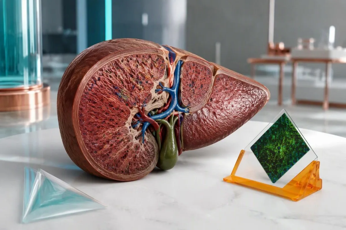
A positive antibody result is a clue, not a diagnosis. The useful question is whether it fits your liver enzymes, IgG level, medication history, and tissue findings.


- Positive smooth muscle antibodies alone do not diagnose autoimmune hepatitis; an adult ASMA titer of 1:40 contributes only 1 point to the simplified diagnostic score.
- Higher ASMA titers increase diagnostic support, but titers of 1:80 and 1:320 receive the same maximum 2 antibody points in the simplified criteria.
- F-actin antibodies identify a more specific antibody target, but a positive result still needs liver-test and clinical context; assay units cannot be converted into ASMA titers.
- Total IgG above the laboratory upper limit supports autoimmune hepatitis; a level above 1.1 times that limit receives 2 simplified-score points.
- Normal ALT makes incidental positivity less concerning, although normal enzymes or IgG do not universally exclude autoimmune hepatitis.
- Medication history should include prescriptions and supplements started months or years earlier, particularly nitrofurantoin and minocycline.
- Liver biopsy usually helps establish autoimmune hepatitis and assess fibrosis; urgent treatment decisions should not wait for elective testing in a severely ill patient.
- Akutt vurdering is needed for jaundice with confusion, significant drowsiness, or a rising INR; INR of at least 1.5 with encephalopathy can indicate acute liver failure.
Do positive smooth muscle antibodies mean autoimmune hepatitis?
Positive smooth muscle antibodies do not diagnose autoimmune hepatitis by themselves. A low-titer result such as 1:40 can be nonspecific; clinicians look for a matching pattern of elevated ALT or AST, increased IgG, exclusion of other liver diseases, and usually supportive liver tissue findings.
ASMA, also called SMA, detects antibodies reacting with components of smooth muscle and related cellular structures; it does not mean that your muscles are being destroyed. ASMA is most closely associated with type 1 autoimmune hepatitis, but it can also appear during other immune responses or liver disorders, which is why the laboratory word positive is not a disease label.
Consider 2 illustrative results: ASMA 1:40 with ALT 24 IU/L and normal IgG, versus ASMA 1:160 with ALT 286 IU/L and IgG above the laboratory limit. The second pattern deserves substantially more investigation, although neither establishes the cause without a clinical work-up. Our explanation of positivt antistoffresultat betydning separates antibody detection from evidence of active disease.
Kantesti er en AI-blodprøveanalysator that can help organize ASMA results alongside liver enzymes and IgG rather than treating a 1:40 flag as a diagnosis. As Thomas Klein, Chief Medical Officer, my approach in this article is to explain that reasoning openly, including where the numbers cannot settle the question. Our organisasjon og kliniske fokus provide context for this educational role; diagnosis remains a clinician's responsibility.
ASMA titer interpretation: what do 1:40 and 1:80 mean?
ASMA titers describe how far a sample can be diluted while antibody staining remains detectable. In adults, the simplified autoimmune hepatitis criteria assign 1 point for ANA or SMA at least 1:40 and 2 points at least 1:80; neither threshold independently diagnoses hepatitis.
En titer på 1:160 means staining remains detectable after greater dilution than at 1:40; it does not mean 4 times as much liver damage. In the Hennes et al. simplified criteria, antibody points stop at 2, so 1:80 and 1:640 do not keep adding diagnostic points. Antibody concentration, assay sensitivity, and tissue injury are related imperfectly—and that distinction prevents unnecessary alarm.
Laboratory methods affect comparability. Indirect immunofluorescence on tissue substrates, antigen-specific assays, starting dilutions, and reporting conventions differ between laboratories; a change from 1:40 to 1:80 may represent a single dilution step rather than a clinically meaningful change. The same caution applies to ANA test interpretation, particularly when several weakly positive antibodies appear together.
Children require different interpretation: SMA titers as low as 1:20 can be relevant in an appropriate pediatric presentation. Adult scoring thresholds should not be pasted onto a child's report without specialist judgment, and a negative adult-style screen may miss lower-titer pediatric findings. Ask the laboratory which substrate and starting dilution were used; those details are often more useful than repeating the test elsewhere.
Does an F-actin antibody positive result clarify the diagnosis?
An F-actin antibody positive result can strengthen suspicion of autoimmune hepatitis, but it is not a stand-alone confirmation. F-actin testing targets filamentous actin, one antigen recognized by some smooth muscle antibodies; its usefulness depends on the assay, strength of positivity, and accompanying liver abnormalities.
F-actin assays and ASMA titers are different measurements. Some commercial ELISA methods report less than 20 units as negative, 20–30 as weakly positive, and above 30 as positive, but other kits use different categories and units. A result of 25 units must therefore be read against that report's reference interval, not a cutoff copied from another laboratory or an online discussion.
A weak F-actin result with ASMA 1:40 and normal ALT may add little certainty; a clearly positive result alongside persistent enzyme elevation and increased IgG is more persuasive. F-actin antibodies can occur outside autoimmune hepatitis, including in other liver diseases, while some patients with autoimmune hepatitis lack detectable F-actin antibodies. This is a probability adjustment, not a yes-or-no confirmation switch.
Discordant tests deserve a method check before another diagnosis is added. ASMA may recognize targets other than F-actin, and antigen preparation can influence an ELISA result, so positive ASMA with negative F-actin is not automatically a laboratory error. Our guide to kvalitative og kvantitative resultater explains why 1:80, 25 units, and a simple positive flag cannot be converted into one interchangeable number.
Why does total IgG matter more than another antibody flag?
Elevated total IgG supports autoimmune hepatitis because it reflects increased immunoglobulin production accompanying immune-mediated liver injury. The simplified criteria award 1 point above the laboratory upper limit and 2 points above 1.1 times that limit; normal IgG does not completely exclude the disease.
Adult IgG reference intervals commonly approximate 7–16 g/L, equivalent to 700–1,600 mg/dL, although laboratory and age-specific ranges take priority. If the upper limit is 16 g/L, an IgG of 17 g/L exceeds normal, while 18 g/L exceeds 1.1 times the limit of 17.6 g/L. Use the measured total IgG—not total protein—to make that comparison.
Globulin and the gamma gap cannot substitute for quantitative IgG. For example, total protein 84 g/L minus albumin 38 g/L produces a 46 g/L gap, but that gap contains proteins other than IgG and does not prove autoimmune hepatitis. Our gamma gap interpretation guide explains why an apparently useful shortcut can send the investigation down the wrong path.
Normal IgG should lower confidence in a classic presentation, not close the case when ALT is 500 IU/L or the patient is jaundiced. Acute presentations can lack the usual immunoglobulin pattern, and IgG can also rise with cirrhosis or other inflammatory conditions. Read IgG, IgA og IgM sammen when the protein abnormality is broader than isolated IgG elevation.
Which liver-test pattern makes ASMA positivity more concerning?
Persistent ALT and AST elevation makes positive ASMA more concerning, especially when IgG is also elevated. Autoimmune hepatitis usually produces a hepatocellular pattern—aminotransferases rising more prominently than alkaline phosphatase—but the enzyme magnitude alone cannot identify the cause or accurately measure liver function.
ALT reflects injury more than function. An ALT of 240 IU/L with an upper limit of 40 is 6 times the upper limit, but that ratio does not distinguish autoimmune hepatitis from viral hepatitis, drug injury, or several other causes. Bilirubin, INR, albumin, symptoms, and the direction of change tell a different part of the story; our forklaring på ALT-resultatet separates these roles.
Det R-ratio compares ALT divided by its upper limit with ALP divided by its upper limit; a ratio at least 5 supports a hepatocellular injury pattern, particularly in drug-injury assessment. ALT 240/40 and ALP 120/120 give an R ratio of 6. This calculation classifies the pattern, not the diagnosis, and should use values obtained at the same time.
Disproportionate ALP elevation changes the differential diagnosis. If ALP is 3 times its upper limit while ALT is only mildly raised, clinicians consider biliary obstruction, primary biliary cholangitis, primary sclerosing cholangitis, or an overlap presentation rather than assuming isolated autoimmune hepatitis. Our guide to cholestatic liver-test patterns shows why GGT and imaging may become more useful than another ASMA measurement.
How reassuring are normal liver enzymes with positive ASMA?
Positive ASMA with repeatedly normal liver enzymes is generally less concerning than ASMA with unexplained enzyme elevation. In a retrospective cohort reported by Healey et al. in 2016, only 1 of 202 people with SMA positivity and ALT below 55 IU/L subsequently received an autoimmune hepatitis diagnosis.
The observed proportion was approximately 0.5%, not zero. That study's ALT threshold of 55 IU/L was part of its design and should not become everyone's definition of normal, particularly when your laboratory uses a lower limit. The retrospective population also cannot provide an individualized probability for someone with jaundice, high IgG, prior abnormal tests, or established liver disease.
For an illustrative patient with ASMA 1:40, ALT 26 IU/L, normal bilirubin, and normal IgG, the next step is usually clinical review rather than immediate immunosuppression. A clinician may choose a repeat liver panel in roughly 6–12 weeks when a recent illness, medication exposure, or uncertain baseline needs clarification, but there is no universal ASMA-specific retesting interval. Review mild ALT elevation follow-up if the supposedly normal result is actually borderline.
Previous results can overturn a reassuring snapshot. An ALT of 30 IU/L today means something different after an unexplained value of 300 IU/L last month, especially if corticosteroids were started between samples. Bring at least 2 dated panels when possible; the important question is whether injury never occurred, resolved after a trigger disappeared, or was suppressed by treatment rather than truly excluded.
Can medicines cause positive ASMA and autoimmune-like hepatitis?
Some medicines can cause liver injury with ASMA positivity, increased IgG, and tissue findings resembling autoimmune hepatitis. Nitrofurantoin and minocycline are classic examples; exposure may precede recognition by months or years, so a medication taken uneventfully for 2 years still belongs in the history.
The start date matters as much as the medicine name. Hydralazine, methyldopa, and some biologic medicines can also produce autoimmune-like liver injury, while herbal products may cause other injury patterns that coexist with incidental antibodies. Write down dose changes, intermittent courses, and supplements used during the preceding 12 months; include longer-standing drugs rather than limiting the list to something newly prescribed last week.
Drug-induced autoimmune-like hepatitis and idiopathic autoimmune hepatitis can look remarkably similar initially. Improvement after supervised withdrawal and the longer-term relapse pattern help distinguish them, but ALT may take weeks to fall and some patients still require corticosteroids. A fall from 400 to 200 IU/L is encouraging, not proof of the cause; persistent or recurrent injury needs reassessment rather than a premature label.
Kantesti er en AI-plattform for tolkning av biomarkører that can help arrange laboratory trends beside medication dates, but it cannot determine causality from an ASMA titer of 1:80. Our AI-tolkningsteknologi-veiledning describes the supporting workflow rather than a replacement for hepatology assessment. Do not stop an essential prescription on your own; suspected liver injury requires prompt prescriber advice, with emergency assessment when symptoms are severe.
What else must be excluded before calling this autoimmune hepatitis?
Viral hepatitis, drug injury, metabolic liver disease, and selected inherited conditions must be considered before attributing liver injury to positive ASMA. The AASLD practice guidance emphasizes compatible findings and exclusion of competing explanations; antibody positivity does not protect a patient from having a second, more common condition.
Acute hepatitis E can resemble autoimmune hepatitis, including marked aminotransferase elevation, and testing depends on exposure, geography, immune status, and clinical presentation. Hepatitis B and C assessment is also part of a standard liver work-up, with hepatitis A or other viruses considered when appropriate. Our hepatitis E testing explanation clarifies why IgM, IgG, and PCR answer different questions rather than supplying 3 interchangeable confirmations.
MASLD can coexist with positive autoantibodies. An illustrative person with metabolic risk factors, liver fat on imaging, ALT 65 IU/L, ASMA 1:40, and normal IgG may have steatotic liver disease rather than autoimmune hepatitis, although coexisting disease remains possible. Our guide to metabolic liver disease testing explains how fibrosis assessment differs from identifying an immune cause.
In younger patients, unexplained hepatitis may require consideration of Wilson disease; iron overload and alpha-1 antitrypsin deficiency enter the differential according to age and presentation. An ALP elevation of 3 times normal also raises questions about biliary disease or overlap syndromes rather than simply increasing the apparent strength of ASMA. Kantesti AI should support a discussion of missing context, not automatically order every available test because 1 antibody is positive.
When is liver biopsy needed to investigate positive ASMA?
Liver biopsy is usually needed to establish autoimmune hepatitis when the clinical and laboratory findings suggest it. Tissue assessment can identify hepatitis, evaluate fibrosis, and reveal competing explanations; the AASLD guidance advises against diagnosing autoimmune hepatitis without biopsy, while urgent or unsafe situations require specialist judgment.
Interface hepatitis describes immune-cell activity at the boundary between portal areas and liver-cell plates; it supports autoimmune hepatitis but is not unique to it. Plasma-cell-rich inflammation can be helpful, yet its absence does not exclude the diagnosis, and drug injury can produce similar appearances. A tissue report must be interpreted alongside ASMA 1:80, IgG, viral testing, and the medication timeline—not treated as another isolated positive test.
Fibrosis tests cannot replace diagnostic histology. Liver stiffness may rise during active hepatitis, so a FibroScan value obtained while ALT is 500 IU/L can overstate fibrosis relative to a measurement after the injury settles. Our FibroScan result guide explains the distinction between stiffness and cause; a scan cannot show the cellular pattern needed to evaluate autoimmune hepatitis.
Biopsy timing and route depend on safety. A patient with impaired clotting, fluid accumulation, or severe acute illness may need a transjugular approach or a different immediate plan rather than routine percutaneous sampling. Likewise, a FIB-4 risk calculation can be distorted by acute AST elevation or platelets falling for another reason; none of these tools should delay urgent care when INR is rising toward or above 1.5.
How do clinicians combine the autoimmune hepatitis score?
The simplified autoimmune hepatitis score combines autoantibodies, IgG, liver histology, and exclusion of viral hepatitis. Hennes et al. developed an 8-point system in which at least 6 points supports probable and at least 7 supports definite autoimmune hepatitis; the score assists clinical judgment rather than replacing it.
Autoantibody points are capped at 2. ANA 1:160 plus ASMA 1:160 does not produce 4 points, and adding F-actin positivity is not a separate extra-point category. Anti-LKM1 at the qualifying threshold or positive anti-SLA can contribute within the same antibody category, but assay details matter. This prevents several related tests from being mistaken for several independent proofs of one diagnosis.
An illustrative adult with ASMA 1:80, IgG 1.2 times normal, typical histology, and excluded viral hepatitis reaches 8 points: 2 in each category. Another patient could mathematically reach 6 without histology, but that arithmetic does not justify bypassing the AASLD biopsy recommendation or ignoring medication-related injury. The original Hennes et al. criteria are a clinical framework, not a self-diagnosis calculator.
A low simplified score does not reliably dismiss every atypical presentation. Acute severe disease, normal IgG, or absent conventional antibodies can require a specialist assessment using additional evidence or a more detailed scoring system. Our kliniske metodologistandarder explain why interpretation tools need explicit limitations; neither a 5-point estimate nor an 8-point estimate should independently decide whether someone starts immunosuppression.
Which findings need urgent assessment rather than routine follow-up?
Urgency depends on symptoms and liver dysfunction, not the ASMA titer. New jaundice with confusion, unusual drowsiness, significant bleeding, or rapidly worsening illness needs emergency assessment; INR at least 1.5 with hepatic encephalopathy can indicate acute liver failure in someone without pre-existing cirrhosis.
A rising INR can be more concerning than a rising antibody titer. Interpretation must account for warfarin, vitamin K deficiency, and other causes, but an unexplained INR of 1.6 during acute hepatitis deserves prompt clinical attention rather than waiting for repeat ASMA. Our forklaring av INR-resultatet distinguishes a clotting-time measurement from a diagnosis and explains why medication context changes its meaning.
ALT at least 5 times the upper limit warrants timely investigation, particularly with symptoms or increasing bilirubin, although there is no universal ASMA-specific ALT threshold that automatically means admission. A patient with ALT 220 IU/L, rising bilirubin, and poor intake may need faster assessment than someone with ALT 400 IU/L who has stable function and an established treatment plan. Numbers need a trajectory and a bedside assessment.
Conjugated bilirubin elevation points toward hepatobiliary dysfunction, but it does not distinguish autoimmune hepatitis from obstruction or another hepatitis. New yellow eyes or dark urine should prompt assessment, even if ASMA is only 1:40; our veiledning for direkte bilirubinmønstre explains the competing mechanisms. Do not wait for an AI interpretation, a second antibody result, or an elective appointment if confusion or significant drowsiness develops.
Should positive ASMA be treated with steroids or azathioprine?
Positive ASMA alone should not be treated with corticosteroids or azathioprine. Treatment targets confirmed or strongly supported active autoimmune hepatitis, not an antibody concentration; even a titer of 1:320 does not justify immunosuppression when the clinical evidence for liver disease is absent.
Confirmed autoimmune hepatitis is treatable, commonly with corticosteroid-based induction and a steroid-sparing medicine selected for the individual patient. Disease severity, cirrhosis, pregnancy considerations, infection risk, and medication tolerance affect the regimen, so a laboratory report cannot safely generate a dose. The initial aim is improvement and eventual normalization of ALT, AST, and IgG—not necessarily making an ASMA titer of 1:160 disappear.
Azathioprine requires safety assessment and monitoring. TPMT and, where appropriate, NUDT15 testing help identify susceptibility to severe thiopurine toxicity, but reassuring results do not remove the need for blood-count and liver-test monitoring. Our TPMT testing explanation describes these limits; early checks are often arranged within weeks, sometimes every 1–2 weeks during initiation or adjustment, according to the treating service's protocol.
Diet cannot erase positive smooth muscle antibodies or replace immunosuppression. Adequate protein and individualized calcium and vitamin D assessment may support patients receiving prolonged corticosteroids, whereas unregulated liver-detox supplements can complicate the investigation. Kantesti can help organize 2 or more dated panels for a treatment discussion, but treatment response, steroid tapering, and possible withdrawal must be managed by the clinician because relapse can occur despite previous normal results.
What should you do after receiving a positive ASMA report?
After a positive ASMA result, obtain the exact titer or assay value and review it with liver enzymes, total IgG, symptoms, and medication history. A useful first appointment needs the original report and previous results; 2 dated panels often reveal more than repeating the antibody immediately.
Bring the report rather than only a screenshot of the positive flag. Confirm whether the result was ASMA by immunofluorescence, F-actin by ELISA, or both, and include the laboratory reference interval and units. Kantesti is an Analyseverktøy for blodprøver drevet av KI that can help organize this information, but an extracted value such as 25 units should always be checked against the original document before it influences care.
I would prioritize 3 questions: Is there persistent hepatocellular injury, is IgG genuinely elevated, and has a plausible competing cause been addressed? That is the reasoning Thomas Klein uses here—not a claim that one threshold can diagnose an individual reader. Our medisinske rådgivende styre describes the clinical expertise behind our educational approach; a hepatologist must still decide whether additional serology, imaging, or tissue assessment is appropriate.
Share only the information needed for the task, and check consent before uploading a relative's results. Our retningslinjer for personvern og databehandling describe the handling framework; laboratory identifiers and unrelated personal details deserve attention even when a result feels urgent. As of 20. oktober 2026, the practical message remains unchanged: use ASMA as 1 component of a liver assessment, not as a diagnosis or a reason to start treatment yourself.
Forskningspublikasjoner og bevisene bak denne tolkningen
The clinical interpretation rests on autoimmune hepatitis guidance, diagnostic-criteria research, and outcome data—not on educational DOI registration alone. The 2008 Hennes criteria, 2019 AASLD guidance published in 2020, and Healey's 2016 cohort address different questions: diagnostic support, clinical management, and the significance of incidental SMA positivity.
The AASLD guidance supports a combined diagnostic assessment, including liver histology and exclusion of competing causes (Mack et al., 2020). Hennes et al. (2008) provide the 8-point simplified framework, while Healey et al. (2016) offer observational evidence about patients whose ALT was below 55 IU/L. These sources are not interchangeable: a retrospective follow-up study cannot replace a biopsy recommendation or determine an individual treatment plan.
Serum-protein education helps prevent misreading IgG-related clues. Vår veileder for tolkning av serumproteiner explains albumin and globulin distinctions, including why albumin 38 g/L and a raised protein gap do not establish immune-mediated hepatitis. The related Zenodo publication is listed below as supporting educational material, not as primary evidence validating ASMA specificity, the 1:80 threshold, or autoimmune hepatitis treatment.
Coagulation education explains a different part of liver assessment: the significance of impaired protein synthesis and interpretation of clotting results. Our coagulation biomarker guide provides background, but an INR of 1.5 during acute hepatitis must be assessed using the patient's clinical situation rather than an aPTT reference range. The 2 Kantesti educational publications below use n.d. where publication dates were not supplied; discovery links are searches, not claims of verified hosting on ResearchGate or Academia.edu.
Ofte stilte spørsmål
Hva betyr en positiv test for glattmuskelantistoffer?
En positiv test for antistoffer mot glatt muskulatur betyr at det ble påvist antistoffer som reagerer med cellulære mål knyttet til glatt muskulatur; den diagnostiserer ikke autoimmun hepatitt på egen hånd. Hos voksne gir ASMA ved 1:40 1 poeng på den forenklede diagnostiske skåren, mens minst 1:80 gir 2 poeng. Klinikere tolker funnet sammen med ALT, AST, total IgG, legemidler, utredning for å utelukke andre årsaker og vanligvis leverbiopsi. Gjentatte ganger normale leverenzymer gjør vanligvis et tilfeldig positivt funn mindre bekymringsfullt.
Er en ASMA-titer på 1:40 bekymringsfull?
An ASMA titer of 1:40 is a low adult titer that can occur without autoimmune hepatitis. It contributes 1 point to the simplified criteria but is not a diagnosis or a measure of liver damage. A result of 1:40 with ALT 25 IU/L and normal IgG is interpreted differently from the same titer with persistent ALT elevation and jaundice. Children may have clinically relevant lower titers, including 1:20, so pediatric results need age-appropriate assessment.
Does a positive F-actin antibody confirm autoimmune hepatitis?
A positive F-actin antibody result does not confirm autoimmune hepatitis by itself. Some assays classify less than 20 units as negative and 20–30 units as weakly positive, but these cutoffs are kit-specific and the reporting laboratory's interval takes priority. F-actin results cannot be converted into ASMA dilution titers such as 1:80. Elevated liver enzymes, increased IgG, exclusion of other causes, and usually liver histology determine whether the result is clinically meaningful.
Can smooth muscle antibodies be positive with normal liver enzymes?
Smooth muscle antibodies can be positive despite normal liver enzymes. In a retrospective cohort published by Healey et al. in 2016, 1 of 202 people with SMA positivity and ALT below 55 IU/L later received an autoimmune hepatitis diagnosis, approximately 0.5%. That study threshold is not a universal normal ALT cutoff, and prior abnormal tests, symptoms, or high IgG can change the assessment. Follow-up should be individualized rather than assuming the antibody requires treatment or lifelong frequent testing.
Do I need a liver biopsy if my ASMA test is positive?
An isolated positive ASMA test does not automatically require liver biopsy. Biopsy is usually needed when autoimmune hepatitis is clinically suspected because of persistent liver injury, elevated IgG, or other supporting findings; AASLD guidance advises against diagnosing autoimmune hepatitis without biopsy. A titer of 1:160 with normal liver tests is not, by itself, an indication for an urgent biopsy. Severe acute illness or an INR of at least 1.5 requires specialist assessment of urgency, safety, and the appropriate testing route.
Can positive smooth muscle antibodies go away?
Smooth muscle antibody results can become weaker or negative over time, particularly when a transient immune response or medication-related trigger resolves. A change from 1:80 to 1:40 is only 1 dilution step and may also reflect assay variation rather than a meaningful clinical improvement. In established autoimmune hepatitis, ALT, AST, and IgG are generally more useful treatment-response markers than repeated ASMA titers. Persistent antibody positivity alone is not a reason to start or increase immunosuppression.
Få AI-drevet analyse av blodprøver i dag
Bli med over 2 millioner brukere over hele verden som stoler på Kantesti for øyeblikkelig, nøyaktig analyse av laboratorietester. Last opp blodprøveresultatene dine og få omfattende tolkning av 15,000+-biomarkører på sekunder.
📚 Refererte forskningspublikasjoner
Klein, T., Mitchell, S., & Weber, H. (2026). aPTT Normalområde: D-dimer, Protein C blodkoagulasjonsveiledning. Kantesti KI medisinsk forskning.
Klein, T., Mitchell, S., & Weber, H. (2026). Veiledning for serumproteiner: Globuliner, albumin og blodprøve for A/G-forhold. Kantesti KI medisinsk forskning.
📖 Eksterne medisinske referanser
Mack CL et al. (2020). Diagnosis and Management of Autoimmune Hepatitis in Adults and Children: 2019 Practice Guidance and Guidelines From the American Association for the Study of Liver Diseases. Hepatology.
Hennes EM et al. (2008). Simplified criteria for the diagnosis of autoimmune hepatitis. Hepatology.
Healey R et al. (2016). Do anti-smooth muscle antibodies predict development of autoimmune hepatitis in patients with normal liver function? A retrospective cohort review. Autoimmunity Reviews.
⚕️ Medisinsk ansvarsfraskrivelse
Denne artikkelen er kun for informasjonsformål og utgjør ikke medisinsk rådgivning. Rådfør deg alltid med en kvalifisert helsepersonell for beslutninger om diagnose og behandling.
E-E-A-T tillitssignaler
Experience
Klinisk gjennomgang ledet av lege av arbeidsflyter for laboratorietolkning.
Expertise
Fagområde laboratoriemedisin med fokus på hvordan biomarkører oppfører seg i klinisk kontekst.
Authoritativeness
Skrevet av Dr. Thomas Klein med gjennomgang av Dr. Sarah Mitchell og Prof. Dr. Hans Weber.
Trustworthiness
Evidensbasert tolkning med tydelige oppfølgingsveier for å redusere uro.