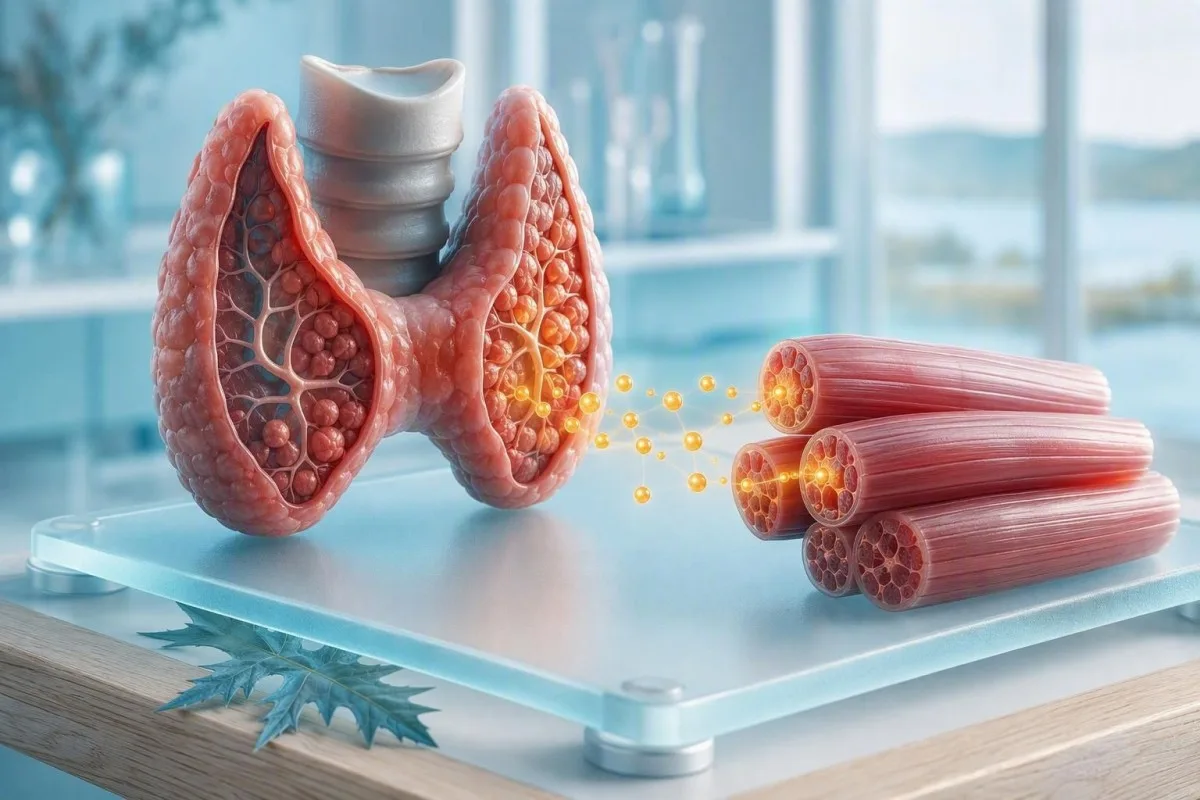
Zems brīvais T3 līmenis var būt saistīts ar nogurumu, sliktu garastāvokli, muskuļu smagumu un aukstuma sajūtu, taču tas biežāk atspoguļo slimību vai enerģijas deficītu, nevis vairogdziedzera mazspēju. Apkārtējie TSH un brīvā T4 rezultāti parasti ir svarīgāki nekā brīvais T3 atsevišķi.


- Zems brīvais T3 bieži neizraisa unikālus simptomus; nogurumam, sliktam garastāvoklim un nepanesamībai pret slodzi ir daudz biežāki izskaidrojumi.
- Tipiska pieaugušo diapazons ir aptuveni 2,0-4,4 pg/mL (3,1-6,8 pmol/L), taču tikai ziņojuma laboratorijas diapazons ir derīgs salīdzinājums.
- Normāls TSH plus normāls brīvais T4 ar zemu brīvo T3 parasti norāda uz slimību, nepietiekamu uzturu vai medikamentu ietekmi, nevis primāru hipotireozi.
- Akūta saslimšana var pazemināt T3 24-48 stundu laikā, samazinot T4 pārvēršanos T3; tas bieži ir atgriezenisks atveseļošanās laikā.
- Kaloriju ierobežojums aptuveni 800-1200 kcal/dienā vai strauja svara zudums var pazemināt T3 bez pastāvīgas vairogdziedzera mazspējas.
- Centrāla hipotireoze tiek aizdomas, kad brīvais T4 ir zems un TSH ir zems, normāls vai tikai nedaudz paaugstināts; tikai brīvais T3 nevar to noteikt.
- Piesardzība ārstēšanā jautājumi: profesionālās vadlīnijas neiesaka rutīnas lietošanu liotironīna, ja ir izolēts zems T3 rādījums ārpus speciālista uzraudzības.
- Atkārtota pārbaude visnoderīgākais ir pēc atveseļošanās, stabila uztura un medikamentu pārskatīšanas, parasti 6–8 nedēļas pēc būtiskām vai nozīmīgām izmaiņām vai pielāgošanas vai atjaunošanas vai uzlabošanas vai korekcijas vai korekcijas vai korekcijas vai korekcijas vai korekcijas vai korekcijas vai korekcijas vai korekcijas vai korekcijas vai korekcijas vai korekcijas vai korekcijas vai korekcijas vai korekcijas vai korekcijas vai korekcijas vai korekcijas vai korekcijas vai korekcijas vai korekcijas vai korekcijas vai korekcijas vai korekcijas vai korekcijas vai korekcijas vai korekcijas vai korekcijas vai korekcijas vai korekcijas vai korekcijas vai korekcijas vai korekcijas vai korekcijas vai korekcijas vai korekcijas vai korekcijas vai korekcijas vai korekcijas vai korekcijas vai korekcijas vai korekcijas vai korekcijas vai korekcijas vai korekcijas vai korekcijas vai korekcijas vai korekcijas vai korekcijas vai korekcijas vai korekcijas vai korekcijas vai korekcijas vai korekcijas vai korekcijas vai korekcijas vai korekcijas vai korekcijas vai korekcijas vai korekcijas vai korekcijas vai korekcijas vai korekcijas vai korekcijas vai korekcijas vai korekcijas vai korekcijas vai korekcijas vai korekcijas vai korekcijas vai korekcijas vai korekcijas vai korekcijas vai korekcijas vai korekcijas vai korekcijas vai korekcijas vai korekcijas vai korekcijas vai korekcijas vai korekcijas vai korekcijas vai korekcijas vai korekcijas vai korekcijas vai korekcijas vai korekcijas vai korekcijas vai korekcijas vai korekcijas vai korekcijas vai korekcijas vai korekcijas vai korekcijas vai korekcijas vai korekcijas vai korekcijas vai korekcijas vai korekcijas vai korekcijas vai korekcijas vai korekcijas vai korekcijas vai korekcijas vai korekcijas vai korekcijas vai korekcijas vai korekcijas vai korekcijas vai korekcijas vai korekcijas vai korekcijas vai korekcijas vai korekcijas vai korekcijas vai korekcijas vai korekcijas vai korekcijas vai korekcijas vai korekcijas vai korekcijas vai korekcijas vai korekcijas vai korekcijas vai korekcijas vai korekcijas vai korekcijas vai korekcijas vai korekcijas vai korekcijas vai korekcijas vai korekcijas vai korekcijas vai korekcijas vai korekcijas vai korekcijas vai korekcijas vai korekcijas vai korekcijas vai korekcijas vai korekcijas vai korekcijas vai korekcijas vai korekcijas vai korekcijas vai korekcijas vai korekcijas vai korekcijas vai korekcijas vai korekcijas vai korekcijas vai korekcijas vai korekcijas vai korekcijas vai korekcijas vai korekcijas vai korekcijas vai korekcijas vai korekcijas vai korekcijas vai korekcijas vai korekcijas vai korekcijas vai korekcijas vai korekcijas vai korekcijas vai korekcijas vai korekcijas vai korekcijas vai korekcijas vai korekcijas vai korekcijas vai korekcijas vai korekcijas vai korekcijas vai korekcijas vai korekcijas vai korekcijas vai korekcijas vai korekcijas vai korekcijas vai korekcijas vai korekcijas vai korekcijas vai korekcijas vai korekcijas vai korekcijas vai korekcijas vai korekcijas vai korekcijas vai korekcijas vai korekcijas vai korekcijas vai korekcijas vai korekcijas vai korekcijas vai korekcijas vai korekcijas vai korekcijas vai korekcijas vai korekcijas vai korekcijas vai korekcijas vai korekcijas vai korekcijas vai korekcijas vai korekcijas vai korekcijas vai korekcijas vai korekcijas vai korekcijas vai korekcijas vai korekcijas vai korekcijas vai korekcijas vai korekcijas vai korekcijas vai korekcijas vai korekcijas vai korekcijas vai korekcijas vai korekcijas vai korekcijas vai korekcijas vai korekcijas vai korekcijas vai korekcijas vai korekcijas vai korekcijas vai korekcijas vai korekcijas vai korekcijas vai korekcijas vai korekcijas vai korekcijas vai korekcijas vai korekcijas vai korekcijas vai korekcijas vai korekcijas vai korekcijas vai korekcijas vai korekcijas vai korekcijas vai korekcijas vai korekcijas vai korekcijas vai korekcijas vai korekcijas vai korekcijas vai korekcijas vai korekcijas vai korekcijas vai korekcijas vai korekcijas vai korekcijas vai korekcijas vai korekcijas vai korekcijas vai korekcijas vai korekcijas vai korekcijas vai korekcijas vai korekcijas vai korekcijas vai korekcijas vai korekcijas vai korekcijas vai korekcijas vai korekcijas vai korekcijas vai korekcijas vai korekcijas vai korekcijas vai korekcijas vai korekcijas vai korekcijas vai korekcijas vai korekcijas vai korekcijas vai korekcijas vai korekcijas vai korekcijas vai korekcijas vai korekcijas vai korekcijas vai korekcijas vai korekcijas vai korekcijas vai korekcijas vai korekcijas vai korekcijas vai korekcijas vai korekcijas vai korekcijas vai korekcijas vai korekcijas vai korekcijas vai korekcijas vai korekcijas vai korekcijas vai korekcijas vai korekcijas vai korekcijas vai korekcijas vai korekcijas vai korekcijas vai korekcijas vai korekcijas vai korekcijas vai korekcijas vai korekcijas vai korekcijas vai korekcijas vai korekcijas vai korekcijas vai korekcijas vai korekcijas vai korekcijas vai korekcijas vai korekcijas vai korekcijas vai korekcijas vai korekcijas vai korekcijas vai korekcijas vai korekcijas vai korekcijas vai korekcijas vai korekcijas vai korekcijas vai korekcijas vai korekcijas vai korekcijas vai korekcijas vai korekcijas vai korekcijas vai korekcijas vai korekcijas vai korekcijas vai korekcijas vai korekcijas vai korekcijas vai korekcijas vai korekcijas vai korekcijas vai korekcijas vai korekcijas vai korekcijas vai korekcijas vai korekcijas vai korekcijas vai korekcijas vai korekcijas vai korekcijas vai korekcijas vai korekcijas vai korekcijas vai korekcijas vai korekcijas vai korekcijas vai korekcijas vai korekcijas vai korekcijas vai korekcijas vai korekcijas vai korekcijas vai korekcijas vai korekcijas vai korekcijas vai korekcijas vai korekcijas vai korekcijas vai korekcijas vai korekcijas vai korekcijas vai korekcijas vai korekcijas vai korekcijas vai korekcijas vai korekcijas vai korekcijas vai korekcijas vai korekcijas vai korekcijas vai korekcijas vai korekcijas vai korekcijas vai korekcijas vai korekcijas vai korekcijas vai korekcijas vai korekcijas vai korekcijas vai korekcijas vai korekcijas vai korekcijas vai korekcijas vai korekcijas vai korekcijas vai korekcijas vai korekcijas vai korekcijas vai korekcijas vai korekcijas vai korekcijas vai korekcijas vai korekcijas vai korekcijas vai korekcijas vai korekcijas vai korekcijas vai korekcijas vai korekcijas vai korekcijas vai korekcijas vai korekcijas vai korekcijas vai korekcijas vai korekcijas vai korekcijas vai korekcijas vai korekcijas vai korekcijas vai korekcijas vai korekcijas vai korekcijas vai korekcijas vai korekcijas vai korekcijas vai korekcijas vai korekcijas vai korekcijas vai korekcijas vai korekcijas vai korekcijas vai korekcijas vai korekcijas vai korekcijas vai korekcijas vai korekcijas vai korekcijas vai korekcijas vai korekcijas vai korekcijas vai korekcijas vai korekcijas vai korekcijas vai korekcijas vai korekcijas vai korekcijas vai korekcijas vai korekcijas vai korekcijas vai korekcijas vai korekcijas vai korekcijas vai korekcijas vai korekcijas vai korekcijas vai korekcijas vai korekcijas vai korekcijas vai korekcijas vai korekcijas vai korekcijas vai korekcijas vai korekcijas vai korekcijas vai korekcijas vai korekcijas vai korekcijas vai korekcijas vai korekcijas vai korekcijas vai korekcijas vai korekcijas vai korekcijas vai korekcijas vai korekcijas vai korekcijas vai korekcijas vai korekcijas vai korekcijas vai korekcijas vai korekcijas vai korekcijas vai korekcijas vai korekcijas vai korekcijas vai korekcijas vai korekcijas vai korekcijas vai korekcijas vai korekcijas vai korekcijas vai korekcijas vai korekcijas vai korekcijas vai korekcijas vai korekcijas vai korekcijas vai korekcijas vai korekcijas vai korekcijas vai korekcijas vai korekcijas vai korekcijas vai korekcijas vai korekcijas vai korekcijas vai korekcijas vai korekcijas vai korekcijas vai korekcijas vai korekcijas vai korekcijas vai korekcijas vai korekcijas vai korekcijas vai korekcijas vai korekcijas vai korekcijas vai korekcijas vai korekcijas vai korekcijas vai korekcijas vai korekcijas vai korekcijas vai korekcijas vai korekcijas vai korekcijas vai korekcijas vai korekcijas vai korekcijas vai korekcijas vai korekcijas vai korekcijas vai korekcijas vai korekcijas vai korekcijas vai korekcijas vai korekcijas vai korekcijas vai korekcijas vai korekcijas vai korekcijas vai korekcijas vai korekcijas vai korekcijas vai korekcijas vai korekcijas vai korekcijas vai korekcijas vai korekcijas vai korekcijas vai korekcijas vai korekcijas vai korekcijas vai korekcijas vai korekcijas vai korekcijas vai korekcijas vai korekcijas vai korekcijas vai korekcijas vai korekcijas vai korekcijas vai korekcijas vai korekcijas vai korekcijas vai korekcijas vai korekcijas vai korekcijas vai korekcijas vai korekcijas vai korekcijas vai korekcijas vai korekcijas vai korekcijas vai korekcijas vai korekcijas vai korekcijas vai korekcijas vai korekcijas vai korekcijas vai korekcijas vai korekcijas vai korekcijas vai korekcijas vai korekcijas vai korekcijas vai korekcijas vai korekcijas vai korekcijas vai korekcijas vai korekcijas vai korekcijas vai korekcijas vai korekcijas vai korekcijas vai korekcijas vai korekcijas vai korekcijas vai korekcijas vai korekcijas vai korekcijas vai korekcijas vai korekcijas vai korekcijas vai korekcijas vai korekcijas vai korekcijas vai korekcijas vai korekcijas vai korekcijas vai korekcijas vai korekcijas vai korekcijas vai korekcijas vai korekcijas vai korekcijas vai korekcijas vai korekcijas vai korekcijas vai korekcijas vai korekcijas vai korekcijas vai korekcijas vai korekcijas vai korekcijas vai korekcijas vai korekcijas vai korekcijas vai korekcijas vai korekcijas vai korekcijas vai korekcijas vai korekcijas vai korekcijas vai korekcijas vai korekcijas vai korekcijas vai korekcijas vai korekcijas vai korekcijas vai korekcijas vai korekcijas vai korekcijas vai korekcijas vai korekcijas vai korekcijas vai korekcijas vai korekcijas vai korekcijas vai korekcijas vai korekcijas vai korekcijas vai korekcijas vai korekcijas vai korekcijas vai korekcijas vai korekcijas vai korekcijas vai korekcijas vai korekcijas vai korekcijas vai korekcijas vai korekcijas vai korekcijas vai korekcijas vai korekcijas vai korekcijas vai korekcijas vai korekcijas vai korekcijas vai korekcijas vai korekcijas vai korekcijas vai korekcijas vai korekcijas vai korekcijas vai korekcijas vai korekcijas vai korekcijas vai korekcijas vai korekcijas vai korekcijas vai korekcijas vai korekcijas vai korekcijas vai korekcijas vai korekcijas vai korekcijas vai korekcijas vai korekcijas vai korekcijas vai korekcijas vai korekcijas vai korekcijas vai korekcijas vai korekcijas vai korekcijas vai korekcijas vai korekcijas vai korekcijas vai korekcijas vai korekcijas vai korekcijas vai korekcijas vai korekcijas vai korekcijas vai korekcijas vai korekcijas vai korekcijas vai korekcijas vai korekcijas vai korekcijas vai korekcijas vai korekcijas vai korekcijas vai korekcijas vai korekcijas vai korekcijas vai korekcijas vai korekcijas vai korekcijas vai korekcijas vai korekcijas vai korekcijas vai korekcijas vai korekcijas vai korekcijas vai korekcijas vai korekcijas vai korekcijas vai korekcijas vai korekcijas vai korekcijas vai korekcijas vai korekcijas vai korekcijas vai korekcijas vai korekcijas vai korekcijas vai korekcijas vai korekcijas vai korekcijas vai korekcijas vai korekcijas vai korekcijas vai korekcijas vai korekcijas vai korekcijas vai korekcijas vai korekcijas vai korekcijas vai korekcijas vai korekcijas vai korekcijas vai korekcijas vai korekcijas vai korekcijas vai korekcijas vai korekcijas vai korekcijas vai korekcijas vai korekcijas vai korekcijas vai korekcijas vai korekcijas vai korekcijas vai korekcijas vai korekcijas vai korekcijas vai korekcijas vai korekcijas vai korekcijas vai korekcijas vai korekcijas vai korekcijas vai korekcijas vai korekcijas vai korekcijas vai korekcijas vai korekcijas vai korekcijas vai korekcijas vai korekcijas vai korekcijas vai korekcijas vai korekcijas vai korekcijas vai korekcijas vai korekcijas vai korekcijas vai korekcijas vai korekcijas vai korekcijas vai korekcijas vai korekcijas vai korekcijas vai korekcijas vai korekcijas vai korekcijas vai korekcijas vai korekcijas vai korekcijas vai korekcijas vai korekcijas vai korekcijas vai korekcijas vai korekcijas vai korekcijas vai korekcijas vai korekcijas vai korekcijas vai korekcijas vai korekcijas vai korekcijas vai korekcijas vai korekcijas vai korekcijas vai korekcijas vai korekcijas vai korekcijas vai korekcijas vai korekcijas vai korekcijas vai korekcijas vai korekcijas vai korekcijas vai korekcijas vai korekcijas vai korekcijas vai korekcijas vai korekcijas vai korekcijas vai korekcijas vai korekcijas vai korekcijas vai korekcijas vai korekcijas vai korekcijas vai korekcijas vai korekcijas vai korekcijas vai korekcijas vai korekcijas vai korekcijas vai korekcijas vai korekcijas vai korekcijas vai korekcijas vai korekcijas vai korekcijas vai korekcijas vai korekcijas vai korekcijas vai korekcijas vai korekcijas vai korekcijas vai korekcijas vai korekcijas vai korekcijas vai korekcijas vai korekcijas vai korekcijas vai korekcijas vai korekcijas vai korekcijas vai korekcijas vai korekcijas vai korekcijas vai korekcijas vai korekcijas vai korekcijas vai korekcijas vai korekcijas vai korekcijas vai korekcijas vai korekcijas vai korekcijas vai korekcijas vai korekcijas vai korekcijas vai korekcijas vai korekcijas vai korekcijas vai korekcijas vai korekcijas vai korekcijas vai korekcijas vai korekcijas vai korekcijas vai korekcijas vai korekcijas vai korekcijas vai korekcijas vai korekcijas vai korekcijas vai korekcijas vai korekcijas vai korekcijas vai korekcijas vai korekcijas vai korekcijas vai korekcijas vai korekcijas vai korekcijas vai korekcijas vai korekcijas vai korekcijas vai korekcijas vai korekcijas vai korekcijas vai korekcijas vai korekcijas vai korekcijas vai korekcijas vai korekcijas vai korekcijas vai korekcijas vai korekcijas vai korekcijas vai korekcijas vai korekcijas vai korekcijas vai korekcijas vai korekcijas vai korekcijas vai korekcijas vai korekcijas vai korekcijas vai korekcijas vai korekcijas vai korekcijas vai korekcijas vai korekcijas vai korekcijas vai korekcijas vai korekcijas vai korekcijas vai korekcijas vai korekcijas vai korekcijas vai korekcijas vai korekcijas vai korekcijas vai korekcijas vai korekcijas vai korekcijas vai korekcijas vai korekcijas vai korekcijas vai korekcijas vai korekcijas vai korekcijas vai korekcijas vai korekcijas vai korekcijas vai korekcijas vai korekcijas vai korekcijas vai korekcijas vai korekcijas vai korekcijas vai korekcijas vai korekcijas vai korekcijas vai korekcijas vai korekcijas vai korekcijas vai korekcijas vai korekcijas vai korekcijas vai korekcijas vai korekcijas vai korekcijas vai korekcijas vai korekcijas vai korekcijas vai korekcijas vai korekcijas vai korekcijas vai korekcijas vai korekcijas vai korekcijas vai korekcijas vai korekcijas vai korekcijas vai korekcijas vai korekcijas vai korekcijas vai korekcijas vai korekcijas vai korekcijas vai korekcijas vai korekcijas vai korekcijas vai korekcijas vai korekcijas vai korekcijas vai korekcijas vai korekcijas vai korekcijas vai korekcijas vai korekcijas vai korekcijas vai korekcijas vai korekcijas vai korekcijas vai korekcijas vai korekcijas vai korekcijas vai korekcijas vai korekcijas vai korekcijas vai korekcijas vai korekcijas vai korekcijas vai korekcijas vai korekcijas vai korekcijas vai korekcijas vai korekcijas vai korekcijas vai korekcijas vai korekcijas vai korekcijas vai korekcijas vai korekcijas vai korekcijas vai korekcijas vai korekcijas vai korekcijas vai korekcijas vai korekcijas vai korekcijas vai korekcijas vai korekcijas vai korekcijas vai korekcijas vai korekcijas vai korekcijas vai korekcijas vai korekcijas vai korekcijas vai korekcijas vai korekcijas vai korekcijas vai korekcijas vai korekcijas vai korekcijas vai korekcijas vai korekcijas vai korekcijas vai korekcijas vai korekcijas vai korekcijas vai korekcijas vai korekcijas vai korekcijas vai korekcijas vai korekcijas vai korekcijas vai korekcijas vai korekcijas vai korekcijas vai korekcijas vai korekcijas vai korekcijas vai korekcijas vai korekcijas vai korekcijas vai korekcijas vai korekcijas vai korekcijas vai korekcijas vai korekcijas vai korekcijas vai korekcijas vai korekcijas vai korekcijas vai korekcijas vai korekcijas vai korekcijas vai korekcijas vai korekcijas vai korekcijas vai korekcijas vai korekcijas vai korekcijas vai korekcijas vai korekcijas vai korekcijas vai korekcijas vai korekcijas vai korekcijas vai korekcijas vai korekcijas vai korekcijas vai korekcijas vai korekcijas vai korekcijas vai korekcijas vai korekcijas vai korekcijas vai korekcijas vai korekcijas vai korekcijas vai korekcijas vai korekcijas vai korekcijas vai korekcijas vai korekcijas vai korekcijas vai korekcijas vai korekcijas vai korekcijas vai korekcijas vai korekcijas vai korekcijas vai korekcijas vai korekcijas vai korekcijas vai korekcijas vai korekcijas vai korekcijas vai korekcijas vai korekcijas vai korekcijas vai korekcijas vai korekcijas vai korekcijas vai korekcijas vai korekcijas vai korekcijas vai korekcijas vai korekcijas vai korekcijas vai korekcijas vai korekcijas vai korekcijas vai korekcijas vai korekcijas vai korekcijas vai korekcijas vai korekcijas vai korekcijas vai korekcijas vai korekcijas vai korekcijas vai korekcijas vai korekcijas vai korekcijas vai korekcijas vai korekcijas vai korekcijas vai korekcijas vai korekcijas vai korekcijas vai korekcijas vai korekcijas vai korekcijas vai korekcijas vai korekcijas vai korekcijas vai korekcijas vai korekcijas vai korekcijas vai korekcijas vai korekcijas vai korekcijas vai korekcijas vai korekcijas vai korekcijas vai korekcijas vai korekcijas vai korekcijas vai korekcijas vai korekcijas vai korekcijas vai korekcijas vai korekcijas vai korekcijas vai korekcijas vai korekcijas vai korekcijas vai korekcijas vai korekcijas vai korekcijas vai korekcijas vai korekcijas vai korekcijas vai korekcijas vai korekcijas vai korekcijas vai korekcijas vai korekcijas vai korekcijas vai korekcijas vai korekcijas vai korekcijas vai korekcijas vai korekcijas vai korekcijas vai korekcijas vai korekcijas vai korekcijas vai korekcijas vai korekcijas vai korekcijas vai korekcijas vai korekcijas vai korekcijas vai korekcijas vai korekcijas vai korekcijas vai korekcijas vai korekcijas vai korekcijas vai korekcijas vai korekcijas vai korekcijas vai korekcijas vai korekcijas vai korekcijas vai korekcijas vai korekcijas vai korekcijas vai korekcijas vai korekcijas vai korekcijas vai korekcijas vai korekcijas vai korekcijas vai korekcijas vai korekcijas vai korekcijas vai korekcijas vai korekcijas vai korekcijas vai korekcijas vai korekcijas vai korekcijas vai korekcijas vai korekcijas vai korekcijas vai korekcijas vai korekcijas vai korekcijas vai korekcijas vai korekcijas vai korekcijas vai korekcijas vai korekcijas vai korekcijas vai korekcijas vai korekcijas vai korekcijas vai korekcijas vai korekcijas vai korekcijas vai korekcijas vai korekcijas vai korekcijas vai korekcijas vai korekcijas vai korekcijas vai korekcijas vai korekcijas vai korekcijas vai korekcijas vai korekcijas vai korekcijas vai korekcijas vai korekcijas vai korekcijas vai korekcijas vai korekcijas vai korekcijas vai korekcijas vai korekcijas vai korekcijas vai korekcijas vai korekcijas vai korekcijas vai korekcijas vai korekcijas vai korekcijas vai korekcijas vai korekcijas vai korekcijas vai korekcijas vai korekcijas vai korekcijas vai korekcijas vai korekcijas vai korekcijas vai korekcijas vai korekcijas vai korekcijas vai korekcijas vai korekcijas vai korekcijas vai korekcijas vai korekcijas vai korekcijas vai korekcijas vai korekcijas vai korekcijas vai korekcijas vai korekcijas vai korekcijas vai korekcijas vai korekcijas vai korekcijas vai korekcijas vai korekcijas vai korekcijas vai korekcijas vai korekcijas vai korekcijas vai korekcijas vai korekcijas vai korekcijas vai korekcijas vai korekcijas vai korekcijas vai korekcijas vai korekcijas vai korekcijas vai korekcijas vai korekcijas vai korekcijas vai korekcijas vai korekcijas vai korekcijas vai korekcijas vai korekcijas vai korekcijas vai korekcijas vai korekcijas vai korekcijas vai korekcijas vai korekcijas vai korekcijas vai korekcijas vai korekcijas vai korekcijas vai korekcijas vai korekcijas vai korekcijas vai korekcijas vai korekcijas vai korekcijas vai korekcijas vai korekcijas vai korekcijas vai korekcijas vai korekcijas vai korekcijas vai korekcijas vai korekcijas vai korekcijas vai korekcijas vai korekcijas vai korekcijas vai korekcijas vai korekcijas vai korekcijas vai korekcijas vai korekcijas vai korekcijas vai korekcijas vai korekcijas vai korekcijas vai korekcijas vai korekcijas vai korekcijas vai korekcijas vai korekcijas vai korekcijas vai korekcijas vai korekcijas vai korekcijas vai korekcijas vai korekcijas vai korekcijas vai korekcijas vai korekcijas vai korekcijas vai korekcijas vai korekcijas vai korekcijas vai korekcijas vai korekcijas vai korekcijas vai korekcijas vai korekcijas vai korekcijas vai korekcijas vai korekcijas vai korekcijas vai korekcijas vai korekcijas vai korekcijas vai korekcijas vai korekcijas vai korekcijas vai korekcijas vai korekcijas vai korekcijas vai korekcijas vai korekcijas vai korekcijas vai korekcijas vai korekcijas vai korekcijas vai korekcijas vai korekcijas vai korekcijas vai korekcijas vai korekcijas vai korekcijas vai korekcijas vai korekcijas vai korekcijas vai korekcijas vai korekcijas vai korekcijas vai korekcijas vai korekcijas vai korekcijas vai korekcijas vai korekcijas vai korekcijas vai korekcijas vai korekcijas vai korekcijas vai korekcijas vai korekcijas vai korekcijas vai korekcijas vai korekcijas vai korekcijas vai korekcijas vai korekcijas vai korekcijas vai korekcijas vai korekcijas vai korekcijas vai korekcijas vai korekcijas vai korekcijas vai korekcijas vai korekcijas vai korekcijas vai korekcijas vai korekcijas vai korekcijas vai korekcijas vai korekcijas vai korekcijas vai korekcijas vai korekcijas vai korekcijas vai korekcijas vai korekcijas vai korekcijas vai korekcijas vai korekcijas vai korekcijas vai korekcijas vai korekcijas vai korekcijas vai korekcijas vai korekcijas vai korekcijas vai korekcijas vai korekcijas vai korekcijas vai korekcijas vai korekcijas vai korekcijas vai korekcijas vai korekcijas vai korekcijas vai korekcijas vai korekcijas vai korekcijas vai korekcijas vai korekcijas vai korekcijas vai korekcijas vai korekcijas vai korekcijas vai korekcijas vai korekcijas vai korekcijas vai korekcijas vai korekcijas vai korekcijas vai korekcijas vai korekcijas vai korekcijas vai korekcijas vai korekcijas vai korekcijas vai korekcijas vai korekcijas vai korekcijas vai korekcijas vai korekcijas vai korekcijas vai korekcijas vai korekcijas vai korekcijas vai korekcijas vai korekcijas vai korekcijas vai korekcijas vai korekcijas vai korekcijas vai korekcijas vai korekcijas vai korekcijas vai korekcijas vai korekcijas vai korekcijas vai korekcijas vai korekcijas vai korekcijas vai korekcijas vai korekcijas vai korekcijas vai korekcijas vai korekcijas vai korekcijas vai korekcijas vai korekcijas vai korekcijas vai korekcijas vai korekcijas vai korekcijas vai korekcijas vai korekcijas vai korekcijas vai korekcijas vai korekcijas vai korekcijas vai korekcijas vai korekcijas vai korekcijas vai korekcijas vai korekcijas vai korekcijas vai korekcijas vai korekcijas vai korekcijas vai korekcijas vai korekcijas vai korekcijas vai korekcijas vai korekcijas vai korekcijas vai korekcijas vai korekcijas vai korekcijas vai korekcijas vai korekcijas vai korekcijas vai korekcijas vai korekcijas vai korekcijas vai korekcijas vai korekcijas vai korekcijas vai korekcijas vai korekcijas vai korekcijas vai korekcijas vai korekcijas vai korekcijas vai korekcijas vai korekcijas vai korekcijas vai korekcijas vai korekcijas vai korekcijas vai korekcijas vai korekcijas vai korekcijas vai korekcijas vai korekcijas vai korekcijas vai korekcijas vai korekcijas vai korekcijas vai korekcijas vai korekcijas vai korekcijas vai korekcijas vai korekcijas vai korekcijas vai korekcijas vai korekcijas vai korekcijas vai korekcijas vai korekcijas vai korekcijas vai korekcijas vai korekcijas vai korekcijas vai korekcijas vai korekcijas vai korekcijas vai korekcijas vai korekcijas vai korekcijas vai korekcijas vai korekcijas vai korekcijas vai korekcijas vai korekcijas vai korekcijas vai korekcijas vai korekcijas vai korekcijas vai korekcijas vai korekcijas vai korekcijas vai korekcijas vai korekcijas vai korekcijas vai korekcijas vai korekcijas vai korekcijas vai korekcijas vai korekcijas vai korekcijas vai korekcijas vai korekcijas vai korekcijas vai korekcijas vai korekcijas vai korekcijas vai korekcijas vai korekcijas vai korekcijas vai korekcijas vai korekcijas vai korekcijas vai korekcijas vai korekcijas vai korekcijas vai korekcijas vai korekcijas vai korekcijas vai korekcijas vai korekcijas vai korekcijas vai korekcijas vai korekcijas vai korekcijas vai korekcijas vai korekcijas vai korekcijas vai korekcijas vai korekcijas vai korekcijas vai korekcijas vai korekcijas vai korekcijas vai korekcijas vai korekcijas vai korekcijas vai korekcijas vai korekcijas vai korekcijas vai korekcijas vai korekcijas vai korekcijas vai korekcijas vai korekcijas vai korekcijas vai korekcijas vai korekcijas vai korekcijas vai korekcijas vai korekcijas vai korekcijas vai korekcijas vai korekcijas vai korekcijas vai korekcijas vai korekcijas vai korekcijas vai korekcijas vai korekcijas vai korekcijas vai korekcijas vai korekcijas vai korekcijas vai korekcijas vai korekcijas vai korekcijas vai korekcijas vai korekcijas vai korekcijas vai korekcijas vai korekcijas vai korekcijas vai korekcijas vai korekcijas vai korekcijas vai korekcijas vai korekcijas vai korekcijas vai korekcijas vai korekcijas vai korekcijas vai korekcijas vai korekcijas vai korekcijas vai korekcijas vai korekcijas vai korekcijas vai korekcijas vai korekcijas vai korekcijas vai korekcijas vai korekcijas vai korekcijas vai korekcijas vai korekcijas vai korekcijas vai korekcijas vai korekcijas vai korekcijas vai korekcijas vai korekcijas vai korekcijas vai korekcijas vai korekcijas vai korekcijas vai korekcijas vai korekcijas vai korekcijas vai korekcijas vai korekcijas vai korekcijas vai korekcijas vai korekcijas vai korekcijas vai korekcijas vai korekcijas vai korekcijas vai korekcijas vai korekcijas vai korekcijas vai korekcijas vai korekcijas vai korekcijas vai korekcijas vai korekcijas vai korekcijas vai korekcijas vai korekcijas vai korekcijas vai korekcijas vai korekcijas vai korekcijas vai korekcijas vai korekcijas vai korekcijas vai korekcijas vai korekcijas vai korekcijas vai korekcijas vai korekcijas vai korekcijas vai korekcijas vai korekcijas vai korekcijas vai korekcijas vai korekcijas vai korekcijas vai korekcijas vai korekcijas vai korekcijas vai korekcijas vai korekcijas vai korekcijas vai korekcijas vai korekcijas vai korekcijas vai korekcijas vai korekcijas vai korekcijas vai korekcijas vai korekcijas vai korekcijas vai korekcijas vai korekcijas vai korekcijas vai korekcijas vai korekcijas vai korekcijas vai korekcijas vai korekcijas vai korekcijas vai korekcijas vai korekcijas vai korekcijas vai korekcijas vai korekcijas vai korekcijas vai korekcijas vai korekcijas vai korekcijas vai korekcijas vai korekcijas vai korekcijas vai korekcijas vai korekcijas vai korekcijas vai korekcijas vai korekcijas vai korekcijas vai korekcijas vai korekcijas vai korekcijas vai korekcijas vai korekcijas vai korekcijas vai korekcijas vai korekcijas vai korekcijas vai korekcijas vai korekcijas vai korekcijas vai korekcijas vai korekcijas vai korekcijas vai korekcijas vai korekcijas vai korekcijas vai korekcijas vai korekcijas vai korekcijas vai korekcijas vai korekcijas vai korekcijas vai korekcijas vai korekcijas vai korekcijas vai korekcijas vai korekcijas vai korekcijas vai korekcijas vai korekcijas vai korekcijas vai korekcijas vai korekcijas vai korekcijas vai korekcijas vai korekcijas vai korekcijas vai korekcijas vai korekcijas vai korekcijas vai korekcijas vai korekcijas vai korekcijas vai korekcijas vai korekcijas vai korekcijas vai korekcijas vai korekcijas vai korekcijas vai korekcijas vai korekcijas vai korekcijas vai korekcijas vai korekcijas vai korekcijas vai korekcijas vai korekcijas vai korekcijas vai korekcijas vai korekcijas vai korekcijas vai korekcijas vai korekcijas vai korekcijas vai korekcijas vai korekcijas vai korekcijas vai korekcijas vai korekcijas vai korekcijas vai korekcijas vai korekcijas vai korekcijas vai korekcijas vai korekcijas vai korekcijas vai korekcijas vai korekcijas vai korekcijas vai korekcijas vai korekcijas vai korekcijas vai korekcijas vai korekcijas vai korekcijas vai korekcijas vai korekcijas vai korekcijas vai korekcijas vai korekcijas vai korekcijas vai korekcijas vai korekcijas vai korekcijas vai korekcijas vai korekcijas vai korekcijas vai korekcijas vai korekcijas vai korekcijas vai korekcijas vai korekcijas vai korekcijas vai korekcijas vai korekcijas vai korekcijas vai korekcijas vai korekcijas vai korekcijas vai korekcijas vai korekcijas vai korekcijas vai korekcijas vai korekcijas vai korekcijas vai korekcijas vai korekcijas vai korekcijas vai korekcijas vai korekcijas vai korekcijas vai korekcijas vai korekcijas vai korekcijas vai korekcijas vai korekcijas vai korekcijas vai korekcijas vai korekcijas vai korekcijas vai korekcijas vai korekcijas vai korekcijas vai korekcijas vai korekcijas vai korekcijas vai korekcijas vai korekcijas vai korekcijas vai korekcijas vai korekcijas vai korekcijas vai korekcijas vai korekcijas vai korekcijas vai korekcijas vai korekcijas vai korekcijas vai korekcijas vai korekcijas vai korekcijas vai korekcijas vai korekcijas vai korekcijas vai korekcijas vai korekcijas vai korekcijas vai korekcijas vai korekcijas vai korekcijas vai korekcijas vai korekcijas vai korekcijas vai korekcijas vai korekcijas vai korekcijas vai korekcijas vai korekcijas vai korekcijas vai korekcijas vai korekcijas vai korekcijas vai korekcijas vai korekcijas vai korekcijas vai korekcijas vai korekcijas vai korekcijas vai korekcijas vai korekcijas vai korekcijas vai korekcijas vai korekcijas vai korekcijas vai korekcijas vai korekcijas vai korekcijas vai korekcijas vai korekcijas vai korekcijas vai korekcijas vai korekcijas vai korekcijas vai korekcijas vai korekcijas vai korekcijas vai korekcijas vai korekcijas vai korekcijas vai korekcijas vai korekcijas vai korekcijas vai korekcijas vai korekcijas vai korekcijas vai korekcijas vai korekcijas vai korekcijas vai korekcijas vai korekcijas vai korekcijas vai korekcijas vai korekcijas vai korekcijas vai korekcijas vai korekcijas vai korekcijas vai korekcijas vai korekcijas vai korekcijas vai korekcijas vai korekcijas vai korekcijas vai korekcijas vai korekcijas vai korekcijas vai korekcijas vai korekcijas vai korekcijas vai korekcijas vai korekcijas vai korekcijas vai korekcijas vai korekcijas vai korekcijas vai korekcijas vai korekcijas vai korekcijas vai korekcijas vai korekcijas vai korekcijas vai korekcijas vai korekcijas vai korekcijas vai korekcijas vai korekcijas vai korekcijas vai korekcijas vai korekcijas vai korekcijas vai korekcijas vai korekcijas vai korekcijas vai korekcijas vai korekcijas vai korekcijas vai korekcijas vai korekcijas vai korekcijas vai korekcijas vai korekcijas vai korekcijas vai korekcijas vai korekcijas vai korekcijas vai korekcijas vai korekcijas vai korekcijas vai korekcijas vai korekcijas vai korekcijas vai korekcijas vai korekcijas vai korekcijas vai korekcijas vai korekcijas vai korekcijas vai korekcijas vai korekcijas vai korekcijas vai korekcijas vai korekcijas vai korekcijas vai korekcijas vai korekcijas vai korekcijas vai korekcijas vai korekcijas vai korekcijas vai korekcijas vai korekcijas vai korekcijas vai korekcijas vai korekcijas vai korekcijas vai korekcijas vai korekcijas vai korekcijas vai korekcijas vai korekcijas vai korekcijas vai korekcijas vai korekcijas vai korekcijas vai korekcijas vai korekcijas vai korekcijas vai korekcijas vai korekcijas vai korekcijas vai korekcijas vai korekcijas vai korekcijas vai korekcijas vai korekcijas vai korekcijas vai korekcijas vai korekcijas vai korekcijas vai korekcijas vai korekcijas vai korekcijas vai korekcijas vai korekcijas vai korekcijas vai korekcijas vai korekcijas vai korekcijas vai korekcijas vai korekcijas vai korekcijas vai korekcijas vai korekcijas vai korekcijas vai korekcijas vai korekcijas vai korekcijas vai korekcijas vai korekcijas vai korekcijas vai korekcijas vai korekcijas vai korekcijas vai korekcijas vai korekcijas vai korekcijas vai korekcijas vai korekcijas vai korekcijas vai korekcijas vai korekcijas vai korekcijas vai korekcijas vai korekcijas vai korekcijas vai korekcijas vai korekcijas vai korekcijas vai korekcijas vai korekcijas vai korekcijas vai korekcijas vai korekcijas vai korekcijas vai korekcijas vai korekcijas vai korekcijas vai korekcijas vai korekcijas vai korekcijas vai korekcijas vai korekcijas vai korekcijas vai korekcijas vai korekcijas vai korekcijas vai korekcijas vai korekcijas vai korekcijas vai korekcijas vai korekcijas vai korekcijas vai korekcijas vai korekcijas vai korekcijas vai korekcijas vai korekcijas vai korekcijas vai korekcijas vai korekcijas vai korekcijas vai korekcijas vai korekcijas vai korekcijas vai korekcijas vai korekcijas vai korekcijas vai korekcijas vai korekcijas vai korekcijas vai korekcijas vai korekcijas vai korekcijas vai korekcijas vai korekcijas vai korekcijas vai korekcijas vai korekcijas vai korekcijas vai korekcijas vai korekcijas vai korekcijas vai korekcijas vai korekcijas vai korekcijas vai korekcijas vai korekcijas vai korekcijas vai korekcijas vai korekcijas vai korekcijas vai korekcijas vai korekcijas vai korekcijas vai korekcijas vai korekcijas vai korekcijas vai korekcijas vai korekcijas vai korekcijas vai korekcijas vai korekcijas vai korekcijas vai korekcijas vai korekcijas vai korekcijas vai korekcijas vai korekcijas vai korekcijas vai korekcijas vai korekcijas vai korekcijas vai korekcijas vai korekcijas vai korekcijas vai korekcijas vai korekcijas vai korekcijas vai korekcijas vai korekcijas vai korekcijas vai korekcijas vai korekcijas vai korekcijas vai korekcijas vai korekcijas vai korekcijas vai korekcijas vai korekcijas vai korekcijas vai korekcijas vai korekcijas vai korekcijas vai korekcijas vai korekcijas vai korekcijas vai korekcijas vai korekcijas vai korekcijas vai korekcijas vai korekcijas vai korekcijas vai korekcijas vai korekcijas vai korekcijas vai korekcijas vai korekcijas vai korekcijas vai korekcijas vai korekcijas vai korekcijas vai korekcijas vai korekcijas vai korekcijas vai korekcijas vai korekcijas vai korekcijas vai korekcijas vai korekcijas vai korekcijas vai korekcijas vai korekcijas vai korekcijas vai korekcijas vai korekcijas vai korekcijas vai korekcijas vai korekcijas vai korekcijas vai korekcijas vai korekcijas vai korekcijas vai korekcijas vai korekcijas vai korekcijas vai korekcijas vai korekcijas vai korekcijas vai korekcijas vai korekcijas vai korekcijas vai korekcijas vai korekcijas vai korekcijas vai korekcijas vai korekcijas vai korekcijas vai korekcijas vai korekcijas vai korekcijas vai korekcijas vai korekcijas vai korekcijas vai korekcijas vai korekcijas vai korekcijas vai korekcijas vai korekcijas vai korekcijas vai korekcijas vai korekcijas vai korekcijas vai korekcijas vai korekcijas vai korekcijas vai korekcijas vai korekcijas vai korekcijas vai korekcijas vai korekcijas vai korekcijas vai korekcijas vai korekcijas vai korekcijas vai korekcijas vai korekcijas vai korekcijas vai korekcijas vai korekcijas vai korekcijas vai korekcijas vai korekcijas vai korekcijas vai korekcijas vai korekcijas vai korekcijas vai korekcijas vai korekcijas vai korekcijas vai korekcijas vai korekcijas vai korekcijas vai korekcijas vai korekcijas vai korekcijas vai korekcijas vai korekcijas vai korekcijas vai korekcijas vai korekcijas vai korekcijas vai korekcijas vai korekcijas vai korekcijas vai korekcijas vai korekcijas vai korekcijas vai korekcijas vai korekcijas vai korekcijas vai korekcijas vai korekcijas vai korekcijas vai korekcijas vai korekcijas vai korekcijas vai korekcijas vai korekcijas vai korekcijas vai korekcijas vai korekcijas vai korekcijas vai korekcijas vai korekcijas vai korekcijas vai korekcijas vai korekcijas vai korekcijas vai korekcijas vai korekcijas vai korekcijas vai korekcijas vai korekcijas vai korekcijas vai korekcijas vai korekcijas vai korekcijas vai korekcijas vai korekcijas vai korekcijas vai korekcijas vai korekcijas vai korekcijas vai korekcijas vai korekcijas vai korekcijas vai korekcijas vai korekcijas vai korekcijas vai korekcijas vai korekcijas vai korekcijas vai korekcijas vai korekcijas vai korekcijas vai korekcijas vai korekcijas vai korekcijas vai korekcijas vai korekcijas vai korekcijas vai korekcijas vai korekcijas vai korekcijas vai korekcijas vai korekcijas vai korekcijas vai korekcijas vai korekcijas vai korekcijas vai korekcijas vai korekcijas vai korekcijas vai korekcijas vai korekcijas vai korekcijas vai korekcijas vai korekcijas vai korekcijas vai korekcijas vai korekcijas vai korekcijas vai korekcijas vai korekcijas vai korekcijas vai korekcijas vai korekcijas vai korekcijas vai korekcijas vai korekcijas vai korekcijas vai korekcijas vai korekcijas vai korekcijas vai korekcijas vai korekcijas vai korekcijas vai korekcijas vai korekcijas vai korekcijas vai korekcijas vai korekcijas vai korekcijas vai korekcijas vai korekcijas vai korekcijas vai korekcijas vai korekcijas vai korekcijas vai korekcijas vai korekcijas vai korekcijas vai korekcijas vai korekcijas vai korekcijas vai korekcijas vai korekcijas vai korekcijas vai korekcijas vai korekcijas vai korekcijas vai korekcijas vai korekcijas vai korekcijas vai korekcijas vai korekcijas vai korekcijas vai korekcijas vai korekcijas vai korekcijas vai korekcijas vai korekcijas vai korekcijas vai korekcijas vai korekcijas vai korekcijas vai korekcijas vai korekcijas vai korekcijas vai korekcijas vai korekcijas vai korekcijas vai korekcijas vai korekcijas vai korekcijas vai korekcijas vai korekcijas vai korekcijas vai korekcijas vai korekcijas vai korekcijas vai korekcijas vai korekcijas vai korekcijas vai korekcijas vai korekcijas vai korekcijas vai korekcijas vai korekcijas vai korekcijas vai korekcijas vai korekcijas vai korekcijas vai korekcijas vai korekcijas vai korekcijas vai korekcijas vai korekcijas vai korekcijas vai korekcijas vai korekcijas vai korekcijas vai korekcijas vai korekcijas vai korekcijas vai korekcijas vai korekcijas vai korekcijas vai korekcijas vai korekcijas vai korekcijas vai korekcijas vai korekcijas vai korekcijas vai korekcijas vai korekcijas vai korekcijas vai korekcijas vai korekcijas vai korekcijas vai korekcijas vai korekcijas vai korekcijas vai korekcijas vai korekcijas vai korekcijas vai korekcijas vai korekcijas vai korekcijas vai korekcijas vai korekcijas vai korekcijas vai korekcijas vai korekcijas vai korekcijas vai korekcijas vai korekcijas vai korekcijas vai korekcijas vai korekcijas vai korekcijas vai korekcijas vai korekcijas vai korekcijas vai korekcijas vai korekcijas vai korekcijas vai korekcijas vai korekcijas vai korekcijas vai korekcijas vai korekcijas vai korekcijas vai korekcijas vai korekcijas vai korekcijas vai korekcijas vai korekcijas vai korekcijas vai korekcijas vai korekcijas vai korekcijas vai korekcijas vai korekcijas vai korekcijas vai korekcijas vai korekcijas vai korekcijas vai korekcijas vai korekcijas vai korekcijas vai korekcijas vai korekcijas vai korekcijas vai korekcijas vai korekcijas vai korekcijas vai korekcijas vai korekcijas vai korekcijas vai korekcijas vai korekcijas vai korekcijas vai korekcijas vai korekcijas vai korekcijas vai korekcijas vai korekcijas vai korekcijas vai korekcijas vai korekcijas vai korekcijas vai korekcijas vai korekcijas vai korekcijas vai korekcijas vai korekcijas vai korekcijas vai korekcijas vai korekcijas vai korekcijas vai korekcijas vai korekcijas vai korekcijas vai korekcijas vai korekcijas vai korekcijas vai korekcijas vai korekcijas vai korekcijas vai korekcijas vai korekcijas vai korekcijas vai korekcijas vai korekcijas vai korekcijas vai korekcijas vai korekcijas vai korekcijas vai korekcijas vai korekcijas vai korekcijas vai korekcijas vai korekcijas vai korekcijas vai korekcijas vai korekcijas vai korekcijas vai korekcijas vai korekcijas vai korekcijas vai korekcijas vai korekcijas vai korekcijas vai korekcijas vai korekcijas vai korekcijas vai korekcijas vai korekcijas vai korekcijas vai korekcijas vai korekcijas vai korekcijas vai korekcijas vai korekcijas vai korekcijas vai korekcijas vai korekcijas vai korekcijas vai korekcijas vai korekcijas vai korekcijas vai korekcijas vai korekcijas vai korekcijas vai korekcijas vai korekcijas vai korekcijas vai korekcijas vai korekcijas vai korekcijas vai korekcijas vai korekcijas vai korekcijas vai korekcijas vai korekcijas vai korekcijas vai korekcijas vai korekcijas vai korekcijas vai korekcijas vai korekcijas vai korekcijas vai korekcijas vai korekcijas vai korekcijas vai korekcijas vai korekcijas vai korekcijas vai korekcijas vai korekcijas vai korekcijas vai korekcijas vai korekcijas vai korekcijas vai korekcijas vai korekcijas vai korekcijas vai korekcijas vai korekcijas vai korekcijas vai korekcijas vai korekcijas vai korekcijas vai korekcijas vai korekcijas vai korekcijas vai korekcijas vai korekcijas vai korekcijas vai korekcijas vai korekcijas vai korekcijas vai korekcijas vai korekcijas vai korekcijas vai korekcijas vai korekcijas vai korekcijas vai korekcijas vai korekcijas vai korekcijas vai korekcijas vai korekcijas vai korekcijas vai korekcijas vai korekcijas vai korekcijas vai korekcijas vai korekcijas vai korekcijas vai korekcijas vai korekcijas vai korekcijas vai korekcijas vai korekcijas vai korekcijas vai korekcijas vai korekcijas vai korekcijas vai korekcijas vai korekcijas vai korekcijas vai korekcijas vai korekcijas vai korekcijas vai korekcijas vai korekcijas vai korekcijas vai korekcijas vai korekcijas vai korekcijas vai korekcijas vai korekcijas vai korekcijas vai korekcijas vai korekcijas vai korekcijas vai korekcijas vai korekcijas vai korekcijas vai korekcijas vai korekcijas vai korekcijas vai korekcijas vai korekcijas vai korekcijas vai korekcijas vai korekcijas vai korekcijas vai korekcijas vai korekcijas vai korekcijas vai korekcijas vai korekcijas vai korekcijas vai korekcijas vai korekcijas vai korekcijas vai korekcijas vai korekcijas vai korekcijas vai korekcijas vai korekcijas vai korekcijas vai korekcijas vai korekcijas vai korekcijas vai korekcijas vai korekcijas vai korekcijas vai korekcijas vai korekcijas vai korekcijas vai korekcijas vai korekcijas vai korekcijas vai korekcijas vai korekcijas vai korekcijas vai korekcijas vai korekcijas vai korekcijas vai korekcijas vai korekcijas vai korekcijas vai korekcijas vai korekcijas vai korekcijas vai korekcijas vai korekcijas vai korekcijas vai korekcijas vai korekcijas vai korekcijas vai korekcijas vai korekcijas vai korekcijas vai korekcijas vai korekcijas vai korekcijas vai korekcijas vai korekcijas vai korekcijas vai korekcijas vai korekcijas vai korekcijas vai korekcijas vai korekcijas vai korekcijas vai korekcijas vai korekcijas vai korekcijas vai korekcijas vai korekcijas vai korekcijas vai korekcijas vai korekcijas vai korekcijas vai korekcijas vai korekcijas vai korekcijas vai korekcijas vai korekcijas vai korekcijas vai korekcijas vai korekcijas vai korekcijas vai korekcijas vai korekcijas vai korekcijas vai korekcijas vai korekcijas vai korekcijas vai korekcijas vai korekcijas vai korekcijas vai korekcijas vai korekcijas vai korekcijas vai korekcijas vai korekcijas vai korekcijas vai korekcijas vai korekcijas vai korekcijas vai korekcijas vai korekcijas vai korekcijas vai korekcijas vai korekcijas vai korekcijas vai korekcijas vai korekcijas vai korekcijas vai korekcijas vai korekcijas vai korekcijas vai korekcijas vai korekcijas vai korekcijas vai korekcijas vai korekcijas vai korekcijas vai korekcijas vai korekcijas vai korekcijas vai korekcijas vai korekcijas vai korekcijas vai korekcijas vai korekcijas vai korekcijas vai korekcijas vai korekcijas vai korekcijas vai korekcijas vai korekcijas vai korekcijas vai korekcijas vai korekcijas vai korekcijas vai korekcijas vai korekcijas vai korekcijas vai korekcijas vai korekcijas vai korekcijas vai korekcijas vai korekcijas vai korekcijas vai korekcijas vai korekcijas vai korekcijas vai korekcijas vai korekcijas vai korekcijas vai korekcijas vai korekcijas vai korekcijas vai korekcijas vai korekcijas vai korekcijas vai korekcijas vai korekcijas vai korekcijas vai korekcijas vai korekcijas vai korekcijas vai korekcijas vai korekcijas vai korekcijas vai korekcijas vai korekcijas vai korekcijas vai korekcijas vai korekcijas vai korekcijas vai korekcijas vai korekcijas vai korekcijas vai korekcijas vai korekcijas vai korekcijas vai korekcijas vai korekcijas vai korekcijas vai korekcijas vai korekcijas vai korekcijas vai korekcijas vai korekcijas vai korekcijas vai korekcijas vai korekcijas vai korekcijas vai korekcijas vai korekcijas vai korekcijas vai korekcijas vai korekcijas vai korekcijas vai korekcijas vai korekcijas vai korekcijas vai korekcijas vai korekcijas vai korekcijas vai korekcijas vai korekcijas vai korekcijas vai korekcijas vai korekcijas vai korekcijas vai korekcijas vai korekcijas vai korekcijas vai korekcijas vai korekcijas vai korekcijas vai korekcijas vai korekcijas vai korekcijas vai korekcijas vai korekcijas vai korekcijas vai korekcijas vai korekcijas vai korekcijas vai korekcijas vai korekcijas vai korekcijas vai korekcijas vai korekcijas vai korekcijas vai korekcijas vai korekcijas vai korekcijas vai korekcijas vai korekcijas vai korekcijas vai korekcijas vai korekcijas vai korekcijas vai korekcijas vai korekcijas vai korekcijas vai korekcijas vai korekcijas vai korekcijas vai korekcijas vai korekcijas vai korekcijas vai korekcijas vai korekcijas vai korekcijas vai korekcijas vai korekcijas vai korekcijas vai korekcijas vai korekcijas vai korekcijas vai korekcijas vai korekcijas vai korekcijas vai korekcijas vai korekcijas vai korekcijas vai korekcijas vai korekcijas vai korekcijas vai korekcijas vai korekcijas vai korekcijas vai korekcijas vai korekcijas vai korekcijas vai korekcijas vai korekcijas vai korekcijas vai korekcijas vai korekcijas vai korekcijas vai korekcijas vai korekcijas vai korekcijas vai korekcijas vai korekcijas vai korekcijas vai korekcijas vai korekcijas vai korekcijas vai korekcijas vai korekcijas vai korekcijas vai korekcijas vai korekcijas vai korekcijas vai korekcijas vai korekcijas vai korekcijas vai korekcijas vai korekcijas vai korekcijas vai korekcijas vai korekcijas vai korekcijas vai korekcijas vai korekcijas vai korekcijas vai korekcijas vai korekcijas vai korekcijas vai korekcijas vai korekcijas vai korekcijas vai korekcijas vai korekcijas vai korekcijas vai korekcijas vai korekcijas vai korekcijas vai korekcijas vai korekcijas vai korekcijas vai korekcijas vai korekcijas vai korekcijas vai korekcijas vai korekcijas vai korekcijas vai korekcijas vai korekcijas vai korekcijas vai korekcijas vai korekcijas vai korekcijas vai korekcijas vai korekcijas vai korekcijas vai korekcijas vai korekcijas vai korekcijas vai korekcijas vai korekcijas vai korekcijas vai korekcijas vai korekcijas vai korekcijas vai korekcijas vai korekcijas vai korekcijas vai korekcijas vai korekcijas vai korekcijas vai korekcijas vai korekcijas vai korekcijas vai korekcijas vai korekcijas vai korekcijas vai korekcijas vai korekcijas vai korekcijas vai korekcijas vai korekcijas vai korekcijas vai korekcijas vai korekcijas vai korekcijas vai korekcijas vai korekcijas vai korekcijas vai korekcijas vai korekcijas vai korekcijas vai korekcijas vai korekcijas vai korekcijas vai korekcijas vai korekcijas vai korekcijas vai korekcijas vai korekcijas vai korekcijas vai korekcijas vai korekcijas vai korekcijas vai korekcijas vai korekcijas vai korekcijas vai korekcijas vai korekcijas vai korekcijas vai korekcijas vai korekcijas vai korekcijas vai korekcijas vai korekcijas vai korekcijas vai korekcijas vai korekcijas vai korekcijas vai korekcijas vai korekcijas vai korekcijas vai korekcijas vai korekcijas vai korekcijas vai korekcijas vai korekcijas vai korekcijas vai korekcijas vai korekcijas vai korekcijas vai korekcijas vai korekcijas vai korekcijas vai korekcijas vai korekcijas vai korekcijas vai korekcijas vai korekcijas vai korekcijas vai korekcijas vai korekcijas vai korekcijas vai korekcijas vai korekcijas vai korekcijas vai korekcijas vai korekcijas vai korekcijas vai korekcijas vai korekcijas vai korekcijas vai korekcijas vai korekcijas vai korekcijas vai korekcijas vai korekcijas vai korekcijas vai korekcijas vai korekcijas vai korekcijas vai korekcijas vai korekcijas vai korekcijas vai korekcijas vai korekcijas vai korekcijas vai korekcijas vai korekcijas vai korekcijas vai korekcijas vai korekcijas vai korekcijas vai korekcijas vai korekcijas vai korekcijas vai korekcijas vai korekcijas vai korekcijas vai korekcijas vai korekcijas vai korekcijas vai korekcijas vai korekcijas vai korekcijas vai korekcijas vai korekcijas vai korekcijas vai korekcijas vai korekcijas vai korekcijas vai korekcijas vai korekcijas vai korekcijas vai korekcijas vai korekcijas vai korekcijas vai korekcijas vai korekcijas vai korekcijas vai korekcijas vai korekcijas vai korekcijas vai korekcijas vai korekcijas vai korekcijas vai korekcijas vai korekcijas vai korekcijas vai korekcijas vai korekcijas vai korekcijas vai korekcijas vai korekcijas vai korekcijas vai korekcijas vai korekcijas vai korekcijas vai korekcijas vai korekcijas vai korekcijas vai korekcijas vai korekcijas vai korekcijas vai korekcijas vai korekcijas vai korekcijas vai korekcijas vai korekcijas vai korekcijas vai korekcijas vai korekcijas vai korekcijas vai korekcijas vai korekcijas vai korekcijas vai korekcijas vai korekcijas vai korekcijas vai korekcijas vai korekcijas vai korekcijas vai korekcijas vai korekcijas vai korekcijas vai korekcijas vai korekcijas vai korekcijas vai korekcijas vai korekcijas vai korekcijas vai korekcijas vai korekcijas vai korekcijas vai korekcijas vai korekcijas vai korekcijas vai korekcijas vai korekcijas vai korekcijas vai korekcijas vai korekcijas vai korekcijas vai korekcijas vai korekcijas vai korekcijas vai korekcijas vai korekcijas vai korekcijas vai korekcijas vai korekcijas vai korekcijas vai korekcijas vai korekcijas vai korekcijas vai korekcijas vai korekcijas vai korekcijas vai korekcijas vai korekcijas vai korekcijas vai korekcijas vai korekcijas vai korekcijas vai korekcijas vai korekcijas vai korekcijas vai korekcijas vai korekcijas vai korekcijas vai korekcijas vai korekcijas vai korekcijas vai korekcijas vai korekcijas vai korekcijas vai korekcijas vai korekcijas vai korekcijas vai korekcijas vai korekcijas vai korekcijas vai korekcijas vai korekcijas vai korekcijas vai korekcijas vai korekcijas vai korekcijas vai korekcijas vai korekcijas vai korekcijas vai korekcijas vai korekcijas vai korekcijas vai korekcijas vai korekcijas vai korekcijas vai korekcijas vai korekcijas vai korekcijas vai korekcijas vai korekcijas vai korekcijas vai korekcijas vai korekcijas vai korekcijas vai korekcijas vai korekcijas vai korekcijas vai korekcijas vai korekcijas vai korekcijas vai korekcijas vai korekcijas vai korekcijas vai korekcijas vai korekcijas vai korekcijas vai korekcijas vai korekcijas vai korekcijas vai korekcijas vai korekcijas vai korekcijas vai korekcijas vai korekcijas vai korekcijas vai korekcijas vai korekcijas vai korekcijas vai korekcijas vai korekcijas vai korekcijas vai korekcijas vai korekcijas vai korekcijas vai korekcijas vai korekcijas vai korekcijas vai korekcijas vai korekcijas vai korekcijas vai korekcijas vai korekcijas vai korekcijas vai korekcijas vai korekcijas vai korekcijas vai korekcijas vai korekcijas vai korekcijas vai korekcijas vai korekcijas vai korekcijas vai korekcijas vai korekcijas vai korekcijas vai korekcijas vai korekcijas vai korekcijas vai korekcijas vai korekcijas vai korekcijas vai korekcijas vai korekcijas vai korekcijas vai korekcijas vai korekcijas vai korekcijas vai korekcijas vai korekcijas vai korekcijas vai korekcijas vai korekcijas vai korekcijas vai korekcijas vai korekcijas vai korekcijas vai korekcijas vai korekcijas vai korekcijas vai korekcijas vai korekcijas vai korekcijas vai korekcijas vai korekcijas vai korekcijas vai korekcijas vai korekcijas vai korekcijas vai korekcijas vai korekcijas vai korekcijas vai korekcijas vai korekcijas vai korekcijas vai korekcijas vai korekcijas vai korekcijas vai korekcijas vai korekcijas vai korekcijas vai korekcijas vai korekcijas vai korekcijas vai korekcijas vai korekcijas vai korekcijas vai korekcijas vai korekcijas vai korekcijas vai korekcijas vai korekcijas vai korekcijas vai korekcijas vai korekcijas vai korekcijas vai korekcijas vai korekcijas vai korekcijas vai korekcijas vai korekcijas vai korekcijas vai korekcijas vai korekcijas vai korekcijas vai korekcijas vai korekcijas vai korekcijas vai korekcijas vai korekcijas vai korekcijas vai korekcijas vai korekcijas vai korekcijas vai korekcijas vai korekcijas vai korekcijas vai korekcijas vai korekcijas vai korekcijas vai korekcijas vai korekcijas vai korekcijas vai korekcijas vai korekcijas vai korekcijas vai korekcijas vai korekcijas vai korekcijas vai korekcijas vai korekcijas vai korekcijas vai korekcijas vai korekcijas vai korekcijas vai korekcijas vai korekcijas vai korekcijas vai korekcijas vai korekcijas vai korekcijas vai korekcijas vai korekcijas vai korekcijas vai korekcijas vai korekcijas vai korekcijas vai korekcijas vai korekcijas vai korekcijas vai korekcijas vai korekcijas vai korekcijas vai korekcijas vai korekcijas vai korekcijas vai korekcijas vai korekcijas vai korekcijas vai korekcijas vai korekcijas vai korekcijas vai korekcijas vai korekcijas vai korekcijas vai korekcijas vai korekcijas vai korekcijas vai korekcijas vai korekcijas vai korekcijas vai korekcijas vai korekcijas vai korekcijas vai korekcijas vai korekcijas vai korekcijas vai korekcijas vai korekcijas vai korekcijas vai korekcijas vai korekcijas vai korekcijas vai korekcijas vai korekcijas vai korekcijas vai korekcijas vai korekcijas vai korekcijas vai korekcijas vai korekcijas vai korekcijas vai korekcijas vai korekcijas vai korekcijas vai korekcijas vai korekcijas vai korekcijas vai korekcijas vai korekcijas vai korekcijas vai korekcijas vai korekcijas vai korekcijas vai korekcijas vai korekcijas vai korekcijas vai korekcijas vai korekcijas vai korekcijas vai korekcijas vai korekcijas vai korekcijas vai korekcijas vai korekcijas vai korekcijas vai korekcijas vai korekcijas vai korekcijas vai korekcijas vai korekcijas vai korekcijas vai korekcijas vai korekcijas vai korekcijas vai korekcijas vai korekcijas vai korekcijas vai korekcijas vai korekcijas vai korekcijas vai korekcijas vai korekcijas vai korekcijas vai korekcijas vai korekcijas vai korekcijas vai korekcijas vai korekcijas vai korekcijas vai korekcijas vai korekcijas vai korekcijas vai korekcijas vai korekcijas vai korekcijas vai korekcijas vai korekcijas vai korekcijas vai korekcijas vai korekcijas vai korekcijas vai korekcijas vai korekcijas vai korekcijas vai korekcijas vai korekcijas vai korekcijas vai korekcijas vai korekcijas vai korekcijas vai korekcijas vai korekcijas vai korekcijas vai korekcijas vai korekcijas vai korekcijas vai korekcijas vai korekcijas vai korekcijas vai korekcijas vai korekcijas vai korekcijas vai korekcijas vai korekcijas vai korekcijas vai korekcijas vai korekcijas vai korekcijas vai korekcijas vai korekcijas vai korekcijas vai korekcijas vai korekcijas vai korekcijas vai korekcijas vai korekcijas vai korekcijas vai korekcijas vai korekcijas vai korekcijas vai korekcijas vai korekcijas vai korekcijas vai korekcijas vai korekcijas vai korekcijas vai korekcijas vai korekcijas vai korekcijas vai korekcijas vai korekcijas vai korekcijas vai korekcijas vai korekcijas vai korekcijas vai korekcijas vai korekcijas vai korekcijas vai korekcijas vai korekcijas vai korekcijas vai korekcijas vai korekcijas vai korekcijas vai korekcijas vai korekcijas vai korekcijas vai korekcijas vai korekcijas vai korekcijas vai korekcijas vai korekcijas vai korekcijas vai korekcijas vai korekcijas vai korekcijas vai korekcijas vai korekcijas vai korekcijas vai korekcijas vai korekcijas vai korekcijas vai korekcijas vai korekcijas vai korekcijas vai korekcijas vai korekcijas vai korekcijas vai korekcijas vai korekcijas vai korekcijas vai korekcijas vai korekcijas vai korekcijas vai korekcijas vai korekcijas vai korekcijas vai korekcijas vai korekcijas vai korekcijas vai korekcijas vai korekcijas vai korekcijas vai korekcijas vai korekcijas vai korekcijas vai korekcijas vai korekcijas vai korekcijas vai korekcijas vai korekcijas vai korekcijas vai korekcijas vai korekcijas vai korekcijas vai korekcijas vai korekcijas vai korekcijas vai korekcijas vai korekcijas vai korekcijas vai korekcijas vai korekcijas vai korekcijas vai korekcijas vai korekcijas vai korekcijas vai korekcijas vai korekcijas vai korekcijas vai korekcijas vai korekcijas vai korekcijas vai korekcijas vai korekcijas vai korekcijas vai korekcijas vai korekcijas vai korekcijas vai korekcijas vai korekcijas vai korekcijas vai korekcijas vai korekcijas vai korekcijas vai korekcijas vai korekcijas vai korekcijas vai korekcijas vai korekcijas vai korekcijas vai korekcijas vai korekcijas vai korekcijas vai korekcijas vai korekcijas vai korekcijas vai korekcijas vai korekcijas vai korekcijas vai korekcijas vai korekcijas vai korekcijas vai korekcijas vai korekcijas vai korekcijas vai korekcijas vai korekcijas vai korekcijas vai korekcijas vai korekcijas vai korekcijas vai korekcijas vai korekcijas vai korekcijas vai korekcijas vai korekcijas vai korekcijas vai korekcijas vai korekcijas vai korekcijas vai korekcijas vai korekcijas vai korekcijas vai korekcijas vai korekcijas vai korekcijas vai korekcijas vai korekcijas vai korekcijas vai korekcijas vai korekcijas vai korekcijas vai korekcijas vai korekcijas vai korekcijas vai korekcijas vai korekcijas vai korekcijas vai korekcijas vai korekcijas vai korekcijas vai korekcijas vai korekcijas vai korekcijas vai korekcijas vai korekcijas vai korekcijas vai korekcijas vai korekcijas vai korekcijas vai korekcijas vai korekcijas vai korekcijas vai korekcijas vai korekcijas vai korekcijas vai korekcijas vai korekcijas vai korekcijas vai korekcijas vai korekcijas vai korekcijas vai korekcijas vai korekcijas vai korekcijas vai korekcijas vai korekcijas vai korekcijas vai korekcijas vai korekcijas vai korekcijas vai korekcijas vai korekcijas vai korekcijas vai korekcijas vai korekcijas vai korekcijas vai korekcijas vai korekcijas vai korekcijas vai korekcijas vai korekcijas vai korekcijas vai korekcijas vai korekcijas vai korekcijas vai korekcijas vai korekcijas vai korekcijas vai korekcijas vai korekcijas vai korekcijas vai korekcijas vai korekcijas vai korekcijas vai korekcijas vai korekcijas vai korekcijas vai korekcijas vai korekcijas vai korekcijas vai korekcijas vai korekcijas vai korekcijas vai korekcijas vai korekcijas vai korekcijas vai korekcijas vai korekcijas vai korekcijas vai korekcijas vai korekcijas vai korekcijas vai korekcijas vai korekcijas vai korekcijas vai korekcijas vai korekcijas vai korekcijas vai korekcijas vai korekcijas vai korekcijas vai korekcijas vai korekcijas vai korekcijas vai korekcijas vai korekcijas vai korekcijas vai korekcijas vai korekcijas vai korekcijas vai korekcijas vai korekcijas vai korekcijas vai korekcijas vai korekcijas vai korekcijas vai korekcijas vai korekcijas vai korekcijas vai korekcijas vai korekcijas vai korekcijas vai korekcijas vai korekcijas vai korekcijas vai korekcijas vai korekcijas vai korekcijas vai korekcijas vai korekcijas vai korekcijas vai korekcijas vai korekcijas vai korekcijas vai korekcijas vai korekcijas vai korekcijas vai korekcijas vai korekcijas vai korekcijas vai korekcijas vai korekcijas vai korekcijas vai korekcijas vai korekcijas vai korekcijas vai korekcijas vai korekcijas vai korekcijas vai korekcijas vai korekcijas vai korekcijas vai korekcijas vai korekcijas vai korekcijas vai korekcijas vai korekcijas vai korekcijas vai korekcijas vai korekcijas vai korekcijas vai korekcijas vai korekcijas vai korekcijas vai korekcijas vai korekcijas vai korekcijas vai korekcijas vai korekcijas vai korekcijas vai korekcijas vai korekcijas vai korekcijas vai korekcijas vai korekcijas vai korekcijas vai korekcijas vai korekcijas vai korekcijas vai korekcijas vai korekcijas vai korekcijas vai korekcijas vai kore.
Kādi zema brīvā T3 simptomi var un ko nevar izskaidrot
Simptomi zema brīvā T3 var ietvert lēnu atveseļošanos pēc slodzes, muskuļu sāpes, zemu motivāciju, aizcietējumus, jutīgumu pret aukstumu un kognitīvo miglu, taču neviens no tiem nav pietiekami specifisks, lai diagnosticētu hipotireozi. Savā klīniskajā praksē Thomas Klein, MD, ir redzējis pacientus, kuri par katru simptomu vaino brīvo T3 2,6 pg/mL, lai gan dzelzs deficīts, miega trūkums, depresija vai nesena diēta bija darbības ziņā svarīgāki cēloņi.
Brīvais T3 ir bioloģiski aktīvā hormona frakcija, kas cirkulē ārpus saistošajiem proteīniem, taču tas veido nelielu un svārstīgu vairogdziedzera fizioloģijas daļu. Lielākā daļa laboratoriju norāda aptuveni 2.0-4.4 pg/mL vai 3.1-6.8 pmol/L pieaugušajiem, savukārt daži Eiropas testi nosaka zemāku robežu ap 2,3 pg/mL; salīdziniet tikai ar diapazonu, kas norādīts blakus jūsu rezultātam mūsu brīvā T3 diapazona ceļvedis.
Muskuļu sūdzības ir ticamas, ja vairogdziedzera darbība ir patiešām zema, jo T3 ietekmē mitohondriālos enzīmus, glikogēna izmantošanu un miozīna ekspresiju. Pacientam, kuram attīstās simetriska augšstilbu vājums, palēnināti refleksi, sausa āda, bradikardija zem 60 sitieniem minūtē, un pieaugošs TSH, ir pārliecinošāks hipotireozes modelis nekā personai ar nogurušām kājām pēc trim intensīvām treniņu sesijām.
Garastāvokļa simptomiem ir nepieciešama tāda pati atturība. Zems garastāvoklis, trauksmainas pārdomas un koncentrēšanās problēmas pārklājas ar vairogdziedzera slimībām, taču viens zems brīvais T3 nevar atšķirt endokrīno slimību no smagas depresijas, B12 deficīta, anēmijas vai hroniska miega ierobežojuma; ar garastāvokli saistītas asins analīžu norādes var palīdzēt izstrādāt plašāku pārskatu.
Brīvā T3 testu interpretācija sākas ar TSH un brīvo T4
Brīvā T3 testa interpretācija ir drošākā, ja vispirms tiek nolasīti TSH un brīvā T4. Augsts TSH ar zemu brīvo T4 atbalsta primāro hipotireozi, savukārt zems brīvais T3 ar normālu TSH un brīvo T4 parasti nenorāda.
Neārstētiem pieaugušajiem TSH, kas pārsniedz laboratorijas augšējo robežu, bieži 4,0–4,5 mIU/l, kā arī zems brīvais T4, norāda uz primāro hipotireozi daudz uzticamāk nekā brīvais T3. Amerikas Vairogdziedzera asociācijas vadlīnijas, ko vada Jonklaas, iesaka izmantot TSH kā galveno marķieri lielākajai daļai primārās hipotireozes ārstēšanas (Jonklaas et al., 2014); skatiet mūsu vienkāršās valodas ceļvedi par vairogdziedzera analīzes saīsinājumiem.
Normāls TSH nav universāla tīrība. Hipofīzes vai hipotalāma slimības var izraisīt centrālu hipotireozi, kur brīvais T4 ir zems, bet TSH ir zems, normāls vai nedaudz paaugstināts un bioloģiski vājš; šis modelis prasa klīnisko novērtējumu, īpaši galvassāpju, redzeslauka izmaiņu, zema nātrija līmeņa vai citu hipofīzes hormonu anomāliju gadījumā.
Kantesti ir AI asins analīžu analizators kas nolasa brīvo T3 blakus TSH, brīvajam T4, medikamentiem un iepriekšējām vērtībām, nevis ārstē atsevišķu sarkano karodziņu kā diagnozi. Šī atšķirība novērš biežu un dārgu kļūdu: pieņemšanu, ka normāls TSH automātiski padara katru noguruma simptomu par ne-medicīnisku.
Kāpēc slimība var izraisīt zemu T3 ar normālu TSH
Zems T3 ar normālu TSH bieži rodas akūtu vai hronisku sistēmisku slimību laikā, jo organisms mazāk pārvērš T4 par T3. Šis modelis tiek saukts par neirotīrības slimību sindromu vai eitireoīdu slimības sindromu, un tas automātiski nenozīmē, ka vairogdziedzeris ir atteicies.
Iekaisuma citokīni, samazināts kaloriju patēriņš, kortizola pieaugums un mainīta deiodināzes aktivitāte var pazemināt T3 līmeni 24–48 stundām nozīmīgas slimības. Intensīvās aprūpes nodaļā zems T3 bieži parādās pirms TSH izmaiņām; Fliers un kolēģi to apraksta kā adaptīvu un dažkārt nediagnostisku vielmaiņas reakciju, nevis vienkāršu vairogdziedzera mazspēju (Fliers et al., 2015).
Svarīgāka ir smaguma pakāpe nekā viens atskaites punkts. Personai ar pneimoniju, CRP līmenis 120 mg/L, albumīns 28 g/L un brīvais T3 zem normas robežas problēma ir fundamentāli atšķirīga nekā līdzīgi labi jūtošam ambulatorajam pacientam ar tādu pašu T3 vērtību; mūsu apskats eitireoīdā slimības sindroms izskaidro, kāpēc atveseļošanās testēšana bieži ir gudrāka nekā tūlītēja hormonu ārstēšana.
Kantesti AI norāda uz slimības modeli vairogdziedzera analīzē kā turpmākās darbības kontekstu, nevis kā izrakstīšanas pamudinājumu. Ja zems T3 pavada drudzis, elpas trūkums, apjukums, sāpes krūtīs, ģībonis vai skābekļa piesātinājums zem 92%, steidzama problēma ir klīniskā slimības pašas izvērtēšana, nevis mēģinājums koriģēt laboratorisko rādītāju.
Kaloriju ierobežojums ir biežs zema brīvā T3 cēlonis
Kaloriju ierobežošana var pazemināt brīvo T3 pat tad, ja vairogdziedzeris ir strukturāli normāls. Izmaiņas īpaši bieži rodas pēc strauja svara zuduma, ilgstošas badošanās, izturības treniņiem ar zemu enerģijas pieejamību vai diētām, kas nodrošina pārāk maz ogļhidrātu un kopējā proteīna.
Kontrolētos badošanās pētījumos cirkulējošais T3 var samazināties par aptuveni 30–50% 1–2 nedēļu laikā, kamēr apgrieztais T3 paaugstinās un TSH var palikt normāls. Tas ir enerģijas taupīšanas signāls: mazāk aktīvais vairogdziedzera hormons samazina atpūtas enerģijas patēriņu, kas ir kaitinoši svara zuduma laikā, bet nav pierādījums tam, ka personai nepieciešamas vairogdziedzera zāles.
Es to bieži redzu skrējējiem, kuri palielinājuši nobraukumu, ēdot 1400 kcal dienā, pēc tam ziņojot par aukstām rokām, sliktu atveseļošanos, izlaistām menstruācijām vai samazinātu libido. Šajā situācijā kopā ar vairogdziedzera analīzēm izvērtējiet enerģijas patēriņu, feritīnu, D vitamīnu, CBC un traumu vēsturi; mūsu sportistu RED-S laboratorijas ceļvedis aptver plašāku modeli.
Nemēģiniet “labot” ar diētu saistītu zemu T3 ar jodu, brūnaļģēm vai pašrocīgi izrakstītu liotironīnu. Pietiekamas enerģijas pieejamības atjaunošana, bieži vien vairāk nekā 6–12 nedēļās, parasti ir pirmā intervence, savukārt pārmērīgs joda daudzums var pasliktināt autoimūnu vairogdziedzera slimību; praktiska pārtikas konteksta informācija ir mūsu vairogdziedzera uztura rakstā.
Zāles un piedevas, kas ietekmē brīvā T3 rezultātus
Glikokortikoīdi, dopamīns, amiodarons un daži kontrastvielas var samazināt T4 uz T3 pārveidošanos vai nomākt TSH. Tāpēc zāles var radīt zema brīvā T3 modeli bez primāras hipotireozes, un tās nekad nedrīkst pārtraukt bez ārsta ieteikuma.
Prednizona devas ap 20 mg/dienā vai vairāk, deksametazons un dopamīna infūzijas var īslaicīgi pazemināt TSH un T3. Amiodarons satur ievērojamu daudzumu joda un inhibē deiodināzes fermentus, tāpēc tas var paaugstināt brīvo T4, vienlaikus pazeminot T3; šādam nesaskaņotam modelim nepieciešams klīniskais speciālists, kurš zina kardioloģisko indikāciju, nevis nejauša uztura bagātinātāju reakcija.
Biotīns rada citu problēmu: tas parasti rada viltus augsta brīvā T3 un brīvā T4 rezultātus ar viltus zemu TSH jutīgās imūntesta metodēs, nevis patiesu zemu T3. Lielas devas matu un nagu kopšanas līdzekļos bieži vien ir 5,000–10,000 mikrogramus; daudzas laboratorijas iesaka pārtraukt biotīna lietošanu vismaz 48 stundas pirms testēšanas, bet sekojiet laboratorijas vai ārsta norādījumiem.
Kantesti ir AI laboratorijas testa interpretācijas pakalpojumā kas mudina lietotājus reģistrēt vairogdziedzera medikamentu devu, laiku, uztura bagātinātājus un nesen veiktu kontrastvielu izmantošanu attēlveidošanā, kad tiek augšupielādēts rezultāts. Pacientiem, kuri jau lieto levotiroksīnu, zīmola maiņas atkārtotas testēšanas plāns parasti ir noderīgāks nekā brīvā T3 ikdienas meklēšana.
Analīžu ierobežojumi var padarīt brīvo T3 mazāk izšķirošu, nekā šķiet
Brīvā T3 testu metodes ir tehniski mazāk uzticamas zemākā diapazonā nekā TSH parastās ambulatorās pārbaudēs. Proteīnu izmaiņas, smagas slimības, grūtniecība un uz testu specifiska interference var mainīt mērīto brīvo frakciju, nenosakot audu vairogdziedzera darbības atbilstību.
Parastās brīvā T3 imūntestēšanas metodes novērtē ļoti nelielu nesaistītu koncentrāciju, nevis tieši mēra šūnu hormonu darbību. Divas laboratorijas var pamatoti ziņot par dažādām vērtībām no viena un tā paša cilvēka, jo to antivielas, kalibrēšana un atsauces intervāli atšķiras; sekojot tendencei, izmantojiet to pašu laboratoriju, kad vien iespējams.
Grūtniecība palielina tireoksīnu saistošā globulīna daudzumu, bet smaga aknu vai nieru slimība maina proteīnu saistīšanos un metabolismu. Brīvo hormonu imūntestēšanas metodes šajos apstākļos var būt mazāk uzticamas, tāpēc grūtniecības aprūpē bieži uzsver grūtniecības trimestrim specifisku TSH un brīvā T4 interpretāciju; salīdziniet metodes mūsu brīvā T4 pret kopējo T4 ceļvedī.
Rezultāts kļūst ticamāks, ja pirmsanalītiskie apstākļi ir stabili: tā pati laboratorija, rīta paraugu savākšana, ja iespējams, nav akūta drudzis un nav pēkšņu uztura bagātinātāju izmaiņu. 0.2 pg/mL kustība pāri atsauces robežai var būt parasta analītiska un bioloģiska variācija, nevis nozīmīga pasliktināšanās.
Kad zems brīvais T3 norāda uz patiesu vairogdziedzera slimību
Zems brīvā T3 līmenis rada bažas par patiesu vairogdziedzera slimību, ja tas sakrīt ar augstu TSH, zemu brīvā T4 līmeni vai pastāvīgiem simptomiem ar atbalstošiem izmeklēšanas atklājumiem. Hašimoto tireoidīts ir biežs cēlonis, taču antivielas norāda uz risku un cēloni; tās mums nepasaka, cik nopietni būs simptomi.
Pastāvīgi augsts TSH 10 mIU/L parasti ir spēcīgāks ārstēšanas slieksnis nekā izolēts zems T3, īpaši, ja brīvā T4 ir zems. Pozitīvas TPO antivielas atbalsta autoimūnu tireoidītu, taču daži cilvēki gadiem ilgi paliek eitireoīdi; mūsu skaidrojums par TPO antivielām ar normālu TSH palīdz izvairīties no pārmērīgas diagnostikas.
Fizikālās pazīmes pievieno noderīgu varbūtību. Cieta, nesāpīga goitra, palēninātas potītes refleksas, raupji mati, neizskaidrojami paaugstināts LDL un ģimenes anamnēze par autoimūnu vairogdziedzera slimību padara vairogdziedzera skaidrojumu ticamāku, savukārt svārstīgi simptomi ar normālu TSH un brīvo T4 bieži prasa plašāku diferenciāldiagnozi.
Manā pieredzē simptomu smagums un TSH nav precīzi saskaņoti. Pacients ar TSH 18 mIU/L joprojām var justies samērā labi, savukārt cits ar TSH 3.5 mIU/L var saskarties ar dzelzs deficītu, traumu, menopauzi vai bezmiegu; asins analīzēm vajadzētu vadīt sarunu, nevis izbeigt to.
Muskuļu simptomiem nepieciešams vairāk nekā vairogdziedzera skaidrojums
Hipotireoīdu muskuļu simptomi parasti ir pakāpeniski, simetriski un tiem pievienojas citi vairogdziedzera rādītāji. Jauna fokāla vājuma, nokarena seja, grūtības norīt, kolas krāsas urīns vai strauji progresējošas sāpes prasa atšķirīgu un dažreiz steidzamu novērtējumu.
Hipotireoze var mēreni paaugstināt kreatīnkināzi, jo lēnāka muskuļu metabolismā un membrānu atjaunošanās ļauj fermentiem izplūst. CK var sasniegt 1000 SV/L vai vairāk klīniskas slimības gadījumā, taču paaugstināta CK pēc intensīvas fiziskās slodzes, statīnu lietošanas, vīrusu infekcijas vai saspiešanas traumas ir atšķirīga diferenciāldiagnoze; skatiet, kad augsts CK prasa steidzamu palīdzību.
Praktiska klīniska atšķirība ir funkcija. Grūtības atkārtoti piecelties no krēsla, uzkāpt pa kāpnēm vai pacelt priekšmetus virs galvas vairāku mēnešu laikā liecina par proksimālu vājumu un prasa izmeklēšanu; krampji tikai pēc dehidratācijas vai ilgas treniņa biežāk norāda uz šķidrumu, magniju, nātriju, treniņu slodzi vai kondicionēšanu.
Kantesti AI analizē vairogdziedzera vērtības kopā ar CK, feritīnu, hemoglobīnu, elektrolītiem un nieru rādītājiem, kad šie testi ir pieejami. Tās uz modeļiem balstītais rezultāts ir paredzēts, lai atbalstītu klīnisko vizīti, un mūsu medicīniskās validācijas standarti aprakstīsim, kāpēc nekāda automātiska interpretācija nevar aizstāt neiroloģisko izmeklēšanu.
Garastāvoklis, atmiņa, holesterīns un svars ir konteksta norādes
Zems garastāvoklis, lēnāka domāšana, svara pieaugums un augstāks LDL holesterīna līmenis var rasties klīniskas hipotireozes gadījumā, taču tie ir vāji rādītāji, ja TSH un brīvais T4 ir normāli. Kombinācija ir pārliecinošāka nekā viens simptoms vai zems brīvā T3 līmenis.
Vairogdziedzera hormons ietekmē aknu LDL receptoru aktivitāti, tāpēc neārstēta klīniska hipotireoze var paaugstināt LDL holesterīna un triglicerīdu līmeni. Pēkšņs LDL pieaugums par 1 mmol/L (39 mg/dL) kopā ar augstu TSH ir iemesls vairogdziedzera pārbaudei pirms diētas neveiksmes pieņemšanas; mūsu ceļvedis negaidītiem LDL pieaugumiem izskaidro citus biežos cēloņus.
Kognitīvie simptomi prasa kalibrēšanu. Hipotireoze var palēnināt apstrādes ātrumu, taču progresējošu atmiņas zudumu, apmaldīšanos pazīstamās vietās, redzes halucinācijas vai personības pārmaiņas nedrīkst skaidrot ar robežvērtīgu T3; saprātīgā izmeklēšanā var iekļaut CBC, B12, glikozes, medikamentu pārskatīšanu un mērķtiecīgu neiroloģisku novērtējumu.
Zems T3 rezultāts nenozīmē “vairogdziedzera rezistenci”, “virsnieru nogurumu” vai nepieciešamību pēc kombinētajām zālēm. Pierādījumi par T3 pievienošanu pacientiem ar pastāvīgiem simptomiem, neskatoties uz normālu TSH, ir godīgi jaukti, un ATA vadlīnijas neapstiprina rutīnas kombinētu terapiju visiem pacientiem (Jonklaas et al., 2014).
Kad un kā atkārtot zemu brīvā T3 rezultātu
Atkārtojiet zemu brīvā T3 rezultātu pēc tam, kad iespējamais pagaidu izraisītājs ir izzudis, nevis testējiet atkārtoti ik pēc dažām dienām. Pacientiem ar labu stāvokli bez satraucošiem simptomiem, atkārtoti pārbaudot TSH un brīvo T4 ar brīvo T3 apmēram 6-8 nedēļu laikā, bieži vien ir saprātīgi, ja ir stabilizēts kaloriju patēriņš, medikamenti un slimības stāvoklis.
Pārbaudiet agrāk, kad TSH ir ievērojami patoloģisks, brīvais T4 ir zem normas, ir iesaistīta grūtniecība vai simptomi pastiprinās. Pēc levotiroksīna devas maiņas TSH parasti nepieciešams apmēram 6 nedēļas lai sasniegtu jaunu līdzsvaru; testēšana 10. dienā lielākoties mēra pāreju, nevis galīgo efektu.
Reģistrējiet detaļas, kas maina interpretāciju: infekcija pēdējo 2 nedēļu laikā, miega trūkums, gavēņa ilgums, alkohola patēriņš, jauni uztura bagātinātāji, steroīdu iedarbība, devas lietošanas laiks un vai paraugs tika ņemts tajā pašā laboratorijā. Mūsu asins analīžu laika plāna ceļvedis ir noderīga, jo stāsts par asins paņemšanu var būt tikpat informatīvs kā vērtība.
Kantesti AI var novietot jaunu vairogdziedzera paneli blakus iepriekšējām paneļiem un noteikt, vai TSH ir mainījies no 1,8 līdz 5,6 mIU/L vai vai brīvā T3 vien atradās nemainīgā bāzes līnijā. Tas ir īpaši noderīgi cilvēkiem, kuriem ir gadiem ilgi rezultāti, bet nav skaidras laika skalas.
Jautājumi, ko uzdot klīnicistam par zemu brīvo T3
Labākā ārsta vizīte sākas ar pilnu paneli, simptomu laika skalu, medikamentiem un nesenām veselības izmaiņām. Jautājot, vai aina liecina par primāru vairogdziedzera slimību, centrālu hipotireozi, ne-vairogdziedzera slimību vai enerģijas deficītu, tiek iegūta daudz noderīgāka konsultācija nekā jautājot tikai par T3 ārstēšanu.
Atnesiet oriģinālo atskaiti, nevis pārrakstītu ciparu. Vienības ir svarīgas: 1 pg/mL atbilst aptuveni 1,54 pmol/L T3 gadījumā, un acīmredzami zems rezultāts var būt vienību vai atsauces diapazona pārpratums; mūsu ārpus normas rezultātu ceļvedis parāda, kāpēc brīdinājums nav diagnoze.
Noderīgi jautājumi ietver: Vai biju slims vai nepietiekami barojies? Vai brīvais T4 ir zems? Vai TSH ir atbilstošs brīvajam T4? Vai man nepieciešamas TPO antivielas, feritīns, B12, CBC, celia testēšana vai hipofīzes pārbaude? Ārsts var arī jautāt par izmaiņām menstruālajā ciklā, zīdīšanu, galvassāpēm, steroīdu lietošanu un autoimūno slimību ģimenes vēsturi.
Kantesti ir ar AI balstīts asins analīzes rīks izveidots, lai sakārtotu šos jautājumus lielos laboratorijas PDF failos un laika gaitā iegūtos rezultātos. Lasītāji var uzzināt, kā mūsu organizācija veic klīnisko uzraudzību Par mums lapā, taču laboratorijas interpretācija joprojām ir tikai viens medicīniskās diagnozes komponents.
Sarkanās brīdinājuma zīmes: kad zemu T3 nedrīkst ārstēt vienatnē
Meklējiet savlaicīgu medicīnisko novērtēšanu, ja rodas zems vairogdziedzera hormonu līmenis kopā ar apmulsumu, hipotermiju, ģīboni, krūšu simptomiem, nopietnu elpas trūkumu, grūtniecības komplikācijām vai aizdomām par hipofīzes simptomiem. Šīs situācijas prasa klīnisko izmeklēšanu un dažreiz steidzamus testus; tiešsaistes rezultāts nevar droši noteikt to cēloni.
Nopietnas hipotireozes ārkārtas pazīmes var ietvert apziņas traucējumus, ķermeņa temperatūru zem 35°C, ievērojamu pietūkumu, lēnu sirdsdarbību un zemu nātrija līmeni, lai gan tas ir reti. Personai ar šīm pazīmēm nepieciešama neatliekamā palīdzība, īpaši pēc vairogdziedzera operācijas, radiojoda terapijas vai izrakstītās hormonu aizstāšanas terapijas pārtraukšanas.
Hipofīzes sarkanie karogi ietver jaunas pastāvīgas galvassāpes, redzes zudumu sānos, neizskaidrojamu zemu asinsspiedienu, atkārtotu zemu glikozes līmeni vai samazinātu libido kopā ar zemu brīvo T4 un nepaaugstinātu TSH. 2018. gada Eiropas Vairogdziedzera asociācijas vadlīnijas, ko vada Persani, iesaka apstiprināt zemu brīvo T4 vismaz divās noteikšanas reizēs , vienlaikus izslēdzot testu un ne-vairogdziedzera slimību ietekmi (Persani et al., 2018).
Nenovietojiet liotiroīnu no neregulētiem avotiem un nepalieliniet levotiroksīnu, jo brīvais T3 ir zems. Pārmērīgs T3 var izraisīt sirdsklauves, trauksmi, priekškambaru fibrilāciju un kaulu zudumu; ārsti mūsu Medicīnas konsultatīvā padome uzsvars uz uzraudzītu, uz diagnozi balstītu ārstēšanu.
Zema brīvā T3 rezultāta izmantošana, nesasniedzot ideālu skaitli
Zems brīvā T3 rezultāts ir visnoderīgākais kā pavediens, lai jautātu, kas ķermenī mainījies, nevis kā atsevišķs optimizācijas mērķis. No 2026. gada 21. augusta standarta vairogdziedzera prakse lielākajai daļai pieaugušo joprojām galvenokārt balstās uz TSH un brīvo T4 diagnostikai un devas pielāgošanai.
Praktiskā hierarhija ir vienkārša: vispirms identificējiet acīmredzamu hipotireozi vai centrālo hipotireozi; pēc tam meklējiet slimības, kaloriju deficītu un medikamentus; tad izmeklējiet citus simptomu cēloņus. Šī secība novērš biežu prakses kļūdu — ārstēt laboratorijas rezultātu novirzi, kamēr netiek kontrolēta anēmija, miega apnoja, depresija, hroniskas sāpes vai pārmērīga slodze.
Stabila TSH starp aptuveni 0,4 un 4,0 mIU/L un normāls brīvais T4 padara ievērojamu primāro hipotireozi mazāk ticamu, lai gan tas nepadara simptomus par neesošiem. Sekojiet līdzi miegam, pulsam, zarnu darbībai, ķermeņa svaram, pārtikas uzņemšanai un treniņiem līdzās rezultātiem; mūsu longitudinālās laboratorijas izsekošanas ceļvedis izskaidro, kā pamanīt reālu tendenci.
Kantesti AI interpretē vairogdziedzera paneļus, izmantojot savus kontekstuālos noteikumus un tendenču salīdzinājumu, nevis sola diagnozi no viena biomarķiera. Lasītājiem, kuri interesējas par to, kā šī metodoloģija tiek veidota un pārbaudīta, mūsu AI tehnoloģiju ceļvedis apraksta drošības pasākumus; lielākajai daļai pacientu mierīgs, atkārtojams plāns ir noderīgāks nekā ideālas T3 vērtības meklēšana.
Bieži uzdotie jautājumi
Vai zems brīvais T3 var izraisīt trauksmi vai depresiju?
Zems T3 līmenis var pastāvēt kopā ar depresiju, palēninātu domāšanu, zemu motivāciju un dažreiz trauksmi, taču tas neliecina, ka vairogdziedzera hormons ir cēlonis. Klīniski izteikts hipotireoze ir biežāk sastopama, ja brīvā T4 līmenis ir zems un TSH ir virs laboratorijas normas, bieži virs 4.0-4.5 mIU/L. Depresija un trauksme joprojām būtu jāizvērtē atsevišķi, ņemot vērā miegu, medikamentus, vielu lietošanu, B12, dzelzs līmeni un drošības apsvērumus. Steidzama palīdzība garīgās veselības jomā ir nepieciešama, ja rodas domas par pašnāvību, neatkarīgi no vairogdziedzera rezultātiem.
Ko nozīmē zems T3 ar normālu TSH?
Zems T3 līmenis ar normālu TSH visbiežāk norāda uz ne-vairogdziedzera slimību, kaloriju ierobežojumu, atveseļošanos pēc operācijas vai medikamentu ietekmi, nevis uz primāru hipotireozi. Tipisks pieaugušo brīvā T3 diapazons ir aptuveni 2.0-4.4 pg/mL, taču individuālais laboratorijas intervāls nosaka, vai rezultāts ir zems. Ja brīvā T4 līmenis ir arī zems ar normālu vai zemu TSH, klīnicisti apsver centrālo hipotireozi un novērtē hipofīzes kontekstu. Panela atkārtošana pēc atveseļošanās un stabilas uztura nodrošināšanas bieži ir informatīvāka nekā tūlītējas T3 ārstēšanas uzsākšana.
Vai badošanās var pazemināt brīvo T3?
Jā, ilgstoša badošanās un ievērojama kaloriju ierobežošana var pazemināt brīvo T3 līmeni 1–2 nedēļu laikā, dažreiz par 30–50%, kamēr TSH paliek normas robežās. Organisms samazina T4 pārvēršanos par T3 enerģijas deficīta laikā un var palielināt apgriezto T3 kā daļu no vielmaiņas taupīšanas. Īslaicīga nakts badošanās pirms rutīnas laboratorijas analīzēm parasti rada daudz mazāku efektu nekā vairāku dienu badošanās vai strauja svara zudums. Cilvēkiem ar nogurumu, neregulārām menstruācijām, atkārtotām traumām vai sliktu atveseļošanos pēc fiziskām slodzēm vajadzētu apspriest zemu enerģijas pieejamību ar klīnicistu.
Vai man lietot T3, ja mans brīvais T3 ir zems?
Zems T3 līmenis nav pats par sevi iemesls lietot liotironīnu vai citas T3 zāles. 2014. gada Amerikas vairogdziedzera asociācijas vadlīnijas iesaka levotiroksīnu kā standarta ārstēšanu primārai hipotireozei un neiesaka ikdienas kombinēto terapiju visiem pacientiem ar pastāvīgiem simptomiem. Pārmērīga deva T3 var izraisīt sirdsklauves, trīci, bezmiegu, priekškambaru fibrilāciju un kaulu zudumu. Ārstēšanas lēmumiem jābūt balstītiem uz pilnu TSH, brīvā T4, diagnozes, grūtniecības statusa, sirds slimību anamnēzi un speciālista ieteikumiem.
Cik ilgi brīvais T3 atjaunojas pēc slimības?
Brīvais T3 bieži sāk atgūties dienās līdz vairākām nedēļām pēc akūtas slimības, kaloriju deficīta vai medikamentu ietekmes beigām, taču atveseļošanās ir atkarīga no slimības smaguma. Smagu sistēmisku slimību gadījumā T3 var palikt zems, kamēr CRP ir paaugstināts vai uzturs un mobilitāte joprojām ir slikta. Stabiliem ambulatoriem pacientiem TSH, brīvā T4 un brīvā T3 atkārtota analīze aptuveni 6-8 nedēļu laikā ir parasts praktisks intervāls. Pastāvīgi zems brīvā T4 līmenis, paaugstināts TSH vai simptomu pasliktināšanās liecina par nepieciešamību agrāk veikt ārsta apskati.
Vai zems brīvais T3 ir bīstams grūtniecības laikā?
Zems T3 līmenis grūtniecības laikā jāinterpretē, izmantojot trimestrim specifiskus diapazonus, jo saistošie proteīni un testu veiktspēja būtiski mainās. Mātes hipotiroīdisms galvenokārt tiek novērtēts ar TSH un brīvo T4 vai grūtniecībai piemērotu kopējā T4 metodi, nevis tikai ar brīvo T3. Augsts TSH ar zemu brīvo T4 prasa savlaicīgu dzemdniecības vai endokrinoloģisku novērtēšanu, jo neārstēts atklāts hipotiroidisms var ietekmēt mātes un augļa iznākumus. Grūtniecēm nevajadzētu mainīt vai uzsākt vairogdziedzera medikamentu vai joda piedevu lietošanu bez dzemdniecības speciālista norādījumiem.
Iegūstiet AI vadītu asins analīžu analīzi jau šodien
Pievienojieties vairāk nekā 2 miljoniem lietotāju visā pasaulē, kuri uzticas Kantesti tūlītējai, precīzai laboratorijas analīžu interpretācijai. Augšupielādējiet savas asins analīzes rezultātus un dažu sekunžu laikā saņemiet visaptverošu 15,000+ biomarķieru interpretāciju.
📚 Atsauces pētniecības publikācijas
Kleins, T., Mičels, S., un Vēbers, H. (2026). Kantesti LTD. (2026). AI Blood Test Analyzer: 2,5 miljoni veikto testu | Global Health Report 2026. Zenodo. https://doi.org/10.5281/zenodo.18175532. ResearchGate: https://www.researchgate.net/. Academia.edu: https://www.academia.edu/.. Kantesti AI Medicīnas pētījumi.
Kleins, T., Mičels, S., un Vēbers, H. (2026). Kantesti LTD. (2026). RDW asins analīzes: Pilnīgs ceļvedis RDW-CV, MCV un MCHC. Zenodo. https://doi.org/10.5281/zenodo.18202598. ResearchGate: https://www.researchgate.net/. Academia.edu: https://www.academia.edu/.. Kantesti AI Medicīnas pētījumi.
📖 Ārējās medicīniskās atsauces
⚕️ Medicīniskā atruna
Šis raksts ir paredzēts tikai izglītojošiem nolūkiem un nav medicīnisks padoms. Vienmēr konsultējieties ar kvalificētu veselības aprūpes speciālistu, lai pieņemtu lēmumus par diagnostiku un ārstēšanu.
E-E-A-T uzticēšanās signāli
Pieredze
Ārstu vadīta klīniskā laboratorijas interpretācijas darbplūsmu pārskatīšana.
Ekspertīze
Laboratorijas medicīnas fokuss uz to, kā biomarķieri uzvedas klīniskā kontekstā.
Autoritāte
Sagatavojis Dr. Thomas Klein, pārskatījusi Dr. Sarah Mitchell un prof. Dr. Hans Weber.
Uzticamība
Uz pierādījumiem balstīta interpretācija ar skaidriem turpmākās rīcības ceļiem, lai mazinātu trauksmi.