بەڵێ—ئازاری سنگ لە پەریکاردیتدا زۆرجار بە درۆکەوتن خراپتر دەبێت و بە دانیشتن یان پێشەوە چەماندنەوە باشتر دەبێت. بەڵام ئەو شێوازە ناتوانێت جەڵتەی دڵ بسڕێتەوە؛ ئازاری سنگ کە نوێیە و ڕوون نییە پێویستی بە بەھەڵسەنگاندنی پزیشکی خێرا ھەیە.


- ئازاری پەیوەست بە شوێن زۆرجار بە درۆکەوتن خراپتر دەبێت و بە پێشەوە چەماندنەوە باشتر دەبێت، بەڵام ھیچ یەک لەم وەڵامدانەوانە پەریکاردیت بسەلمێنێت یان جەڵتەی دڵ بسڕێتەوە.
- ئازاری پەیوەست بە ھەناسەدان دەتوانێت بە ھەناسەدانی قوڵ یان کۆکە خراپتر بێت؛ گیرانی خوێنبەری سییەکان و نەخۆشییەکانی تری سییەکان دەتوانن ھەمان شێواز بەرھەم بھێنن.
- ئەلامەتەکانی ئێمرجێنسی بگرێتەوە ئازاری سنگ سوو و بەردەوام، کورتھێنان لە ھەناسەدان، بێھۆشبوون، ئارەقەی سارد، یان ئازارێک کە بڵاودەبێتەوە بۆ قۆڵ، چەناکە، یان پشت. ژمارە فریاکەوتنی خۆت لە ناوچەکەتدا بگرە بێ ئەوەی چاوەڕێی ھەموو نیشانەکان بکەیت.
- گۆڕانکارییەکانی ئێێکجی (ECG) کە تایبەتن بە پەریکاردیتى ئاکووت لە ٦٠٪ی حاڵەتەکاندا لە ڕێنمایی ساڵی ٢٠١٥ی ESCدا ڕاپۆرتیان لێدراوە؛ ئێێکجی (ECG) ئاسایی ڕێگەنادات لەو حاڵەتە.
- سۆنۆگرافی دڵ (Echocardiography) شلەی دەوروبەری دڵ و کاریگەرییەکەی لەسەر پڕبوون ھەڵدەسەنگێنێت. بوونی زیاتر لە ٢٠ مم ئاماژەیەکی مەترسیدارییە، نەک سنوورێک کە ھەموو شلەی ژێری ئەو سنوورە بێ زیان بێت.
- تروپۆنین لەسەر ئاستی ٩٩٪ی تایبەت بە پشکنینەکە ئاماژەیە بۆ زیانی ماسولکەی دڵ؛ تەنھا ئەنجامەکە ناتوانێت جەڵتەی دڵ لە میۆکارديت جیا بکاتەوە.
- پشکنینەکانی هەوکردن وەک CRP و ESR پاڵپشتی هەڵسەنگاندن و بەدواداچوون دەکەن، بەڵام ئەنجامی ئاسایی ناتوانن بە تەنهایی pericarditis ڕەت بکەنەوە.
- دووبارە پەیوەندی بە شێوەیەکی گشتی نزیکەی ١ هەفتە کات دەبات بۆ نەخۆشە گونجاوەکان؛ خراپبوونی نیشانەکان پێویستی بە دووبارە هەڵسەنگاندنەوەی زووترە.
بۆچی ئازاری سنگ لە پەریکاردیتدا بە درۆکەوتن خراپتر دەبێت؟
ئازاری سنگ لە pericarditis دا زۆرجار بە دانیشتن خراپتر دەبێت و کەمتر دەبێتەوە بە دانیشتن یان پێشەوە چەماندن. گۆڕانکارییەکان لە پۆزیشندا پەیوەندی میکانیکی نێوان pericardiumی وروژاو و پێکهاتەکانی نزیک دەگۆڕن، بەڵام ئاسانکاری لە یەک پۆزیشندا ئاماژەیەکە بۆ دیاریکردنی نەخۆشی، نەک بەڵگەی ئەوەی کە جەڵتەی دڵ ئەستەمە.
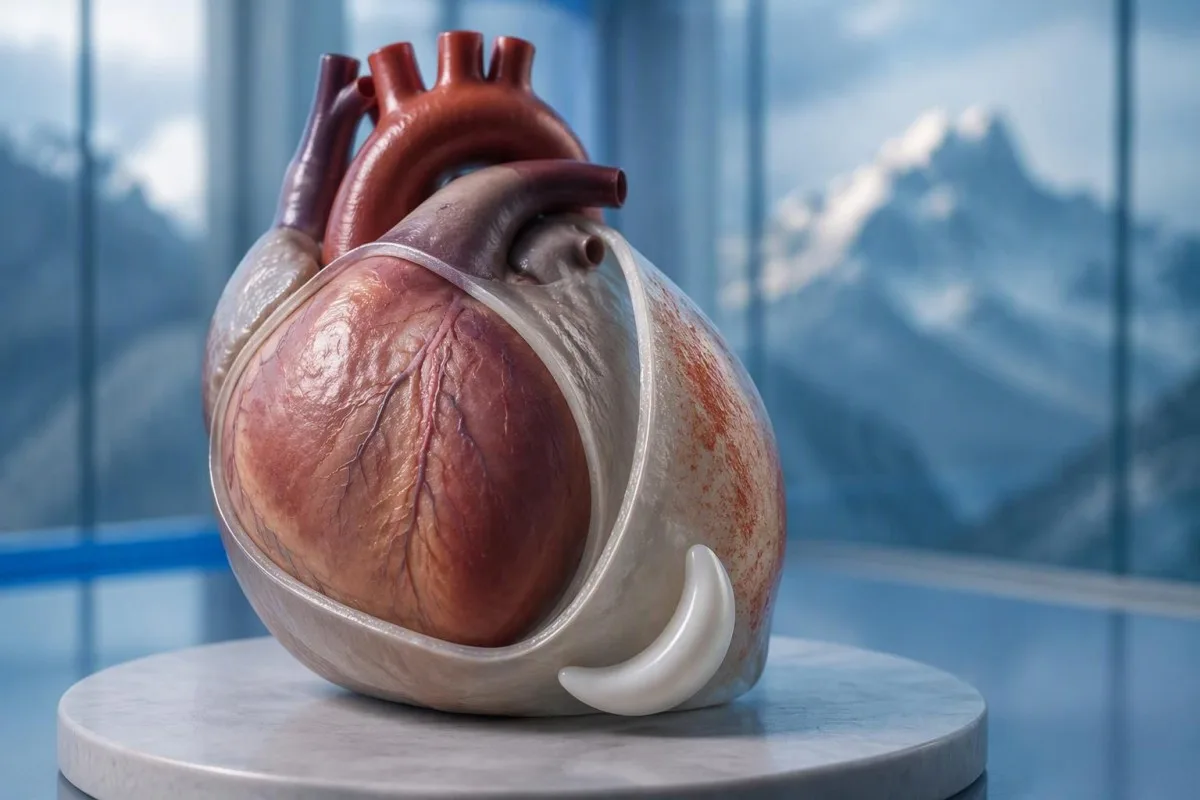
Ew pericardium ساکێکی دوو قاتە کە دڵ دەوری داوە، و ڕووی دەرەوەی پەردەکە پەیوەندی هەستەوەری ئازاری هەیە کە ماسولکەی دڵ خۆی بەو شێوەیە بەشداری ناکات. ڕوونکردنەوە ناسراوەکە کە قاتەکان تەنها “لەلەرە دەکەن” کورتکراوەتەوە: جووڵە، وروژانی شانەکانی دەوروبەر، و وەڵامی هەستی تاکەکەسی کاریگەری لەسەر ئازارەکە هەیە.
کەسێکی تەمەن ٤٢ ساڵ کە ئازارێکی تیژ دوای نەخۆشییەکی هەناسە دروست دەکات و ناتوانێت بە ئاسوودەیی بخەوێت، هاوڕەنگی pericarditis هەیە. من هێشتا لەسەر دەستپێکی ورد، ماندووبوون، هەناسە توندی، و مێژووی دڵ و خوێنبەرەکان دەپرسم پێش ئەوەی دەستنیشان بکەم؛ تەمەن و سەرمابوونێکی دوایی نەخۆشی coronary یان pulmonary embolism ناڕەوێنێتەوە.
Kantestî yek e Analyzerê testa xwînê ya AI کە دەتوانێت یارمەتی ڕوونکردنەوەی ئەنجامی CRP و troponin بدات دوای هەڵسەنگاندنی کلینیکی؛ ناتوانێت بڕیار بدات ئایا ئازاری سنگی پۆزیشنی سەلامەتە. وەک Thomas Klein، من ئەم جیاوازییە بە سادەیی ڕوون دەکەمەوە: ١ نیشانەی دڵنیایی نابێت هەرگیز لەسەر سکیوتێکی شاراوەی سنگدا زاڵ بێت؛ زانیاری دەربارەی سەندەی ئێمە ڕۆڵی پەروەردەیی ئێمە ڕوون دەکاتەوە.
ئایا ئازاری پەریکاردیت بە ھەناسەدانی قوڵ خراپتر دەبێت؟
ئازاری Pericarditis بە شێوەیەکی گشتی بە هەناسەیەکی قوڵ، کۆکە، یان هەندێکجار قوتدانیشدا خراپتر دەبێت. پزیشکان ئازاری پەیوەست بە هەناسە ناودەبەن pleuritic، بەڵام ئەو وەسفە شێوازێکی ئازار دیاری دەکات نەک یەک نەخۆشی دیاریکراو؛ سییەکان، قەفەزی سنگ، و pericardium هەموویان دەتوانن دروستی بکەن.
هەناسەدانێکی قوڵ دیافراگم دەجوڵێنێت و پەیوەندییەکانی ناو قەفەزی سنگ دەگۆڕێت، کە دەتوانێت پەردەی pericardialی وروژاو تووش بکات. پەیوەندییەکان کە دەگرێتەوە CNSphrenic، کە بە شێوەیەکی سەرەکی لە C3–C5, ، یارمەتی ڕوونکردنەوەی هۆکارێک دەدات کە بۆچی هەندێک نەخۆش ناڕەحەتی گوازراوە لە نزیک شان یان شانەکانی قونداردا هەست دەکەن لەبری تەنها پشت سنگی.
ئازارێک کە بە هەر هەناسەیەکدا دەگیرێت پێویستی بە هەڵسەنگاندنێکی فراوانتر هەیە کاتێک بە پەلە دەست پێ دەکات، بە تایبەتی لەگەڵ هەناسە توندی یان قاچێکی ئاوساودا. pulmonary embolism دەتوانێت هەمان سکیوت بەبێ هەستی فشاری کلاسیک دروست بکات؛ تەنانەت ئۆکسجینی 98% ناتوانێت بە تەنهایی ڕەتی بکاتەوە.
هەستیاری کاتێک پزیشکێک لە یەک بەشی سنگ دەگرێت، سەرچاوەیەکی دیوارەی سنگ زیاتر گونجاو دەکات، بەڵام ڕەتکردنەوەیەکی absolute coronary دابین ناکات. ڕوونکردنەوەمان لەسەر پشکنینەکان بۆ ئازاری شاراوە نیشانە تاقیگەیی پشتگیریکەر لەو بارودۆخانە جیا دەکاتەوە کە پێویستیان بە پشکنین و وێنەگرتن هەیە.
چ نیشانەکانی تری پەریکاردیت گرنگن؟
نیشانەکانی Pericarditis دەتوانێت تا، ماندووبوون، هەناسە توندی، و لێدانی دڵی خێراتر لەگەڵ ئازاری سنگدا بگرێتەوە. هیچ یەکێکیان ناچار نین، و کۆمەڵکردنیان گرنگترە لە هەر یەک نیشانە؛ لاوازی دیار، بێهێزی، یان گۆڕانی هەناسەی خراپتر گرنگی هەڵسەنگاندنەکە دەگۆڕێت.
تەمپرەچێرێک لەسەر 38°C is a higher-risk feature in the 2015 ESC pericardial guideline, particularly when the cause is uncertain. A patient with 37.4°C and considerable pain may still need urgent assessment, however; temperature thresholds help clinicians organize risk rather than define a safe home-care boundary.
A fast pulse can reflect pain, fever, dehydration, an arrhythmia, or impaired cardiac filling. I would interpret a resting pulse of 110 beats/min differently in someone comfortable and well hydrated than in someone pale and faint; our ڕێنمایی تاقیکردنەوەی palpitations explains why laboratory results are only part of that assessment.
Breathlessness when lying flat is not automatically the same thing as chest pain when lying flat. Someone who needs 3 pillows because they cannot breathe comfortably may have congestion, an effusion, or another cardiopulmonary problem; the distinction is covered in our ڕێسەی هەناسە تنگی (shortness-of-breath) یەکەمەکانمان دەڵێت کە چی لە یاسای لابۆراتۆری دەتوانێت و چی ناتوانێت ڕەت بکاتەوە., but active breathing difficulty should not wait for online interpretation.
کەی ئازاری سنگ لەگەڵ شوێندا پێویستی بە چارەسەری بەپەلە ھەیە؟
Call emergency services for severe, persistent, or rapidly worsening chest pain, especially with breathlessness, fainting, cold sweating, or pain spreading to the arm, jaw, or back. In the UK call 999 or 112; elsewhere use your local emergency number, and do not drive yourself.
Sudden maximal pain, collapse, new confusion, or coughing up blood also warrants emergency assessment because the differential includes pulmonary embolism and aortic emergencies. Do not wait 5 or 15 minutes to see whether leaning forward fixes the problem; concerning accompanying symptoms make a waiting experiment unsafe.
New unexplained chest pain that is not severe still deserves prompt, usually same-day, medical assessment rather than a routine appointment several weeks away. The 2021 AHA/ACC chest pain guideline emphasizes structured assessment and discourages using supposedly “atypical” symptoms to dismiss acute coronary syndrome (Gulati et al., 2021).
Stop exertion while arranging help, and follow the emergency dispatcher’s instructions about any medication rather than starting treatment for presumed pericarditis. Our پشکنینەکانی خوێن بۆ ئازاری سنگ لە حاڵەتی لەناکاو article explains why 1 laboratory result cannot replace an emergency ECG, examination, and appropriately timed testing.
پزیشکان چۆن پەریکاردیت گوماناوی ھەڵدەسەنگێنن؟
Clinicians assess suspected pericarditis by combining the history, examination, ECG, echocardiography, and selected blood tests. The widely taught 2015 ESC framework requires at least 2 of 4 findings: typical pain, a pericardial rub, characteristic ECG changes, or a new or worsening pericardial effusion (Adler et al., 2015).
Sharp pain, pain worse lying down, and relief leaning forward are not 3 independent diagnostic criteria; they are different features of the same symptom. This distinction prevents a persuasive story from being mistaken for confirmation, especially when the ECG and imaging have not yet been assessed.
A pericardial friction rub may be transient, so a clinician may listen again after an initial examination rather than treating 1 quiet examination as decisive. Vital signs, signs of poor circulation, heart failure, and the history of recent procedures or systemic illness can matter more urgently than whether the rub is audible.
Ew 2025 ESC myocarditis and pericarditis guideline gives contemporary imaging and overlapping myocardial involvement a central place in assessment, so the older checklist is not a complete care pathway (Schulz-Menger et al., 2025). Our clinical standards information should be read as documentation about laboratory interpretation—not evidence that software can reproduce this bedside evaluation.
لە پەریکاردیتدا، ئێێکجی (ECG) چی نیشان دەدات؟
An ECG may show widespread ST-segment elevation and PR-segment depression in acute pericarditis, but these changes are not always present. The 2015 ESC guideline reports characteristic ECG changes in up to 60% of cases, so a normal tracing does not exclude pericarditis (Adler et al., 2015).
A ECG ی 12-دەستەواژە (12-lead ECG) records electrical activity from several perspectives; clinicians assess which leads are affected, reciprocal changes, rhythm, and comparison with previous tracings. “The ST segment looks elevated” is not enough to separate pericarditis, early repolarization, myocardial infarction, and other causes without the clinical setting.
A heart attack more often produces changes corresponding to a coronary territory, whereas pericarditis may produce a broader pattern—but neither rule is absolute. ECG abnormalities can evolve over کاتژمێر, and repeating the tracing during recurrent or changing symptoms may reveal information absent from the first recording.
Electrolyte disturbances can add another layer of confusion, particularly when kidney disease or medication effects are present. A potassium concentration around 6.0 mmol/L can be clinically urgent depending on context, and our potassium and ECG guide explains why ECG interpretation should never be detached from the rest of the assessment.
بۆچی سۆنۆگرافی دڵ (echocardiography) بەکاردەھێنرێت بۆ پەریکاردیت؟
Echocardiography checks for fluid around the heart, ventricular function, and signs that fluid is impairing cardiac filling. A normal echocardiogram does not exclude uncomplicated pericarditis; an effusion greater than 20 mm is a recognized high-risk feature, not the definition of every dangerous effusion.
The 2015 ESC guideline describes effusions as small below 10 mm, moderate at 10–20 mm, and large above 20 mm, using the echo-free space on imaging. These measurements help communicate size, but the distribution of fluid and its hemodynamic effects still require interpretation rather than a number-only decision.
Cardiac tamponade occurs when pericardial pressure compromises filling; how quickly fluid accumulates can matter more than its absolute volume. A collection that develops over کاتژمێر may be less well tolerated than a larger, slowly developing collection, and the classic low-pressure, raised-neck-vein, quiet-heart-sound triad is not reliably present in every patient.
Ew 2025 ESC guideline supports integrating echocardiography with other imaging when the diagnosis or extent of involvement remains uncertain (Schulz-Menger et al., 2025). An elevated NT-proBNP may add information about cardiac strain, but 1 value cannot diagnose tamponade; our NT-proBNP interpretation guide explains several alternative reasons for elevation.
ئایا ترۆپۆنین دەتوانێت پەریکاردیت لە جەڵتەی دڵ جیا بکاتەوە؟
Troponin identifies heart muscle injury, but an elevated result alone cannot distinguish a heart attack from myocarditis or overlapping myopericarditis. The relevant threshold is the assay-specific سنووری سەرەوەی ڕێفەرەنس لە سەدەپێنجەی یەکەم (99th percentile), interpreted alongside symptoms, ECG findings, serial measurements, and sometimes imaging.
There is no universal normal troponin number for every laboratory: تروپۆنین I و تروپۆنین T use different assays, and some laboratories apply sex-specific reference limits. If a hypothetical report lists an upper limit of 14 ng/L, a result of 20 ng/L exceeds that particular limit; it does not independently identify the cause, as our troponin I versus T explanation details.
An early normal troponin may precede a detectable rise, which is why emergency departments use validated serial pathways, often with repeat sampling at 1 or 2 hours for suitable high-sensitivity assays. Selected patients can meet validated single-sample rule-out criteria, but that decision depends on assay, symptom timing, ECG, and risk—not a home interpretation of “normal.”
Pericardial symptoms plus raised troponin prompt assessment for myocardial involvement, especially with arrhythmia, breathlessness, or reduced ventricular function. A 30-year-old with a recent viral illness still needs a proper differential; our myocarditis troponin guide explains why neither youth nor the size of the rise settles the diagnosis.
CRP، ESR، و ژمارەی خوێن چی زیاد دەکەن؟
CRP, ESR, and a full blood count can support evidence of inflammation, but they do not confirm pericarditis or exclude a heart attack. Many laboratories use a standard CRP upper reference limit around 5 mg/L, while others use different limits; the printed laboratory range takes precedence.
CRP ـێک لە 45 مگ/ل indicates a substantial inflammatory response in a laboratory with an upper limit of 5 mg/L, but it cannot say whether the source is pericardial, pulmonary, autoimmune, or elsewhere. High-sensitivity CRP measures the same protein at low concentrations; acute illness values should not be interpreted using stable cardiovascular-risk categories.
Kantestî yek e پلاتفۆرمی تێکست/وەشاندنی تاقیکردنی خوێنی AI that can explain a CRP result against the supplied reference range and help organize its trend alongside other reported results. If CRP changes from 45 to 12 mg/L, the direction may support improvement, but persistent symptoms still matter; our WBC and CRP comparison discusses why these markers can disagree.
ESR often changes more slowly than CRP and is influenced by age, anemia, pregnancy, and protein concentrations, so comparing 2 different markers as though they were interchangeable can mislead follow-up. The 2015 ESC guideline treats inflammatory tests as supportive evidence rather than standalone diagnostic criteria (Adler et al., 2015); our causes of elevated ESR adds context for an unexpectedly persistent elevation.
چ ھۆکاران و تایبەتمەندییە مەترسیدارەکان پلانی چاودێری دەگۆڕن؟
Pericarditis may follow a viral illness, occur after cardiac injury, or accompany autoimmune disease, severe kidney disease, tuberculosis, or other conditions. هەستەوە/تێمپرەچەر (Fever) لە سەر 38°C, a large effusion, tamponade, or failure to improve with initial treatment are among the features that can justify hospital assessment and a broader search for causes.
Ew 2015 ESC guideline lists lack of response to anti-inflammatory treatment within 7 ڕۆژ as a major adverse prognostic feature, alongside subacute onset and the imaging findings above (Adler et al., 2015). This is a clinician’s reassessment trigger—not permission to wait a week through worsening pain, fever, or breathlessness.
Autoimmune testing is most useful when the history supplies a reason: recurrent episodes, joint symptoms, rashes, mouth ulcers, or known systemic disease. 1 positive antibody does not prove autoimmune pericarditis; our ڕێنمایی ئەنجامی دژە-dsDNA explains why antibody findings need compatible clinical evidence.
Severe kidney dysfunction can change both the cause of pericarditis and medication safety; an eGFR below 15 mL/min/1.73 m² identifies kidney failure-range function but does not itself diagnose uremic pericarditis. Our urgent low-eGFR symptoms guide is relevant to that background, while chest pain in this setting requires direct medical assessment.
ئایا باشبوونی بە دەرمان پەریکاردیت بسەلمێنێت؟
Improvement with ibuprofen, aspirin, or a change in posture does not confirm pericarditis. For appropriately diagnosed uncomplicated cases, clinicians commonly use an anti-inflammatory medicine with colchicine, often for approximately 3 مانگ after a first episode, but the regimen must account for kidney function, interactions, and the underlying cause.
A reduction in pain from 8 out of 10 to 3 out of 10 is useful clinical information, but several chest-pain causes can respond to analgesia. Do not use medication response as a reason to cancel assessment, and do not start high-dose anti-inflammatory treatment for undiagnosed chest pain without professional advice.
Colchicine requires particular care with kidney or liver impairment and certain interacting medicines, including some antibiotics; taking 2 medicines together can alter exposure even when each prescription is familiar. Corticosteroids are not routine first-line treatment for every uncomplicated case, and selected bacterial or systemic causes need a different strategy rather than simply more pain relief.
Kantesti can help organize laboratory follow-up for a clinician through our ڕێبەری چاودێریی باری دەرمان, but it should not select the treatment or tapering schedule. A plan that includes 1 named prescriber, clear interaction checks, and instructions for worsening symptoms is more useful than a generic dosing chart.
کەی وەرزشکردن سەلامەتە دوای ئەوەی نیشانەکانی پەریکاردیت باشتر بوون؟
Exercise should be restricted during active pericarditis, and return should follow clinical reassessment rather than pain relief alone. The decision changes substantially if troponin, rhythm testing, or imaging suggests myocardial involvement; 1 symptom-free day is not evidence that strenuous activity is safe.
The older 2015 ESC guideline used a minimum ڕێکەوتی 3 مانگ restriction for athletes with isolated pericarditis, while contemporary guidance places greater emphasis on individualized remission and risk assessment. Patients should not borrow an athlete’s timetable—or a myocarditis timetable—without knowing which diagnosis their clinician has established.
For a hypothetical 28-year-old runner, the difference between isolated pericarditis and myocardial involvement can change follow-up testing and return-to-training decisions. I would ask whether symptoms, inflammatory markers, ventricular function, and any relevant rhythm concerns have resolved; a normal resting pulse of 65 beats/min is only one small part of that picture.
Daily activity, physical work, and competitive sport are 3 different workloads, so a written return plan should specify what the patient can actually do. Our ڕێنمای نشانەی خوێنی (blood biomarker guide) can explain reported measurements, but no biomarker page, wearable reading, or AI result constitutes exercise clearance.
ئەگەر ئازاری سنگ گەڕایەوە یان پشکنینەکان ئاسایی بوون؟
Persistent or recurrent chest pain needs reassessment even when an earlier ECG, echocardiogram, or CRP result was normal. Appropriately selected low-risk outpatients are commonly reviewed within about 1 هەفتە, but recurrence with faintness, breathlessness, or worsening pain warrants earlier—and sometimes emergency—assessment.
Residual discomfort, recurrent inflammation, chest-wall pain, and a new unrelated problem can feel similar to the patient, so 2 episodes should not automatically receive the same explanation. Cardiac MRI can sometimes identify pericardial or myocardial involvement when first-line tests leave uncertainty; it is selected for a clinical question, not ordered for every ache.
A follow-up note should record symptom timing, temperature, activity tolerance, medication changes, and the actual laboratory reference ranges. CRP values of 8 and 6 mg/L from different laboratories may not represent a meaningful improvement without knowing the methods and context; our post-discharge result changes guide explains why small numerical changes deserve restraint.
Kantestî yek e ئامێری توێژینەوەی تاقیکردنەوەی خوێن بە پشتبەستن بە AI that can help organize reported results for discussion, but it cannot verify the cause of renewed chest pain from a PDF. My advice here, as Thomas Klein, is to agree on 3 things before leaving follow-up: the next review date, permitted activity, and the symptoms that require immediate help.
سنوورە کلینیکییەکان و بڵاوکراوە توێژینەدییە پەیوەندیدارەکان
Symptoms and laboratory interpretation cannot safely replace an acute chest-pain assessment. لە ٣ی تشرینی یەکەمی ٢٠٢٦, the clinical references below support the distinction between pericardial clues, myocardial injury, and emergency risk; the 2 Zenodo resources are background laboratory guides, not trials validating a pericarditis diagnosis.
Kantesti medical advisory information describes the professional expertise behind our educational work, but readers should not equate a medical title with an examination of their own chest pain. A personal care plan still needs 1 accountable treating clinician or team who can examine the patient, review the ECG, and act on imaging or troponin findings.
Ew Rêzeya Normal a aPTT: D-Dimer, Rêbernameya Mêjkirina Xwînê ya Proteîna C is available as a Zenodo educational publication with DOI 10.5281/zenodo.18262555. Its associated clotting-test interpretation guide is relevant when clinicians consider coagulation questions, but a normal aPTT cannot exclude pulmonary embolism and D-dimer must be used within an appropriate clinical probability pathway.
Ew Rêbernameya Proteînên Serumê: Testa Xwînê ya Globulîn, Albumîn û Rêjeya A/G is a second Zenodo educational publication with DOI 10.5281/zenodo.18316300. The associated serum protein interpretation guide supplies background on systemic laboratory findings; neither albumin nor the A/G ratio independently diagnoses pericarditis, and the discovery links below are searches rather than verified copies of these publications.
Pirsên Pir tên Pirsîn
ئایا ئازاری سنگ لە پەردیلدا بە ڕاکشان خراپتر دەبێت؟
ئازاری سنگ لە پەیکەریشەبوونی دڵ (Pericarditis) زۆرجار بە پاڵکەوتن خراپتر دەبێت و بە دانیشتن یان پشت-پێدانەواندن باشتر دەبێت. ئەو شێوازە ئاماژەدارە، بەڵام تەنها یەک بەشە لە هەڵسەنگاندنی کلینیکی و ناتوانێت جەڵتەی دڵ بسڕێتەوە. پزیشکان مێژووەکە لەگەڵ پشکنین، ئێێکجی، ئەکۆکاردیۆگرافی، و پشکنینەکانی خوێن تێکەڵ دەکەن. ئازاری نوێی سنگ کە ڕوون ناکرێتەوە پێویستی بە هەڵسەنگاندنی خێرا هەیە، لەگەڵ یارمەتی فریاکەوتن بۆ ئازاری توند، کورتەهەڵاتن، بێهێزبوون، یان ئارەقەی سارد.
ئایا ئازاری سنگ کە بە پاڵکەوتنە پێشەوە سووک دەبێت، هۆکارەکەی جەڵتەی دڵ نابێت؟
ئازاری سنگ کە بە پێشەوە چەماندنەوە باشتر دەبێت، مانای ئەوە نییە کە جەڵتەی دڵ نییە. ڕێنمایی ساڵی 2021ی AHA/ACC بۆ ئازاری سنگ پشتگیری لە هەڵسەنگاندنی ڕێکخراو دەکات بەبێ گوێدانە ئەوەی کە نیشانەکان نا classical دەبن. لەوانەیە پێویست بە ECG و پشکنینی بەرز-هەستیاری ترۆپۆنین لە کاتێکی گونجاودا بکرێت، و ئەنجامی سروشتیی سەرەتایی بە شێوەیەکی خۆکار یەکلایی کەرەوە نییە. پەیوەندی بە هێڵی فریاکەوتن بکە بۆ ئازاری توند یان بەردەوام یان هەناسە بڕکێ، بێهۆشبوون، یان ئارەقکردنەوەی سارد.
ئایا دەکرێت پەردیلان (pericarditis) لەگەڵ ECG (کاردیۆگرام) ئاساییدا هەبێت؟
بەڵێ، پەریکاردیتیس دەکرێت لەگەڵ ئی سی جی (ECG) ئاساییدا ڕووبدات. ڕێنماییەکەی ٢٠١٥ی ESC بۆ پەریکاردیتیس گۆڕانکارییە تایبەتەکانی ئی سی جی (ECG) لە نزیکەی ١TP36T حاڵەتی توندییدا ڕاپۆرت کردووە، بە واتای ئەوەی ڕێژەیەکی بەرچاو نیشانەی کلاسیکیان پیشان نەداوە. دۆزینەوەکانی ئی سی جی (ECG) دەتوانن بگۆڕێن، بۆیە پزیشکان لەوانەیە دووبارە ١٢ لید (lead) ئەنجام بدەن کاتێک نیشانەکان دەگۆڕێن. ئی سی جی (ECG) ئاسایی بە تەنیا ناتوانێت پەریکاردیتیس یان هەموو جۆرەکانی جەڵتەی دڵ ڕەتبکاتەوە.
ئایا سۆنارەکانی دڵ هەمیشە پێکەوتنی دەماری دڵ (pericarditis) نیشان دەدەن؟
ئەکۆکاردیۆگرام هەمیشە پەیکەردانی ئاسایی ئاشکرا ناکات چونکە ھەڵئاوسانەکە لەوانەیە ڕووبدات بێ ئەوەی کۆبوونەوەیەکی ئاوی دیار بێت. ئەکۆکاردیۆگرافی بەتایبەتی بەسوودە بۆ پێوانەکردنی شلەی دەوری دڵ، هەڵسەنگاندنی کردارەکانی گەدە، و گەڕان بەدوای پڕبوونی کەم. دابڕانێک کە گەورەتر بێت لە ٢٠ مم بەپێی پۆلێنکردنی ESC ی ٢٠١٥، بەڵام کۆبوونەوە خێراکانی بچووکتر کە هێشتا دروست دەبن دەتوانن مەترسیدار بن. وێنەی زیاتر دەکرێت لەبەرچاو بگیرێت کاتێک پرسیارە کلینیکیەکە چارەسەر نەکراو دەمێنێتەوە.
مانای ترۆپۆنی نۆرمال ئەوەیە ئازاری سنگ جدی نییە؟
ئەنجامێکی ئاسایی ترۆپۆنین مانای ئەوە نییە کە ئازاری سنگ بێ زیانە. کات، سنووری سەدی 99ی تایبەت بە تاقیکردنەوەکە، دۆزینەوەکانی ECG، و هەڵسەنگاندنی گشتی مەترسی دیاری دەکات کە ئەنجامەکە چی ڕەتدەکاتەوە. ڕێگاکانی ترۆپۆنینی پشتڕاستکراوەی بەرز-هەستیاری زۆرجار پێوانەی دووبارە لە 1 یان 2 کاتژمێردا بەکاردەهێنن، هەرچەندە نەخۆشی هەڵبژێردراو دەتوانن مافی ڕێگای دەرکردنی تاک نموونە وەربگرن. ترۆپۆنینیش بەخۆیی گەورەبوونی سییەکان، بارودۆخی ئاۆرتیک، یان پەریکاردیت ڕەتنەکاتەوە.
چەند دەخایەنێت نیشانەکانی پەریکاردیایس بمێنێت؟
نیشانەکانی پەریکاردیتیس لەگەڵ چارەسەری گونجاودا لە ماوەی چەند ڕۆژێک بۆ چەند هەفتەیەک باشتر دەبێت، بەڵام کاتەکە بەپێی هۆکار، دوبارەبوونەوە، و تێوەگلانی ماسولکەی دڵ دەگۆڕێت. کۆڵشیسین بە شێوەیەکی گشتی بۆ ماوەی نزیکەی ٣ مانگ دوای یەکەمین حاڵەتی سادە دەردەکرێت، ئەمەش مانای ئەوە نییە کە ئازار دەبێت لەو ماوەیەدا بەردەوام بێت. نەبوونی باشبوون لە ماوەی ٧ ڕۆژدا لە چارەسەری دژە هەوکردندا هاندەرێکی ناسراوە بۆ دووبارە هەڵسەنگاندن لە ڕێنماییەکانی ٢٠١٥ ESC. ئازاری زیادبوو، کورتنەهاتن، یان بێهۆشبوون پێویستی بە هەڵسەنگاندنی زووتر هەیە نەک چاوەڕوانیکردن بۆ پشکنینی دیاریکراو.
ئەمڕۆ AI-پاوەرد لەسەر تاقیکردنەوەی خوێن بەدەست بهێنە
بە یارمەتی زیاتر لە 2 ملیۆن بەکارهێنەر لە هەموو جیهاندا کە Kantesti دەستپێدەکەن بۆ تاقیکردنەوەی لابراتۆری ڕاست و بەهێز لە کاتێکی کەم. ڕەخنەی تاقیکردنەوەی خوێنت بنێرە و تفسیرێکی تەواو لە 15,000+ نیشانەی زیستی (biomarkers) لە ماوەی چرکەکاندا وەرگرە.
📚 توێژینەوە سەرچاوە پەیوەندیدارەکان
کلاین، ت.، میچێڵ، س.، & وێبەر، ه. (2026). Rêzeya Normal a aPTT: D-Dimer, Rêbernameya Mêjkirina Xwînê ya Proteîna C. Kantesti توێژینەوەی پزیشکی AI.
کلاین، ت.، میچێڵ، س.، & وێبەر، ه. (2026). Rêbernameya Proteînên Serumê: Testa Xwînê ya Globulîn, Albumîn û Rêjeya A/G. Kantesti توێژینەوەی پزیشکی AI.
📖 سەرچاوەی پزیشکی دەرەکی
گولاتی م و هاوکارانی. (٢٠٢١). ڕێنمایی 2021 AHA/ACC/ASE/CHEST/SAEM/SCCT/SCMR بۆ هەڵسەنگاندن و دۆزینەوەی تێکچوونی سینه (Chest Pain). .
Schulz-Menger J et al. (2025). 2025 ESC Guidelines for the management of myocarditis and pericarditis. European Heart Journal.
⚕️ Daxuyaniya Bijîşkî
ئەم مادەیە تەنها بۆ. I think I must continue but user expects all items.
سەنگەری باوەڕپێکردن E-E-A-T
Tecribe
ڕەوی پشکنینی کلینیکی لەلایەن پزیشکەوە بۆ ڕێکخستنی ڕووداوەکانی لێکدانەوەی ئازمایش.
Pisporî
گرێدانی لابراتۆرییەی پزیشکی بەوەی چۆن بایۆمارکەرەکان لە کۆنتێکستی کلینیکی دەگۆڕن.
Desthilatdarî
نووسراوە لەلایەن د. توماس کلاین بە پشکنینی لەلایەن د. سارا میچێڵ و پڕۆف. د. هانس وێبەر.
Bawerî
لێکدانەوە بە بنەمای دڵنیابوون (Evidence-based) بە ڕێڕەوی دوایینەوەی ڕوون بۆ کەمکردنەوەی هەست بە ترس/هەڵوەشاندن.