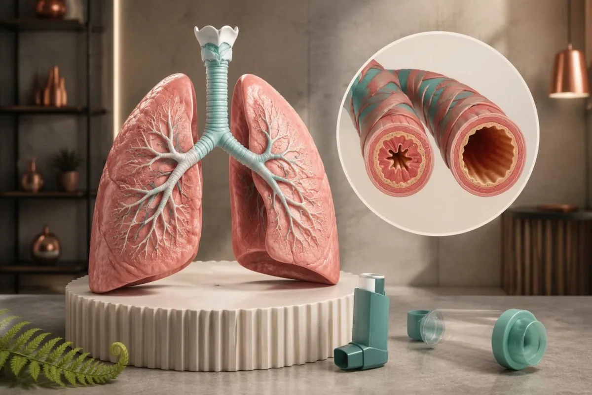
Asthma can cause changing breathlessness, nighttime cough or exercise symptoms—even when nobody hears a wheeze. Breathing tests matter more than an isolated laboratory result.

핵심 요점
- Asthma symptoms include variable wheezing, breathlessness, chest tightness and cough; having 1 symptom does not establish the diagnosis.
- Nighttime waking from asthma symptoms even once in the previous 4 weeks is a reason to review symptom control under the GINA assessment framework.
- Cough without wheezing can occur in cough-variant asthma, but cough lasting more than 8 weeks in adults has several other possible causes.
- Spirometry response of at least 12% and 200 mL in adult FEV1 after a bronchodilator supports variable airflow limitation under commonly used asthma criteria.
- Peak-flow variability averaging more than 10% over 2 weeks supports asthma in adults under GINA criteria; NICE uses a different diagnostic threshold.
- FeNO of at least 50 ppb in adults supports type 2 airway inflammation in the appropriate clinical context, but low FeNO does not exclude asthma.
- 혈중 호산구 above 0.5 × 10^9/L can reflect allergy, asthma or other conditions; a blood result alone cannot confirm asthma.
- 응급 호흡 징후에는 include inability to speak normally, exhaustion, confusion, blue or grey lips, or poor response to the prescribed reliever; do not wait for a test result.
What are the early signs of asthma?
Asthma symptoms are changing episodes of wheezing, breathlessness, chest tightness or cough, often worse at night or after exercise; asthma can occur without obvious wheezing. Spirometry and peak-flow monitoring help demonstrate variable airflow limitation, whereas blood tests alone cannot confirm asthma. Severe breathing difficulty, confusion or a reliever that is not helping requires emergency care.
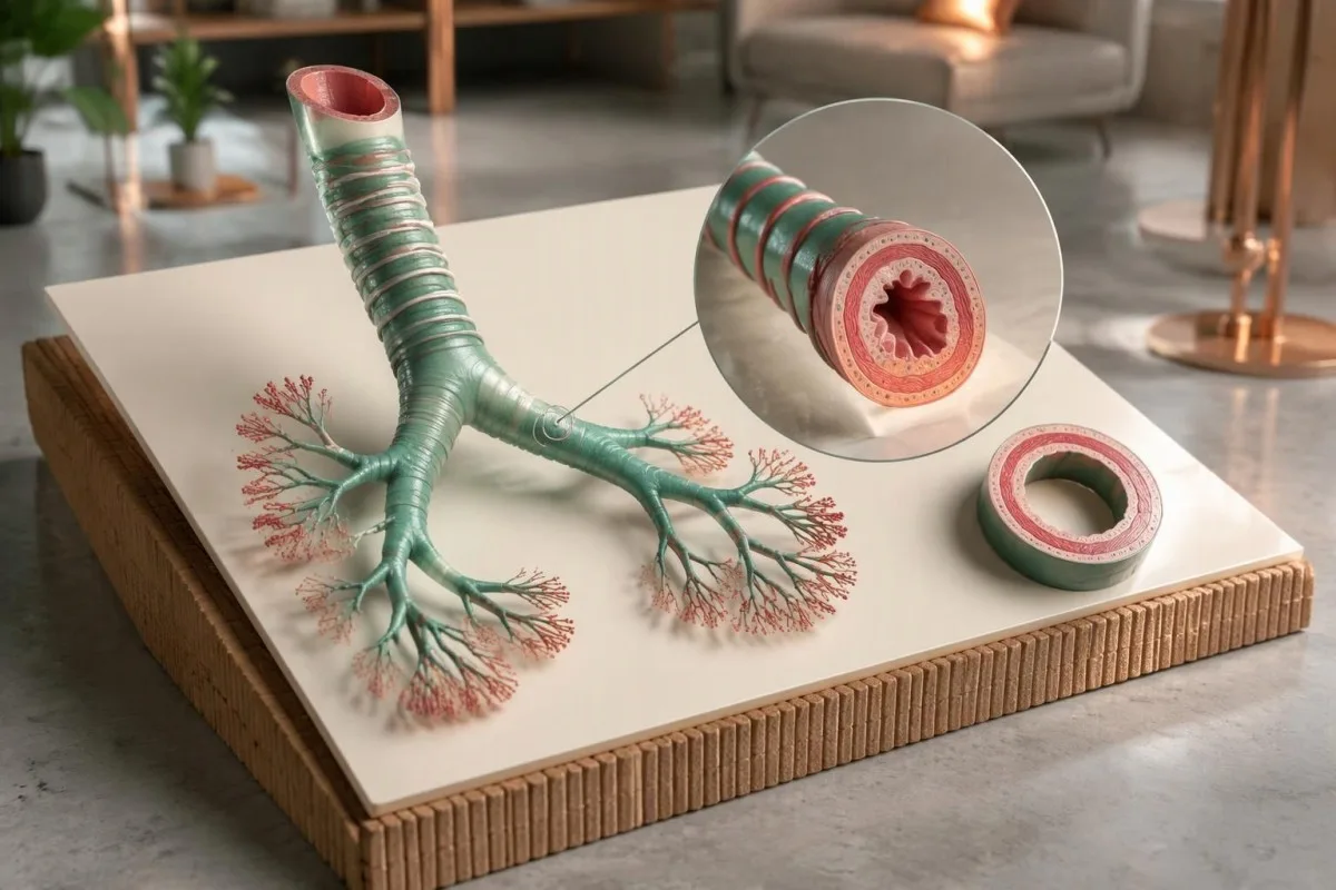
The early signs of asthma often involve a change from your usual breathing rather than a constant symptom: a familiar walk becomes harder, laughter starts a cough, or a cold leaves unusually prolonged chest symptoms. Record the pattern for 2 weeks if symptoms are mild and stable, but seek assessment sooner if breathing is worsening.
Symptom-free intervals do not rule out asthma. A person can have 3 comfortable days followed by tightness after cold-air exercise because airway sensitivity and exposure vary; similarly, a normal chest examination between episodes cannot establish that the airways always function normally.
A hypothetical 34-year-old might report coughing after running, waking twice in a week and feeling entirely well at work. That combination raises suspicion more than one isolated cough, but the next step is a clinical history and objective testing—not assuming that improvement after a borrowed inhaler proves asthma.
칸테스티는 AI 혈액검사 분석기 that helps explain supportive laboratory findings, not a substitute for measuring airflow. Our 조직과 임상 범위 distinguish laboratory interpretation from respiratory diagnosis: even 2 normal laboratory panels cannot exclude an intermittent breathing disorder.
Why are asthma symptoms at night more noticeable?
Asthma symptoms at night can become more noticeable because airway calibre and inflammatory activity follow daily rhythms, while bedroom allergens or reflux may add triggers. Under the GINA 2025 symptom-control assessment, any asthma-related nighttime waking during the previous 4 weeks warrants attention.
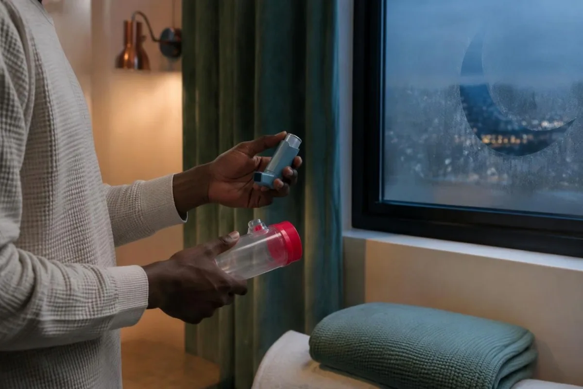
Nighttime cough is not automatically asthma. Cough after lying down with sour taste or regurgitation suggests another possible contributor, and our reflux symptom patterns explain why timing helps; 2 conditions can coexist, so identifying reflux does not necessarily explain every episode of breathlessness.
Repeated waking carries more information than the clock time alone. Write down the number of awakenings over 7 nights, whether you felt tightness or breathlessness, and whether your prescribed reliever helped; a cough at midnight after a meal differs from waking near dawn with difficulty exhaling.
GINA's 2025 strategy assesses daytime symptoms, nighttime waking, activity limitation and, where applicable, short-acting reliever use over 4 weeks. A person who sleeps poorly because of asthma should not interpret a normal daytime oxygen reading as evidence of good control, because oxygen saturation does not measure airway variability.
A bedroom diary can separate exposure from coincidence: note whether symptoms occur at home, away from home or after changing bedding during a 2-week period. Avoid deliberately re-exposing yourself to a suspected trigger; persistent waking deserves a treatment and inhaler-technique review rather than simply adding extra pillows.
Can asthma cause a cough without wheezing?
Asthma cough without wheezing is possible, particularly in cough-variant asthma, where cough may be the main symptom and routine spirometry can be normal. In adults, cough lasting more than 8 weeks requires evaluation for other causes as well as asthma.

Cough-variant asthma and non-asthmatic eosinophilic bronchitis can both cause chronic cough associated with eosinophilic airway inflammation. The distinction matters because the second condition typically lacks the variable airflow limitation or airway hyperresponsiveness characteristic of asthma; an elevated eosinophil count cannot distinguish these 2 diagnoses by itself.
A normal initial breathing test does not close the investigation when the symptom pattern remains convincing. Clinicians may repeat spirometry during symptoms or consider a supervised bronchial challenge, whereas coughing spells with vomiting or a relevant contact history may need whooping cough testing with attention to how many weeks have elapsed.
Medication review can identify an overlooked explanation for cough. An ACE inhibitor used for blood pressure can cause a dry cough even after months of treatment, so bring a complete medication list rather than focusing only on drugs started within the previous 2 weeks; do not stop prescribed treatment without advice.
The absence of wheezing has 2 very different meanings: mild asthma may produce no audible wheeze, while a severe attack may become quiet because too little air is moving. Quiet breathing combined with exhaustion, difficulty speaking or worsening breathlessness is an emergency—not evidence that the attack has ended.
How do exercise-triggered asthma symptoms behave?
Exercise-induced bronchoconstriction causes temporary airway narrowing during or after exercise, often becoming most noticeable within about 5–10 minutes after stopping. Exercise symptoms alone do not prove asthma, because laryngeal obstruction, anaemia and other conditions can also limit breathing.
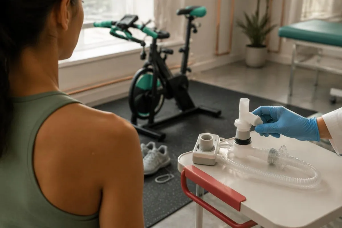
Exercise-related laryngeal obstruction often causes throat tightness or noisy breathing during inhalation at peak effort, then settles relatively quickly after stopping. Bronchoconstriction more often affects expiration and recovery; these are clues rather than absolute rules, and the 2 conditions can occur in the same person.
A supervised exercise challenge measures airflow rather than relying on whether exercise feels difficult. A fall in FEV1 of more than 10% and more than 200 mL after exercise supports asthma in adults under GINA's diagnostic framework; local protocols and criteria for exercise-induced bronchoconstriction may differ.
Reduced exercise tolerance without cough, tightness or variable wheeze deserves a broader assessment. Our iron stores in athletes guide explains why a hypothetical runner with ferritin of 12 ng/mL may need an iron assessment, but that result would not explain away independently documented airway narrowing.
An exercise diary should include intensity, weather and whether symptoms peak during activity or 10 minutes afterward. Once assessed, many people can remain active using a clinician-agreed plan; avoid testing your limits during an unstable period or repeatedly using a reliever without reviewing the underlying treatment.
Which triggers commonly bring on asthma symptoms?
Asthma triggers include respiratory viruses, pollen, dust mites, animal allergens, smoke, air pollution, cold air and exercise. A 2-week exposure diary can suggest patterns, but a suspected trigger is not the same thing as a confirmed allergy or a diagnosis of asthma.
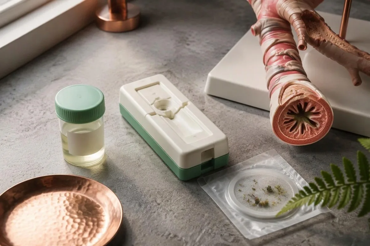
Allergy testing identifies sensitisation, not necessarily the cause of every breathing episode. A positive specific IgE result to 1 allergen becomes clinically useful when exposure reliably matches symptoms; our 알레르기 항원 검사 guide explains why more detailed panels still require a relevant history.
Damp buildings can worsen respiratory symptoms through several mechanisms, including allergen exposure and irritation. There is no single laboratory panel that establishes 'mould-related asthma'; our mould testing limitations distinguish targeted allergy assessment from unsupported broad testing, and symptoms should be evaluated even when 1 panel is normal.
Work-related asthma can appear after months or years of exposure and may improve on weekends or holidays. Record work tasks and breathing measurements across both work and non-work days for an occupational assessment; a specialist may need more than the standard 2-week diagnostic diary, and intentional exposure challenges are unsafe.
Some beta-blockers, including certain eye drops, can worsen bronchoconstriction, while aspirin or other NSAIDs trigger symptoms in a susceptible subgroup rather than everyone with asthma. Review all medications, including over-the-counter products, during 1 appointment; do not stop cardiovascular drugs or attempt a home aspirin challenge.
What else can look like asthma?
Asthma-like symptoms can result from reflux, upper-airway disorders, COPD, anaemia, cardiac disease or respiratory infection. At least 2 separate conditions may contribute, so persistent symptoms despite appropriate asthma treatment should prompt reassessment rather than automatic dose escalation.

Wheeze limited to one area or symptoms beginning suddenly after choking suggest focal airway obstruction rather than typical diffuse asthma. The finding deserves clinical assessment, particularly in a child; a single persistent wheeze has a different diagnostic meaning from episodes that change with several unrelated triggers.
Fixed obstruction on repeated spirometry does not automatically mean COPD, because longstanding asthma can also leave persistent airflow limitation. Smoking history, age and previous lung disease help distinguish causes, while early emphysema or a strong family history may justify alpha-1 antitrypsin assessment rather than assuming 1 diagnostic label explains everything.
Recurrent chest infections, poor growth or a chronic productive cough in a young person should widen the investigation. A sweat chloride result of at least 60 mmol/L supports cystic fibrosis in the appropriate diagnostic setting; values of 30–59 mmol/L require further evaluation rather than being treated as an asthma result.
New breathlessness with chest pressure, fainting or inability to lie comfortably needs timely assessment for non-asthma causes. Even 1 abrupt episode of unexplained breathlessness can be urgent, and attributing symptoms to anxiety before checking relevant respiratory and cardiac possibilities risks missing a treatable condition.
How does spirometry support an asthma diagnosis?
Spirometry measures airflow, including FEV1 and FVC, and can show obstruction that improves after a bronchodilator. An adult FEV1 increase of at least 12% and 200 mL supports variable airflow limitation under commonly used asthma criteria, but a negative test does not exclude intermittent asthma.
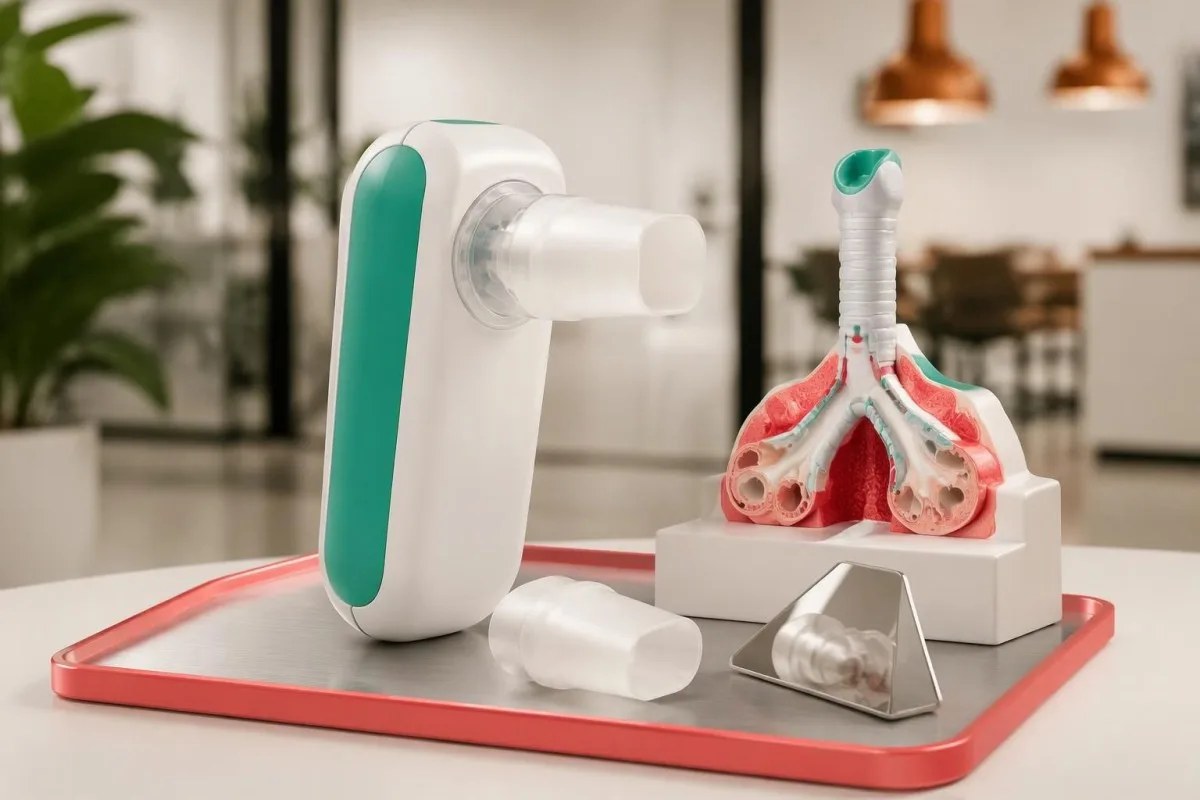
The FEV1/FVC ratio helps identify obstruction, but interpretation should use an age-appropriate lower limit of normal rather than applying 0.70 to every patient. The European Respiratory Society adult diagnostic guideline recommends spirometry and bronchodilator testing within a structured assessment, followed by additional tests when necessary (Louis et al., 2022).
Consider a hypothetical result: FEV1 rises from 1.90 L to 2.18 L after the test medication. The increase is 280 mL and 14.7% of the starting value, meeting the commonly used adult reversibility criterion; the finding supports asthma when the history fits, but it is not a diagnosis detached from clinical context.
Guidelines and laboratory reporting standards do not always express reversibility in the same way. NICE NG245 also accepts an adult FEV1 increase of at least 10% of predicted normal; if predicted FEV1 is 3.40 L, that same 280 mL improvement is only 8.2% of predicted, explaining why 2 reporting conventions can produce different flags.
Testing quality matters: spirometry usually seeks at least 3 acceptable manoeuvres, and adult repeatability commonly requires the best FEV1 values to be within 150 mL. Our breathlessness laboratory guide explains complementary investigations, but no laboratory interpretation can compensate for an unreliable breathing manoeuvre.
How can a peak-flow diary help diagnose asthma?
Peak-flow monitoring can document variable breathing when spirometry is unavailable or initially inconclusive. GINA uses average daily variability above 10% in adults and above 13% in children over 2 weeks as supportive evidence; NICE NG245 uses a 20% diagnostic threshold in its pathway.
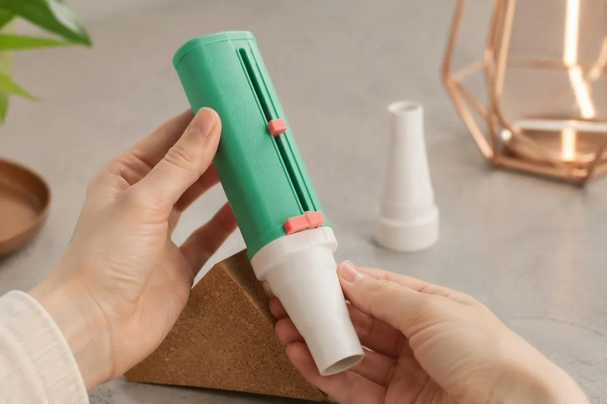
Use the same meter and technique each time, usually measuring morning and evening as instructed. Perform 3 blows per session and record the highest acceptable reading in L/min; note symptoms and medication timing, because taking a bronchodilator before one session but not the other can distort the comparison.
Daily variability can be calculated as the highest minus the lowest reading divided by their mean, multiplied by 100. If a hypothetical day's readings are 380 and 460 L/min, variability is 80/420 × 100, or 19.0%; the diagnostic assessment uses the average across the monitoring period, not that single day's percentage.
Peak-flow action zones and diagnostic variability answer different questions. A personalised plan may use at least 80% of personal best, 50–79%, and below 50% to guide action, whereas diagnosis examines fluctuations over time; a low personal best should not be accepted as normal without checking technique and clinical status.
칸테스티는 AI 혈액검사 결과 해석 플랫폼, not a service for confirming asthma from a 2-week breathing diary. Our AI report safety checks explain why readers should distinguish a recorded value, an automated explanation and a clinician's diagnosis—particularly when different guidelines use different thresholds.
What do FeNO and eosinophils tell you about asthma?
FeNO measures nitric oxide in exhaled breath, while blood eosinophils measure an inflammatory cell population; both can support assessment of type 2 asthma. NICE NG245 uses FeNO of at least 50 ppb in adults and at least 35 ppb in children aged 5–16 within its diagnostic pathways.

The NICE/BTS/SIGN 2024 guideline allows an elevated blood eosinophil count above the laboratory reference range or FeNO of at least 50 ppb to establish an adult diagnosis within its suggestive-history pathway. This does not mean a blood test alone confirms asthma: the recommendation depends on clinical assessment, and GINA places greater emphasis on documenting variable airflow limitation.
A low FeNO reading does not rule out asthma, particularly after inhaled corticosteroid treatment or in non-type-2 disease. Allergic rhinitis can raise FeNO without proving lower-airway asthma, so a hypothetical reading of 60 ppb with nasal symptoms but no compatible breathing history needs a different interpretation from 60 ppb with recurrent nocturnal tightness.
Eosinophil counts above 0.5 × 10^9/L, equivalent to 500 cells/µL, commonly attract attention, although laboratories use different upper limits. Our high eosinophil interpretation explains why persistent counts of at least 1.5 × 10^9/L require broader evaluation for medications, parasites and other disorders—not simply a stronger asthma label.
Steroid timing can change both FeNO and eosinophil results, sometimes making 2 tests taken weeks apart look inconsistent. Our 임상 해석 기준 support reporting such limitations; tell the clinician when you last used inhaled or oral steroids, but never stop treatment just to make a test more likely to turn positive.
Why can't blood tests alone confirm asthma?
Blood tests alone cannot confirm asthma because they do not demonstrate variable airflow limitation or reliably distinguish asthma from every alternative cause of cough. Even 1 markedly abnormal eosinophil or IgE result needs a compatible history and an appropriate diagnostic pathway.
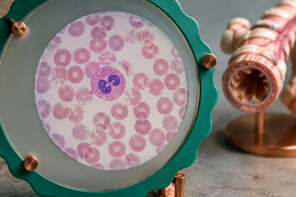
Total IgE and allergen-specific IgE are different measurements: one reflects an overall antibody concentration, while the other tests sensitisation to selected allergens. There is no universal total-IgE cutoff that diagnoses asthma across adults and children; a positive result to 1 allergen may explain sensitisation without proving that exposure causes wheeze.
A CBC may identify anaemia that contributes to breathlessness, and selected inflammatory tests may help assess a suspected infection. Our WBC 및 CRP 비교 explains why these markers can disagree; an elevated CRP of 25 mg/L is not an asthma threshold and cannot independently determine whether antibiotics are needed.
Kantesti AI can help readers compare eosinophil counts, haemoglobin and other reported markers, but it cannot establish variable airflow limitation from a laboratory PDF. A hypothetical eosinophil change from 0.62 to 0.18 × 10^9/L after steroid treatment may reflect treatment exposure rather than the disappearance of asthma.
Routine asthma diagnosis does not require ordering every available inflammatory marker. Bring existing results and a medication timeline covering at least the previous 2 weeks; clinicians can then select tests for a specific question, such as anaemia, allergy or treatment planning, instead of treating a large panel as a substitute for breathing assessment.
Which breathing warning signs require emergency care?
즉시 응급 서비스에 전화하십시오 for severe breathlessness, inability to speak normally, blue or grey lips, confusion, drowsiness, exhaustion or a prescribed reliever that is not helping. If available, peak flow below 50% of personal best or oxygen saturation of 92% or less with acute symptoms adds urgency; do not wait for either measurement.

A quiet or 'silent' chest during worsening breathing can indicate critically reduced air movement, not recovery. Peak flow below 33% of personal best is a life-threatening feature in UK acute-asthma frameworks, but inability to obtain a reading because of severe breathlessness is itself concerning; call the local emergency number, such as 999 or 112 where applicable.
While help is coming, sit upright and use your prescribed reliever according to your written action plan or emergency-dispatch instructions. Do not drive yourself, and do not delay the call to perform repeated peak-flow tests; even 1 episode of confusion or marked exhaustion can signal that self-management is no longer safe.
An oxygen reading does not measure carbon dioxide, and home devices can be inaccurate because of movement, cold fingers or other factors. Our arterial gas interpretation explains why a normal or rising PaCO2 near or above 45 mmHg during severe acute asthma can concern clinicians when vigorous breathing would usually lower it.
Sudden breathlessness with chest pain, coughing up blood or unilateral leg swelling may indicate another emergency; our urgent leg clot signs explain that connection. Breathing difficulty with tongue or throat swelling after an exposure can be anaphylaxis: use a prescribed adrenaline auto-injector immediately and call emergency services, because 1 asthma inhaler does not replace anaphylaxis treatment.
How are asthma symptoms assessed in children?
Children's asthma symptoms may include nighttime cough, wheeze, reduced play or breathlessness rather than a clear description of chest tightness. Objective testing is often possible from about age 5, while younger children usually need a pattern-based assessment and monitored treatment response.
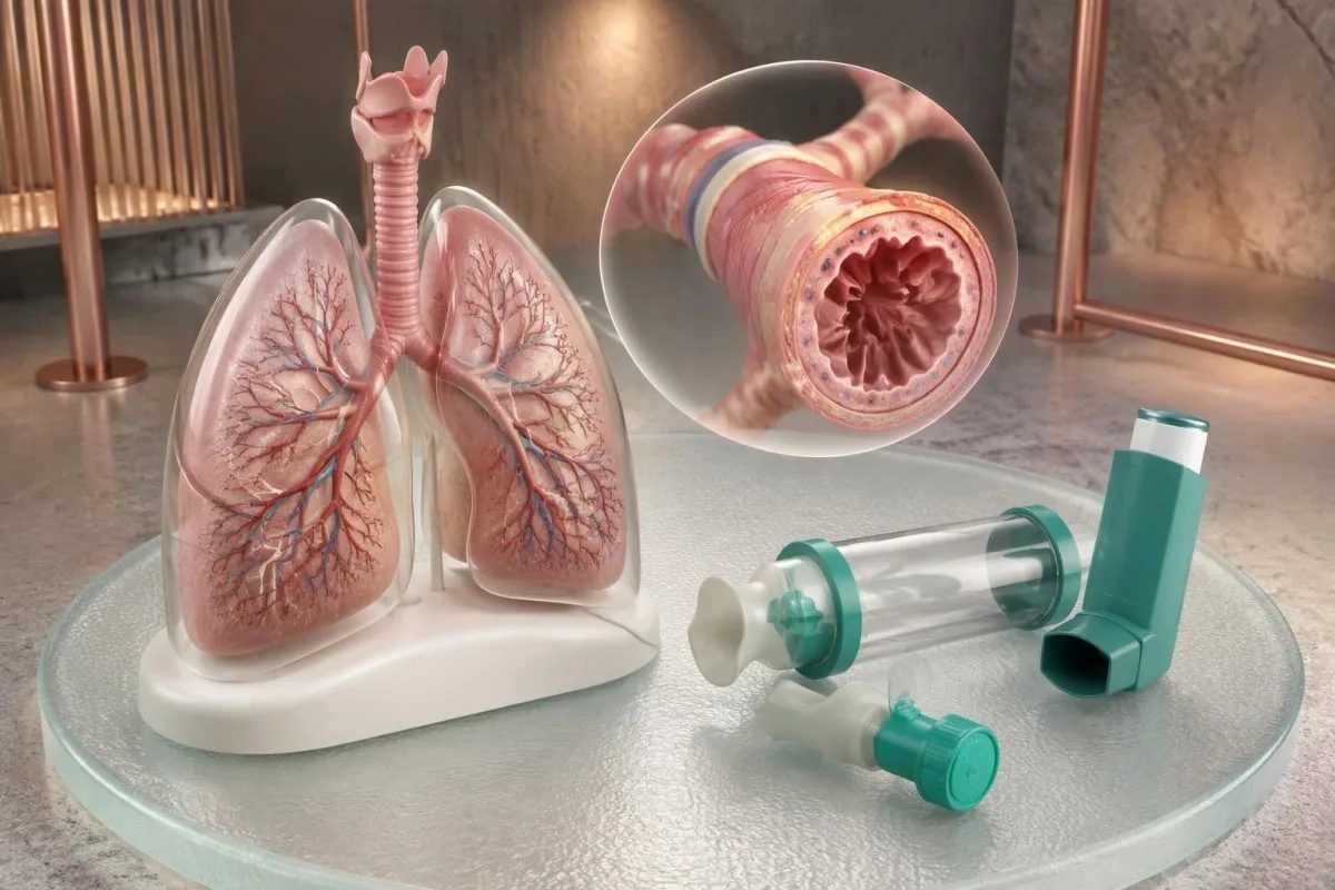
Preschool viral wheeze is not identical to established persistent asthma. Clinicians consider whether symptoms occur only during colds or also between illnesses, along with allergy history and family risk; 2 children who wheeze with the same virus can have different longer-term patterns, so a permanent label should not rest on one episode.
A treatment trial in a child younger than 5 may be assessed over approximately 8–12 weeks under NICE guidance, depending on the presentation. Improvement needs to be interpreted alongside adherence, device technique and natural recovery from a virus; a treatment response is useful evidence, but it is not an isolated laboratory-style confirmation.
Children's spirometry requires age-appropriate interpretation and coaching, not adult thresholds pasted onto smaller lungs. GINA uses an FEV1 increase of at least 12% of predicted in children as a supportive bronchodilator criterion, and a technically poor test may need repetition even if the report prints a reassuring percentage.
우리의 children's laboratory safety guide explains why age-specific ranges matter without replacing respiratory assessment. In a child, marked chest recession, inability to drink or speak normally, unusual sleepiness or worsening breathing warrants urgent help; even 1 such sign outweighs a normal previous test.
What should you bring to an asthma assessment?
Prepare for an asthma assessment by bringing your symptom pattern, medication list, inhaler devices and any previous breathing or laboratory results. A 2-week diary of symptoms, exposures and clinician-requested peak-flow readings is often more useful than a long list of unrelated tests.
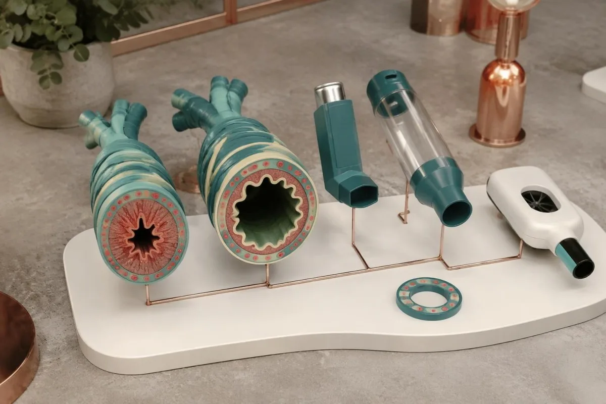
Ask the testing service which inhalers, if any, should be withheld and for how long before spirometry. Instructions vary by medication and test purpose, so do not assume a universal 12-hour rule; if symptoms worsen while following preparation instructions, use needed treatment and contact the service rather than prioritising an ideal test.
Bring the actual inhaler and spacer so a clinician can observe technique rather than relying on a description. Different devices require different inhalation patterns, and 1 correct demonstration can reveal why a prescribed treatment seemed ineffective; poor technique can look like resistant disease even when the medication choice is appropriate.
칸테스티는 AI 기반 혈액검사 분석 도구 that can help explain accompanying laboratory reports while the clinician evaluates breathing symptoms. Our AI 기술 가이드 describes the interpretation workflow, but an estimated processing time of about 60 seconds should never be confused with the time needed to establish a respiratory diagnosis.
Share only the information needed for the assessment, particularly when organising results for another family member. Our 개인정보 및 데이터 관련 정보를 확인하고 explains handling considerations; keep symptom diaries and original reports available for clinical review, and do not let uploading 1 document delay urgent care or a necessary breathing appointment.
Research publications and evidence behind asthma testing
Asthma diagnostic evidence comes primarily from respiratory guidelines and studies of airflow, airway responsiveness and inflammatory markers. The 3 external references below directly support this article; the 2 Zenodo laboratory guides are separate educational resources and are not studies validating asthma diagnosis.
GINA's 2025 strategy links a characteristic symptom history with evidence of variable expiratory airflow limitation, while the 2024 NICE/BTS/SIGN guideline specifies a different sequence of objective tests. The European Respiratory Society adult guideline supplies a third structured approach (Louis et al., 2022); comparing all 3 helps explain why clinicians may reasonably order tests in different sequences.
Kantesti의 RDW 및 적혈구 가이드 provides background when a CBC raises a separate question about anaemia and breathlessness. Its Zenodo DOI identifies that publication, not proof that RDW diagnoses asthma; even 2 abnormal red-cell indices must be interpreted as haematology findings rather than measurements of airway narrowing.
그만큼 신장 비율 해석 가이드 addresses BUN, creatinine and clinical context rather than asthma symptoms. Its separate Zenodo DOI is included for publication transparency, and neither of these 2 laboratory guides establishes the sensitivity, specificity or effectiveness of an asthma diagnostic pathway.
우리의 의료 콘텐츠 표준 distinguish guideline-supported facts from educational examples and product descriptions. Kantesti does not claim that these 2 DOI resources validate asthma detection; uncertain symptoms need clinical assessment, and emergency breathing warning signs require immediate action regardless of how recently a report was interpreted.
자주 묻는 질문
성인 천식의 초기 증상은 무엇인가요?
성인 천식의 초기 징후로는 호흡곤란이 변하는 양상, 가슴 답답함, 쌕쌕거림 또는 밤이나 운동 후 악화되는 기침이 흔히 나타납니다. 증상이 없는 기간이 있다고 해서 천식을 배제할 수는 없습니다. 기류 제한은 날마다 달라질 수 있기 때문입니다. 증상이 경미하고 안정적일 때 2주간 증상 일지를 작성하면 의료진이 증상의 패턴을 파악하는 데 도움이 됩니다. 진단을 뒷받침하려면 폐활량 측정이나 그 밖의 적절한 객관적 검사가 필요하며, 호흡이 악화되면 더 일찍 평가받아야 합니다.
천명음 없이도 천식이 있을 수 있나요?
천식은 들리는 쌕쌕거림 없이도 발생할 수 있습니다. 특히 기침이 주된 증상이거나 진찰 당시 증상이 없을 때 그렇습니다. 성인에게서 기침이 8주 넘게 지속되면 천식과 역류, 약물 효과, 비천식성 호산구성 기관지염을 비롯한 다른 원인에 대한 평가가 필요합니다. 초기 폐활량 측정 결과가 정상이어도 천식 의심이 계속되면 검사를 반복하거나 감독하에 기관지 유발 검사를 시행할 수 있습니다. 심한 호흡곤란 중에 흉부에서 호흡음이 거의 들리지 않는 것은 다른 상황이며, 공기 흐름이 위험할 정도로 감소했음을 나타낼 수 있으므로 응급 처치가 필요합니다.
천식은 왜 밤에 더 심해지나요?
Asthma can worsen at night because airway function follows daily rhythms and bedroom allergens or reflux may add triggers. GINA's symptom-control assessment treats any asthma-related nighttime waking in the previous 4 weeks as a reason to review control. Record awakenings, breathing symptoms and prescribed reliever use rather than assuming every nighttime cough is asthma. Repeated waking should prompt a clinical review, while severe breathing difficulty requires emergency care.
혈액 검사로 천식 여부를 알 수 있나요?
혈액 검사만으로는 천식을 확진할 수 없습니다. 혈액 검사는 기류 제한이 변동하는 양상을 보여 주지 못하며, 호흡기 증상의 다른 모든 원인을 배제하지도 못하기 때문입니다. 호산구 수치가 0.5 × 10^9/L를 초과할 경우 천식, 알레르기, 약물 또는 다른 질환과 관련이 있을 수 있습니다. NICE NG245에서는 병력상 천식이 의심되는 경로에서 성인의 진단 근거로 호산구 증가를 허용하지만, 이는 임상 알고리즘이지 단일 혈액 검사 결과를 해석하는 것이 아닙니다. 임상의는 증상 양상과 현지 지침에 따라 폐활량 측정, FeNO, 최대 호기 유량 모니터링 또는 기관지 유발 검사를 활용할 수도 있습니다.
폐활량 측정 결과가 정상이어도 천식이 있을 수 있나요?
정상 폐활량 측정 결과만으로 천식을 배제할 수는 없습니다. 기도 협착이 간헐적으로 발생하거나 치료로 억제될 수 있기 때문입니다. 임상의는 증상이 있을 때 폐활량 측정을 반복하거나, 2주간 하루 두 번 최대 호기 유량을 측정하도록 하거나, 감독하에 기관지 유발 검사를 요청할 수 있습니다. 일반적으로 사용되는 기준에 따르면, 기관지 확장제 투여 후 성인의 FEV1이 12% 이상이면서 200mL 이상 개선되면 변동성이 있음을 뒷받침합니다. 준비 방법은 약물과 검사에 따라 다르므로, 구체적인 의료진의 지시 없이 흡입제 사용을 중단하지 마십시오.
천식 증상은 언제 응급 상황으로 간주하고 치료해야 하나요?
호흡곤란이 심하거나, 정상적으로 말하기 어렵거나, 기진맥진하거나 혼란스러워지거나, 입술이 파랗거나 회색으로 보이거나, 처방받은 증상완화제가 효과가 없으면 천식 증상은 응급 처치가 필요합니다. 최대호기유속이 개인 최고치의 50% 미만이거나 급성 증상이 있을 때 산소포화도가 92% 이하인 경우에도 우려가 커지지만, 측정값을 확인하느라 응급 조치를 미뤄서는 안 됩니다. 똑바로 앉아 현지 응급 전화번호로 연락하고, 서면 행동 계획이나 상담원의 안내에 따라 처방받은 증상완화제를 사용하세요. 직접 운전하지 말고, 숨이 더 차지면서 가슴에서 숨소리가 거의 나지 않는다면 안심할 일이 아니라 경고 신호로 여기세요.
혈액검사 결과 보는법
검사실 보고서를 업로드하면 Kantesti의 AI가 몇 초 안에 각 바이오마커를 쉬운 말로 설명합니다. 전 세계 200만 명 이상이 사용하고 있습니다.
참고 자료
- Klein, T., Mitchell, S., & Weber, H. (2026). RDW 혈액검사: RDW-CV, MCV 및 MCHC 완전 가이드. Kantesti AI 의학 연구. DOI
- Klein, T., Mitchell, S., & Weber, H. (2026). BUN/크레아티닌 비율 설명: 신장 기능 검사 가이드. Kantesti AI 의학 연구. DOI
- Global Initiative for Asthma (2025). Global Strategy for Asthma Management and Prevention. GINA Strategy Report.
- National Institute for Health and Care Excellence, British Thoracic Society, and Scottish Intercollegiate Guidelines Network (2024). Asthma: diagnosis, monitoring and chronic asthma management (BTS, NICE, SIGN). NICE Guideline NG245.
- Louis R et al. (2022). European Respiratory Society guidelines for the diagnosis of asthma in adults. European Respiratory Journal. DOI
- 200만+분석한 검사
- 127+국가
- 100+언어
의료진 소개


E-E-A-T 신뢰 신호
경험
의사가 주도하는 검사 해석 워크플로 임상 검토.
전문적 지식
임상 맥락에서 바이오마커가 어떻게 거동하는지에 대한 검사실 의학 중심.
권위
Thomas Klein 박사의 의학적 검토를 거쳤으며, Sarah Mitchell 박사와 Hans Weber 교수의 기여가 있었습니다.
신뢰성
경고를 줄이기 위한 명확한 후속 경로가 포함된 근거 기반 해석.


의학적 면책 조항
이 글은 교육 목적만을 위한 것이며 의학적 조언을 구성하지 않습니다. 진단 및 치료 결정에는 항상 자격을 갖춘 의료 전문가와 상담하세요.
게시됨 · 편집팀에 문의
Kantesti Ltd 발행, 잉글랜드 및 웨일스에 등록된 회사, 회사 번호. 17090423, 런던, 영국.
