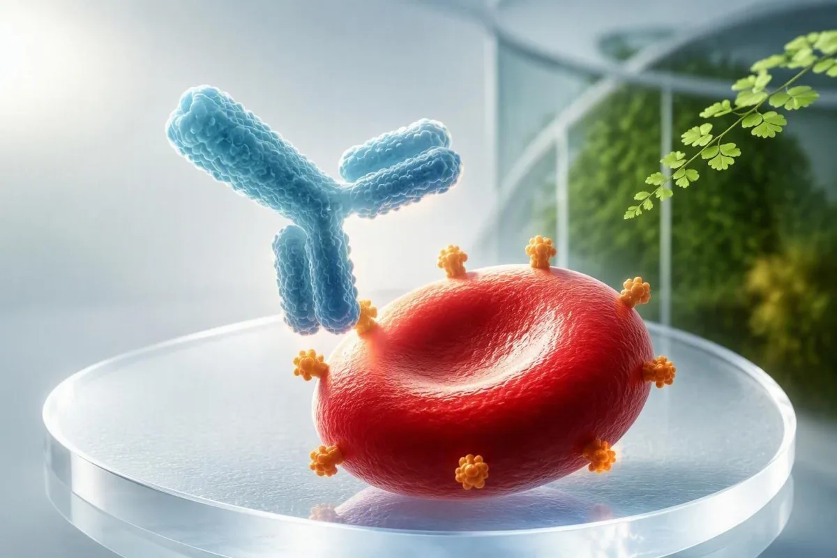
A positive red-cell antibody result tells your maternity team to identify the antibody and measure its significance. Many results never harm a baby, but a small group need carefully timed specialist monitoring.


- ការពិនិត្យស្គ្រីនវិជ្ជមាន means an unexpected red-cell antibody was detected; it does not diagnose fetal anemia by itself.
- Anti-D, anti-c and anti-K are the antibodies most consistently linked with severe fetal anemia in UK pregnancy guidance.
- A titre of 1:16 or 1:32 often triggers fetal-medicine discussion for many antibodies, although each laboratory sets its own critical threshold.
- Anti-D above 4 IU/mL or anti-c above 7.5 IU/mL merits specialist assessment under RCOG guidance.
- Anti-K requires early referral because severe fetal anemia can occur even at a low titre.
- MCA Doppler above 1.5 MoM is the usual non-invasive threshold for investigating moderate or severe fetal anemia.
- Passive anti-D from routine Rh immunoglobulin can remain detectable for roughly 8–12 weeks and is not the same as immune anti-D.
- Paternal antigen testing or fetal DNA testing can show whether the fetus carries the relevant red-cell antigen.
- Do not wait for symptoms: fetal anemia is usually silent in the pregnant person and is found through planned surveillance.
What a Positive Pregnancy Antibody Screen Actually Means
A positive antibody screen in pregnancy means your plasma contains an antibody that reacts with a red-cell antigen you do not have; it is not proof that your baby is affected. The next step is antibody identification, then a decision about whether its level, its trend, and the fetus’s inherited antigen make monitoring necessary.
The test is commonly called an indirect antiglobulin test ឬ indirect Coombs test pregnancy screen. It looks in maternal plasma for antibodies outside the ABO system, whereas the direct antiglobulin test looks for antibody already attached to red cells. Screening is normally done at booking and again around 28 weeks in the UK; the second sample matters because a clinically relevant antibody can arise after an early negative result.
In my practice, the word “positive” creates understandable alarm before anyone has explained the antibody name. A low-level anti-Lea or anti-P1 result often changes very little for the fetus, while anti-D, anti-c, or anti-K changes the pathway immediately. What positive antibody results mean is a useful wider primer, but pregnancy decisions depend on the exact red-cell specificity.
កាន់តេស្ទី គឺជា ឧបករណ៍វិភាគឈាម AI that can organize a maternity laboratory report into plain language, but it cannot determine fetal status or replace the transfusion laboratory and obstetric team. As of September 18, 2026, I advise patients to save the original report, their blood group, prior transfusion history, and the date of every anti-D injection; those four details often resolve apparent contradictions.
Why the screen can change during pregnancy
Red-cell alloantibodies may follow a previous pregnancy, a miscarriage, an ectopic pregnancy, a transfusion, or occasionally an unrecognized fetomaternal bleed. Antibody concentration can remain stable for years, disappear below assay detection, then rise briskly after renewed antigen exposure.
Which Red-Cell Antibodies Can Affect a Fetus?
Anti-D, anti-c, and anti-K are the red-cell antibodies pregnancy teams treat as highest priority because they can cause fetal anemia and newborn jaundice. Anti-E, anti-C, anti-e, anti-Fya, anti-Jka, and anti-S can also cause hemolytic disease, although the average risk is usually lower and less predictable.
Antibodies that cross the placenta are usually IgG; large IgM antibodies generally do not cross in clinically meaningful amounts. Anti-Lea, anti-Leb, anti-M when naturally occurring IgM, and anti-P1 are therefore often reassuring findings, though the transfusion laboratory still records them because they can complicate selecting compatible blood for the mother.
Anti-K behaves differently from anti-D. It can suppress fetal red-cell production as well as shorten circulating red-cell survival, so a modest titre may still matter. RCOG’s Green-top Guideline No. 65 recommends referral once anti-K is detected, rather than waiting for a numerical threshold (RCOG, 2014).
The practical question is never simply “Is an antibody present?” It is “Does the fetus have the target antigen, and is the antibody behaving in a way that could lower fetal hemoglobin?” For related red-cell patterns, our guide to ការធ្វើតេស្ត reticulocyte explains why young red-cell production is central to anemia assessment.
How Laboratories Confirm a Positive Antibody Screen
A pregnancy antibody screen positive result is confirmed by antibody identification and, when relevant, quantitative measurement or titration in the same specialist laboratory. One isolated screen result is not enough to estimate fetal danger because assay method and antibody strength both influence the report.
The laboratory tests maternal plasma against a panel of reagent red cells with known antigen profiles. The pattern of reactions identifies anti-D, anti-c, anti-K, or another specificity; it can also expose multiple antibodies. If the screen is weak or inconsistent, the laboratory may repeat it using a different enhancement medium, because cold-reacting antibodies and nonspecific reactivity can muddy the first result.
For most non-D antibodies, a titre is reported as a doubling dilution such as 1:2, 1:8, 1:16, or 1:32. A rise from 1:4 to 1:16 represents a fourfold increase, not a small change. Results should be compared in the same laboratory where possible, since a 1:16 from one method cannot safely be treated as interchangeable with 1:16 from another.
A prior transfusion can be the missing clue, particularly after surgery or postpartum hemorrhage. I ask specifically about transfusions received abroad, because patients may not realize that a unit given 10 years earlier is relevant. Blood type and reticulocyte markers provide useful context, but the antibody identification card from the blood bank is the document to keep permanently.
Titres, IU/mL and the Numbers That Trigger Referral
Titre thresholds help select monitoring intensity, but they do not directly measure fetal hemoglobin. A critical titre of 1:16 or 1:32 is commonly used for many antibodies in US practice, while UK services quantify anti-D and anti-c in IU/mL and use antibody-specific thresholds.
For anti-D, RCOG advises fetal-medicine referral when the level exceeds 4 IU/mL and identifies levels above 15 IU/mL as associated with a high risk of severe hemolytic disease. For anti-c, referral begins above 7.5 IU/mL, with risk rising substantially above 20 IU/mL. These cutoffs describe population risk; a prior severely affected pregnancy outweighs a seemingly modest current number.
Many maternity units repeat anti-D and anti-c testing every 4 weeks until 28 weeks, then every 2 weeks until delivery. For other antibodies, a repeat at 28 weeks may be enough if there is no history of hemolytic disease and the fetus is not known to be antigen-positive. The ACOG Practice Bulletin on alloimmunization emphasizes that laboratory trends and obstetric history must be read together (ACOG, 2018).
Dr. Thomas Klein’s practical advice is to ask for the actual number and unit, not merely “high” or “low.” A laboratory flag is not a fetal diagnosis, and a falling titre does not guarantee safety once Doppler surveillance has begun. Our article on ការធ្វើតេស្តគុណភាពទល់នឹងបរិមាណ explains why those result formats answer different questions.
Could This Be Passive Anti-D From an Injection?
បាទ—passive anti-D after Rh immunoglobulin can make the antibody screen positive for roughly 8 to 12 weeks, sometimes longer with sensitive assays. Passive anti-D protects against sensitization; immune anti-D means sensitization has already occurred and needs a different plan.
Routine Rh immunoglobulin is often given at 28 weeks in the UK, sometimes as two doses earlier in the third trimester, and after a potentially sensitizing event such as vaginal bleeding, abdominal trauma, or an invasive procedure. A standard UK dose is commonly 1,500 IU at 28–30 weeks, though local schedules vary. The laboratory needs the injection date, dose, and product record before it can interpret anti-D correctly.
No single laboratory test perfectly separates passive anti-D from early immune anti-D. Timing, reaction strength, previous negative screens, and whether the result persists or rises guide the interpretation. In a patient screened 6 days after prophylaxis with weak anti-D and a previously negative booking sample, passive antibody is much more likely; a rising level over several weeks is more concerning.
Do not decline indicated Rh immunoglobulin just because a report says “anti-D detected” unless your maternity unit has confirmed immune anti-D. If there is uncertainty, the transfusion laboratory and fetal-medicine clinician should decide together. Pregnancy test timing and false results is a separate issue, but it is a reminder that test timing can completely change interpretation.
Why Partner Testing Is Offered After Certain Antibodies
Partner antigen testing estimates whether the fetus can inherit the red-cell antigen targeted by the maternal antibody. If the biological father lacks that antigen, the fetus cannot be antigen-positive for it and antibody-related fetal anemia from that specificity is not expected.
A father who is homozygous for the relevant antigen passes it to every fetus; a heterozygous father passes it about 50% of the time. For example, paternal K-negative status makes anti-K unable to affect that fetus, while K-positive status needs zygosity or fetal genotyping to refine the risk. This is a risk calculation, not a test of paternity.
Partner testing should be offered sensitively and never assumed to be straightforward. Some patients prefer direct fetal testing, use donor conception, or cannot access partner testing safely. In those situations, treating the fetus as potentially antigen-positive until non-invasive fetal genotyping is available is clinically sensible.
A positive antibody screen can also affect maternal care if an emergency transfusion becomes necessary. The maternity team should notify the blood bank early so antigen-negative units can be sourced; this is particularly relevant with multiple antibodies. Read our អេម៉ូក្លូប៊ីន និង hematocrit ណែនាំ for why pregnancy anemia still deserves separate attention.
Non-Invasive Fetal Genotyping: What It Can Tell You
Cell-free fetal DNA testing from maternal plasma can determine fetal D, C, c, E, e, and K antigen status without sampling fetal fluid in many specialist services. A fetus reported antigen-negative for the relevant antibody usually does not need surveillance for anemia caused by that antibody.
Fetal genotyping is commonly performed from around 16 សប្តាហ៍ for most Rh antigens and may be feasible from around 20 សប្តាហ៍ for K because of assay performance and local protocol. A result may be inconclusive when fetal DNA fraction is low, especially early in pregnancy or with higher maternal body weight. An inconclusive result is not reassuring; clinicians usually repeat it or monitor as if the fetus is antigen-positive.
កាន់តេស្ទី គឺជា សេវាកម្មបកស្រាយការធ្វើតេស្តរបស់ AI lab that can help patients file serial maternal antibody results by date and unit, but fetal genotyping results require clinician review because test availability and confidence intervals differ between laboratories. Our ស្តង់ដារផ្ទៀងផ្ទាត់ផ្នែកវេជ្ជសាស្ត្រ explain why laboratory source, method, and clinical oversight remain essential.
Invasive testing with amniocentesis or fetal blood sampling is now rarely used solely to establish antigen status when cell-free DNA is available. It still has a role when results conflict, when fetal anemia needs confirmation, or when another diagnostic question justifies the procedure. The shift toward non-invasive testing has reduced procedure-related risk, which most patients find genuinely reassuring.
How MCA Doppler Monitoring Detects Fetal Anemia
Middle cerebral artery peak systolic velocity (MCA-PSV) Doppler is the main non-invasive test for moderate or severe fetal anemia in an antigen-positive at-risk fetus. A value above 1.5 multiples of the median (MoM) prompts urgent fetal-medicine assessment, often including consideration of fetal blood sampling.
Anemic fetal blood is less viscous, so it moves faster through the middle cerebral artery. The sonographer measures the vessel near its origin with the Doppler beam aligned as closely as possible to flow; a poor angle can falsely increase the number. This is why a Doppler scan should be performed in an experienced fetal-medicine unit rather than interpreted from a single still image.
Monitoring commonly starts around 18 to 20 weeks when the fetus is antigen-positive and maternal antibody levels or history justify it. Scans may be weekly or every 1–2 weeks, depending on the antibody, concentration, and prior pregnancy outcome. MCA-PSV is less reliable after about 35 weeks, when delivery may become safer than invasive confirmation in some cases.
The scan is watching a physiological consequence, not the antibody itself. Hydrops—fluid accumulation in at least two fetal compartments—is a late and serious sign, so clinicians aim to intervene long before it appears. For another pregnancy-specific laboratory emergency pattern, see our explanation of HELLP syndrome results.
What Happens If Fetal Anemia Is Suspected
Suspected significant fetal anemia leads to urgent fetal-medicine review, not automatic treatment. The specialist team weighs gestational age, MCA-PSV, ultrasound findings, and previous pregnancy history before recommending fetal blood sampling, intrauterine transfusion, early delivery, or close repeat assessment.
If fetal blood sampling confirms significant anemia, an intrauterine transfusion may be given through the umbilical cord under continuous ultrasound guidance. Donor cells are selected to be antigen-negative for the maternal antibody, CMV-safe according to local policy, irradiated, and crossmatched against maternal plasma. These procedures are concentrated in expert centers because timing and technique matter enormously.
The first transfusion is often performed after 18 weeks, although individual circumstances vary. Repeat transfusions may be needed every 2–4 weeks because donor cells gradually age and the fetus continues to grow. A successful transfusion does not cure maternal alloimmunization; it buys time for safer fetal maturation.
I have seen families focus on the procedure and miss a practical detail: delivery location. A baby at risk of hemolytic disease should be delivered where neonatal bilirubin testing, compatible blood, and intensive care are available if needed. High bilirubin warning patterns explain why newborn jaundice is monitored so closely after birth.
Why a Previous Affected Pregnancy Changes the Plan
A prior baby with anemia, neonatal exchange transfusion, severe jaundice, or an intrauterine transfusion predicts risk better than a single current antibody titre. In a subsequent antigen-positive pregnancy, fetal-medicine teams may begin surveillance earlier even when the first measured antibody level is low.
Immune memory can produce a sharp antibody rise after fetal antigen exposure, sometimes before the routine 28-week screen. Anti-D and anti-c concentrations can lag behind fetal risk, particularly when there was severe disease in a prior pregnancy. This is one reason clinicians ask about phototherapy, neonatal anemia, and transfusions—not just whether a previous pregnancy was “normal.”
Records from a prior delivery can be more useful than a verbal memory of the antibody name. Ask for the newborn discharge letter, direct antiglobulin test, bilirubin peak, hemoglobin nadir, and any transfusion details. A bilirubin level of 20 mg/dL (342 µmol/L) has a very different implication in a well newborn at 4 days than in a rapidly jaundiced infant at 18 hours.
Kantesti AI can preserve a dated trend view for maternal lab reports, yet historic neonatal records should sit alongside it rather than be reduced to a graph. Our មគ្គុទេសក៍លទ្ធផលតាមរយៈពេលវែង (longitudinal result guide) offers practical ideas for organizing those records before the first fetal-medicine appointment.
Why Your Own Transfusion Plan Also Matters
A clinically significant maternal red-cell antibody can delay the supply of compatible blood for the pregnant person, even when the fetus is antigen-negative. Antigen-negative, crossmatch-compatible units may need to be sourced from a regional center, especially with anti-K, anti-Jka, or multiple antibodies.
This issue matters most if there is placenta previa, suspected placenta accreta spectrum, severe anemia, a planned caesarean birth, or any elevated hemorrhage risk. The obstetric and transfusion teams can arrange a current group-and-screen sample and reserve appropriate units. In most uncomplicated pregnancies, no blood is pre-booked, but the antibody record remains visible.
Tell your care team about every known antibody, even if a newer screen is negative. Some antibodies fall below detection yet return after transfusion exposure, and electronic records do not always follow patients across countries. A wallet antibody card or a clear phone photo of the formal laboratory report is surprisingly useful in an emergency.
Maternal hemoglobin affects resilience if blood loss occurs, but it does not tell us whether the fetus is anemic. Iron deficiency can coexist with alloimmunization, which is why ចន្លោះប្រេននីនក្នុងពេលមានផ្ទៃពោះ deserve a separate, early conversation.
Common Misconceptions About a Positive Result
A positive antibody screen does មិនមែន mean you have an autoimmune disease, an infection, or that your baby has the same blood group as you. It means an antibody was detected against a red-cell antigen, and only some antibodies can cross the placenta and affect an antigen-positive fetus.
The phrase “Coombs positive” is often used loosely, which creates confusion. The indirect Coombs test in pregnancy finds free antibodies in plasma; a direct Coombs test on a newborn’s cells may support hemolytic disease after birth, but neither test alone measures anemia severity. These tests answer different clinical questions at different times.
Another misconception is that dietary changes can lower an alloantibody titre. They cannot. Good nutrition supports maternal health, but there is no food, supplement, or detox that removes anti-D, anti-c, or anti-K; be wary of anyone offering one. For safe pregnancy supplement decisions, our lab-guided supplement guide keeps the focus on deficiencies that can actually be corrected.
As Dr. Thomas Klein, I also remind patients that anxiety can make them read every antibody result as a countdown to harm. Most positive screens do not lead to intrauterine treatment, and planned monitoring is what turns a rare serious risk into a managed clinical pathway.
Questions to Ask at Your Next Maternity Appointment
សូមសួររក exact antibody name, current level or titre, previous result, and whether the fetus is known or assumed to be antigen-positive. Those four answers determine nearly every next step after a positive antibody screen in pregnancy.
Useful questions include: Is this passive or immune anti-D? Was my result measured in IU/mL or titre? Do I need paternal testing or fetal genotyping? When is my next sample, and what specific result would trigger MCA Doppler? Write the answers down; the distinction between anti-D and anti-G, for instance, can change whether Rh immunoglobulin remains appropriate.
Ask who will coordinate results after hours and where you should deliver if the fetus becomes at risk. You should also ask whether the blood bank has been notified if you have a planned caesarean birth or a history of postpartum bleeding. Preparing for a useful baseline draw includes simple habits that prevent missing key report details.
កាន់តេស្ទី គឺជា វេទិកាបកស្រាយសូចនាករជីវសាស្ត្រដោយ AI designed to make laboratory language easier to follow across 75+ languages, but a pregnancy antibody screen needs obstetric-led decisions and a transfusion-laboratory safety net. Readers can review how our clinical team works on the ក្រុមប្រឹក្សាប្រឹក្សាវេជ្ជសាស្ត្រ; in this setting, responsible interpretation means knowing exactly when AI should hand the decision back to the specialist.
When to Seek Urgent Care and What Happens After Birth
A positive red-cell antibody screen alone is not usually an emergency, but reduced fetal movements, vaginal bleeding, abdominal trauma, or labour symptoms require prompt maternity assessment regardless of antibody level. After birth, an at-risk newborn may have cord blood testing for blood group, direct antiglobulin test, hemoglobin, and bilirubin.
Fetal anemia itself rarely causes a symptom the pregnant person can feel, which is why keeping Doppler appointments matters. Contact your maternity triage unit the same day for reduced movements; do not wait until the next antibody blood test. Sudden swelling, severe headache, visual changes, or upper abdominal pain have other pregnancy causes and need urgent evaluation too.
Newborn bilirubin can rise quickly in immune hemolysis, sometimes within the first ជៀសវាងការហាត់ប្រាណខ្លាំងសម្រាប់. Treatment may include frequent feeding support, phototherapy, intravenous immunoglobulin in selected cases, or exchange transfusion for severe hyperbilirubinemia; the neonatal team uses age-specific bilirubin thresholds rather than one universal number. Late anemia can occur at 2–6 សប្តាហ៍, so follow-up hemoglobin checks may be arranged even after a seemingly calm discharge.
The best outcome is often quiet and uneventful: an identified low-risk antibody, an antigen-negative fetus, or normal serial Dopplers followed by routine neonatal observation. If you are comparing laboratory reports, our AI technology guide describes the limits of automated interpretation; no app should reassure or alarm you instead of the maternity team when fetal monitoring is due.
សំណួរដែលសួរញឹកញាប់
តើការធ្វើតេស្ដរកអង្គបដិបក្ខវិជ្ជមានមានញឹកញាប់ប៉ុណ្ណាក្នុងពេលមានផ្ទៃពោះ?
ការធ្វើតេស្តរកអង្គបដិបក្ខក្នុងកោសិកាឈាមក្រហមមានលទ្ធផលវិជ្ជមានកើតឡើងប្រមាណ 1% ទៅ 2% នៃការមានផ្ទៃពោះក្នុងក្រុមប្រជាជនដែលទទួលការពិនិត្យជាច្រើនក្នុងប្រទេសចំណូលខ្ពស់ ទោះបីជាអត្រានេះប្រែប្រួលទៅតាមពូជពង្ស ប្រវត្តិនៃការបញ្ចូលឈាម និងការប្រើប្រាស់ថ្នាំ Rh immunoglobulin ក្នុងតំបន់ក៏ដោយ ។ មានតែផ្នែកតូចប៉ុណ្ណោះនៃអង្គបដិបក្ខទាំងនេះដែលអាចបង្កឱ្យមានជំងឺឈាមហូរក្នុងទ Blister របស់គភ៌ និងទារកទើបកើត ។ Anti-D, anti-c, និង anti-K ត្រូវបានត្រួតពិនិត្យយ៉ាងជិតស្និទ្ធបំផុត ដោយសារតែពួកវាមានទំនាក់ទំនងច្បាស់លាស់បំផុតជាមួយនឹងភាពស្លេកស្លាំងរបស់គភ៌ដែលមានសារៈសំខាន់ខាងគ្លីនិក ។ មន្ទីរពិសោធន៍ត្រូវតែកំណត់អត្តសញ្ញាណអង្គបដិបក្ខជាក់លាក់មុនពេលអាចប៉ាន់ប្រមាណហានិភ័យផ្ទាល់ខ្លួនបាន ។.
What antibody titre is dangerous during pregnancy?
For many non-D red-cell antibodies, a titre of 1:16 or 1:32 is treated as a critical threshold for fetal-medicine referral, but the cutoff is laboratory- and antibody-specific. UK guidance uses quantified levels for some antibodies: anti-D above 4 IU/mL and anti-c above 7.5 IU/mL warrant referral, while anti-K warrants referral when first detected. A previous pregnancy affected by fetal anemia overrides a reassuring-looking current titre. Titres estimate risk; MCA Doppler assesses whether fetal anemia may actually be developing.
Can a positive antibody screen be caused by the anti-D injection?
Yes, Rh immunoglobulin can produce passive anti-D on an antibody screen for approximately 8 to 12 weeks after administration. Passive anti-D is expected after prophylaxis and does not mean a person has become sensitized. The transfusion laboratory distinguishes passive from immune anti-D using the injection date, prior screens, reaction strength, and whether the level persists or rises. Never skip recommended Rh immunoglobulin solely because anti-D appears on a report without confirmation from the maternity team.
If my partner is antigen-negative, does my baby need monitoring?
If the biological father is confirmed negative for the specific red-cell antigen, the fetus cannot inherit that antigen and is not at risk from that maternal antibody. For example, a K-negative father means maternal anti-K cannot cause fetal anemia in that pregnancy. Partner testing is optional and must be handled sensitively, because family circumstances vary. Cell-free fetal DNA testing can often provide a direct non-invasive answer from around 16 to 20 weeks, depending on the antigen and local service.
Can red-cell antibodies cause miscarriage?
Red-cell alloantibodies do not usually cause early miscarriage; their main fetal effect is anemia after the placenta transfers IgG antibodies, most often later in pregnancy. Very severe untreated alloimmunization can lead to hydrops or fetal loss, but modern antigen testing, MCA Doppler, and intrauterine transfusion have greatly changed that outlook. A miscarriage or ectopic pregnancy can, however, be a sensitizing event that contributes to antibody formation. Rh immunoglobulin may be recommended after certain early-pregnancy events for RhD-negative people who are not already immune anti-D positive.
Will my baby need treatment after delivery if my antibody screen is positive?
A baby needs treatment after delivery only if they inherited the relevant antigen and show evidence of hemolysis, anemia, or rising bilirubin. Cord blood testing may include blood group, direct antiglobulin test, hemoglobin, and bilirubin, followed by repeat bilirubin measurement within the first 24 hours when risk is present. Phototherapy is common and non-invasive; more intensive treatments are reserved for severe cases. Babies can also develop late anemia at 2 to 6 weeks, so some need outpatient hemoglobin follow-up.
ទទួលការវិភាគឈាមដោយ AI ដែលមានថាមពលថ្ងៃនេះ
ចូលរួមជាមួយអ្នកប្រើប្រាស់ជាង 2 លាននាក់នៅទូទាំងពិភពលោក ដែលទុកចិត្ត Kantesti សម្រាប់ការវិភាគលទ្ធផលតេស្តមន្ទីរពិសោធន៍ភ្លាមៗ និងត្រឹមត្រូវ។ ផ្ទុកឡើងលទ្ធផលពិនិត្យឈាមរបស់អ្នក ហើយទទួលការបកស្រាយយ៉ាងទូលំទូលាយនៃសញ្ញាសម្គាល់ (biomarkers) 15,000+ ក្នុងរយៈពេលប៉ុន្មានវិនាទី។.
📚 ឯកសារស្រាវជ្រាវដែលបានយោង
Klein, T., Mitchell, S., & Weber, H. (2026). ក្រុមស្រាវជ្រាវ Kantesti។ (2026)។ មគ្គុទេសក៍សិក្សាអំពីការសិក្សាជាតិដែក (Iron Studies Guide): TIBC, ការឆ្អែតជាតិដែក (Iron Saturation) និងសមត្ថភាពចង (Binding Capacity)។ Zenodo។..។ ការស្រាវជ្រាវវេជ្ជសាស្ត្រដោយ AI របស់ Kantesti។.
Klein, T., Mitchell, S., & Weber, H. (2026). ក្រុមស្រាវជ្រាវ Kantesti។ (2026)។ ជួរធម្មតា aPTT: D-Dimer, មគ្គុទេសក៍ការកកឈាម Protein C (Protein C Blood Clotting Guide)។ Zenodo។..។ ការស្រាវជ្រាវវេជ្ជសាស្ត្រដោយ AI របស់ Kantesti។.
📖 ឯកសារយោងវេជ្ជសាស្ត្រខាងក្រៅ
សមាគមគ្រូពេទ្យឯកទេសសម្ភព និងរោគស្ត្រីអាមេរិក (American College of Obstetricians and Gynecologists) (2018)។. ACOG Practice Bulletin No. 192: Management of Alloimmunization During Pregnancy.។ Obstetrics & Gynecology។.
Royal College of Obstetricians and Gynaecologists (2014). The Management of Women with Red Cell Antibodies during Pregnancy: Green-top Guideline No. 65. RCOG Green-top Guideline.
Moise KJ Jr (2008). Management of Rhesus Alloimmunization in Pregnancy.។ Obstetrics & Gynecology។.
⚕️ ការបដិសេធផ្នែកវេជ្ជសាស្ត្រ
អត្ថបទនេះមានគោលបំណងសម្រាប់ការអប់រំប៉ុណ្ណោះ ហើយមិនមែនជាការផ្តល់ដំបូន្មានផ្នែកវេជ្ជសាស្ត្រទេ។ សូមពិគ្រោះជាមួយអ្នកផ្តល់សេវាសុខភាពដែលមានសមត្ថភាពជានិច្ច សម្រាប់ការសម្រេចចិត្តអំពីការធ្វើរោគវិនិច្ឆ័យ និងការព្យាបាល។.
សញ្ញាទុកចិត្ត E-E-A-T
បទពិសោធន៍
ការពិនិត្យព្យាបាលដោយវេជ្ជបណ្ឌិតលើដំណើរការការបកស្រាយលទ្ធផលមន្ទីរពិសោធន៍។.
ជំនាញ
ផ្តោតលើវិទ្យាសាស្ត្រមន្ទីរពិសោធន៍ថា សញ្ញាសម្គាល់ (biomarkers) មានឥរិយាបថយ៉ាងដូចម្តេចក្នុងបរិបទព្យាបាល។.
ភាពមានសិទ្ធិអំណាច
សរសេរដោយវេជ្ជបណ្ឌិត Thomas Klein ជាមួយការពិនិត្យឡើងវិញដោយវេជ្ជបណ្ឌិត Sarah Mitchell និងសាស្ត្រាចារ្យវេជ្ជបណ្ឌិត Hans Weber។.
ភាពគួរឱ្យទុកចិត្ត
ការបកស្រាយដោយផ្អែកលើភស្តុតាង ជាមួយផ្លូវបន្តតាមដានច្បាស់លាស់ ដើម្បីកាត់បន្ថយការភ័យខ្លាច។.