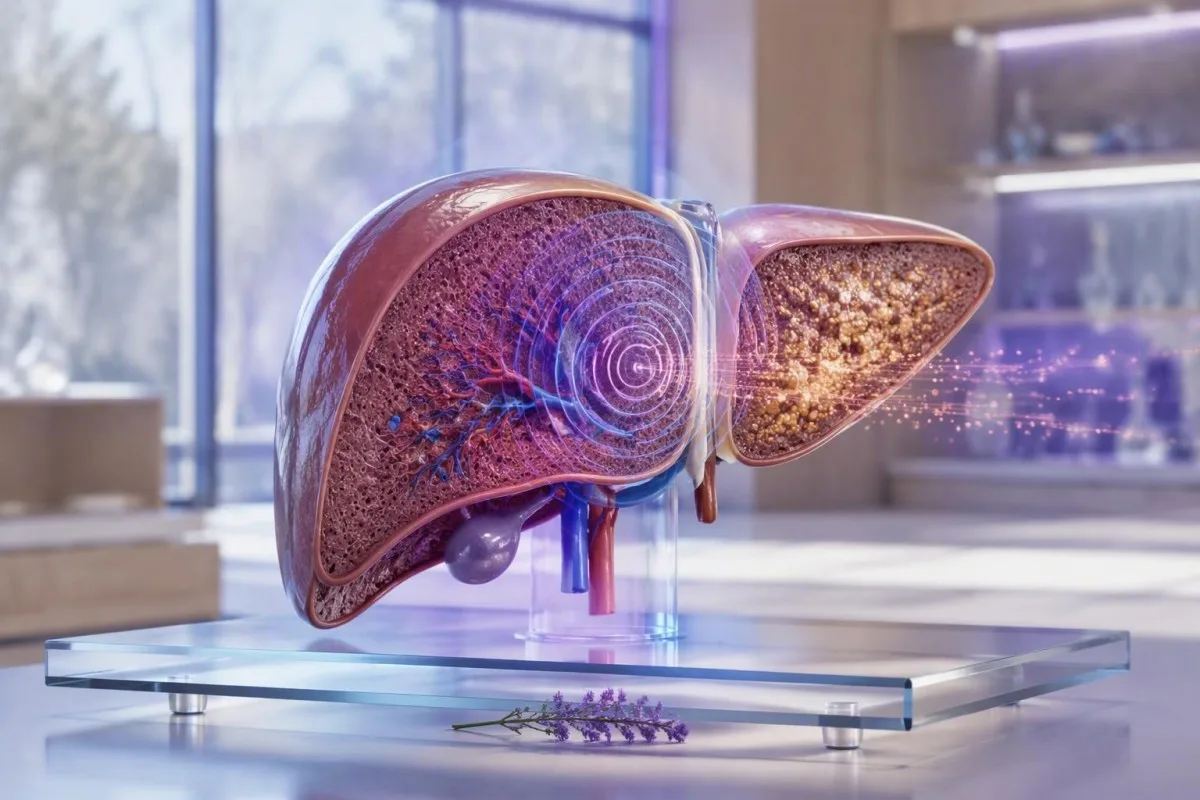
FibroScan-ის შედეგები აფასებს ღვიძლის სიმკვრივეს კპა-ში და ღვიძლის ცხიმს CAP ქულით; ისინი მარტო არ დიაგნოსტირებენ ფიბროზს ან ციროზს. 2026 წლის 28 სექტემბრის მდგომარეობით, ყველაზე სასარგებლო ანგარიში აერთიანებს რიცხვს, სკანირების ხარისხს, მარხვის სტატუსს, ღვიძლის სისხლის ანალიზებს და მიზეზს, რის გამოც ჩაიტარეთ სკანირება.


- ღვიძლის სიმკვრივის გაზომვა 8 კპა-ზე ნაკლები, როგორც წესი, ნაკლებად სავარაუდოს ხდის მოწინავე ფიბროზს მეტაბოლური ღვიძლის დაავადების დროს, როდესაც სკანირება სანდოა.
- FibroScan kPa ქულა 8-12 კპა არის განუსაზღვრელი ზონა, რომელიც ხშირად საჭიროებს სისხლის ანალიზის კონტექსტს ან განმეორებით შეფასებას.
- უფრო მაღალი სიმკვრივე 12-15 კპა-ზე მეტი იწვევს შეშფოთებას მოწინავე ფიბროზის ან ციროზის გამო, მაგრამ ასევე შეიძლება გაზარდოს შუპმა, ჰეპატიტმა, ქოლესტაზმა და კვებამ.
- CAP ქულის მნიშვნელობა დაკავშირებულია ღვიძლის ცხიმთან, არა ნაწიბუროვან ქსოვილთან; დაახლოებით 238, 260 და 290 dB/m არის გავრცელებული კვლევითი ზღვრები მზარდი სტეატოზისთვის.
- უზმოზე ყოფნა მინიმუმ 3 საათის განმავლობაში ტრანსეური ელასტოგრაფიამდე ამცირებს კვებასთან დაკავშირებულ ღვიძლის სიმკვრივის ცრუ მატებას.
- სანდო სკანირებები ჩვეულებრივ მოიცავს მინიმუმ 10 ვალიდურ გაზომვას და IQR/მედიანას თანაფარდობას 30% ან ნაკლებს.
- სასწრაფო გადახედვა გამართლებულია სიყვითლის, დაბნეულობის, სისხლიანი ღებინების, შავი განავლის ან მუცლის შებერილობისას, FibroScan-ის რიცხვის მიუხედავად.
- სპეციალისტის მეთვალყურეობა უფრო მიზანშეწონილია, ვიდრე თვითდიაგნოსტიკა, როდესაც სიმტკიცე არის 8 კპა ან მეტი, თრომბოციტები დაბალია, ან ღვიძლის ფერმენტები რჩება პათოლოგიური.
რას ზომავს FibroScan-ის ანგარიშის ორი მთავარი რიცხვი
FibroScan-ის შედეგები ჩვეულებრივ აჩვენებს ორ განსხვავებულ გაზომვას: ღვიძლის სიმტკიცეს კილოპასკალებში (კპა) და ცხიმის შესუსტებას დეციბელებში მეტრზე (dB/m). უფრო მაღალი კპა მნიშვნელობა შეიძლება ასახავდეს ფიბროზს, მაგრამ ასევე შეიძლება დროებით გაიზარდოს ბოლო კვების, აქტიური ჰეპატიტის, ნაღვლის ბლოკირების ან ღვიძლის გარშემო სითხის წნევის გამო.
ღვიძლის სიმკვრივის გაზომვა იყენებს ვიბრაციით კონტროლირებად ტრანზიენტულ ელასტოგრაფიას: მცირე მექანიკური იმპულსი გადის ღვიძლში და მოწყობილობა აფასებს, თუ რამდენად სწრაფად მოძრაობს ეს ტალღა. უფრო მყარი ქსოვილი უფრო სწრაფად გადასცემს ტალღას. ჩემს კლინიკაში გავრცელებული შეცდომაა 9.5 კპა შედეგის დიაგნოზად მიჩნევა და არა ალბათობის სიგნალად, რომელიც უნდა შემოწმდეს პირის ღვიძლის დაავადების მიზეზთან მიმართებაში.
CAP, ან კონტროლირებადი ატენუაციის პარამეტრი, აფასებს, თუ რამდენად სუსტდება ულტრაბგერითი სიგნალი ღვიძლში გავლისას. CAP იზომება dB/m-ში, ჩვეულებრივ დაახლოებით 100-400 dB/m-ის სკალაზე და ძირითადად ასახავს სტეატოზს. მაღალი CAP ქულა არ ამტკიცებს სტეატოჰეპატიტს, ხოლო დაბალი CAP ქულა არ გამორიცხავს ფიბროზს ალკოჰოლის, ვირუსული ჰეპატიტის, აუტოიმუნური დაავადების ან რკინის ჭარბი დაგროვებისგან.
კანტესტი არის AI სისხლის ტესტის ანალიზატორი რომელიც ათავსებს ღვიძლის ფერმენტებს, თრომბოციტებს, გლუკოზას, ლიპიდებს და რკინის მარკერებს გამოსახულების შედეგების გვერდით, ნაცვლად იმისა, რომ წაიკითხოს რომელიმე ცალ-ცალკე. ეს განსხვავება მნიშვნელოვანია: 7.2 კპა შედეგი ნორმალური თრომბოციტებით და ნორმალური ბილირუბინით ნიშნავს რაღაცას სრულიად განსხვავებულს, ვიდრე 7.2 კპა დაცემული თრომბოციტებით და მომატებული INR-ით. დაიწყეთ FIB-4 გაანგარიშება როდესაც გაქვთ საჭირო ლაბორატორიული მნიშვნელობები.
რატომ ვერ ასახელებს ერთი რიცხვი დაავადებას
FibroScan არ განსაზღვრავს სიმტკიცის მიზეზს. გულის უკმარისობის, მწვავე ალკოჰოლთან დაკავშირებული ჰეპატიტის ან დაბლოკილი სანაღვლე სადინრის მქონე პირს შეიძლება ჰქონდეს კპა მნიშვნელობა დიაპაზონში, რომელიც სხვაგვარად ასოცირდება მნიშვნელოვან ფიბროზთან. ამრიგად, კლინიკური კითხვა არ არის “რა სტადიაზე ვარ?”, არამედ “არის თუ არა ეს შედეგი სანდო, მუდმივი და შეესაბამება თუ არა ჩემს ღვიძლის ფართო შეფასებას?”
როგორ წავიკითხოთ FibroScan kPa ქულა ფიბროზის გადაჭარბებული შეფასების გარეშე
FibroScan კპა ქულა ზომავს წინააღმდეგობას შერეული ტალღის მიმართ, არა უშუალოდ ნაწიბუროვანი ქსოვილის. ბევრ ზრდასრულ ადამიანში მეტაბოლური დისფუნქციით გამოწვეული სტეატოზური ღვიძლის დაავადებით, 8 კპა-ზე ნაკლები სანდო მნიშვნელობა აქვს კარგი უარყოფითი პრედიქტიული ღირებულება მოწინავე ფიბროზის მიმართ.
კპა სკალა არ არის ლინეარული კლინიკური მნიშვნელობით. 5-დან 7 კპა-მდე ცვლილება შეიძლება ასახავდეს ჩვეულებრივ ბიოლოგიურ ან ტექნიკურ ვარიაციას, ხოლო 9-დან 15 კპა-მდე მუდმივი ცვლილება უფრო შემაშფოთებელია - იმ პირობით, რომ იგივე ზონდი, მომზადება და დაავადების კონტექსტი მოქმედებს. EASL-ის მითითებები განიხილავს ღვიძლის სიმტკიცეს 8 კპა-ზე ქვემოთ, როგორც სასარგებლოს კომპენსირებული მოწინავე ქრონიკული ღვიძლის დაავადების გამოსარიცხად რამდენიმე გავრცელებულ სიტუაციაში (EASL, 2021).
სხვადასხვა მდგომარეობას სჭირდება განსხვავებული ზღურბლები. დაუზოგავ ჰეპატიტი C-ის დროს, 7-8 კპა-ის მნიშვნელობები ხშირად გამოიყენებოდა, როგორც შესაძლო მნიშვნელოვანი ფიბროზის ზღურბლები, ხოლო ციროზის ზღურბლები ჩვეულებრივ 12-14 კპა-ის ფარგლებშია. მეტაბოლური ღვიძლის დაავადების დროს, სიმსუქნე და XL ზონდი ცვლის მუშაობას, ამიტომ ჰეპატიტის კვლევის ზღურბლის გადატანა შეიძლება შეცდომაში შეგვიყვანოს.
მე გადავხედე სკანებს გამხდარ პაციენტებში 6.5 კპა მაჩვენებლით და მნიშვნელოვანი მეტაბოლური რისკით, შემდეგ უფრო დიდ პაციენტებში 10 კპა მაჩვენებლით, რომლებიც მიღებული იყო ცუდი ხარისხის M-ზონდის მცდელობებით. არცერთი არ უნდა იყოს შემთხვევით იარლიყი. ფართო კლინიკური კონტექსტისთვის, იხილეთ ჩვენი MASLD ტესტირების გზამკევი.
რატომ არის ერთეული კპა
კილოპასკალი არის წნევის ერთეული. FibroScan გარდაქმნის ტალღის გავრცელების მონაცემებს ქსოვილის სიმტკიცის შეფასებად, ამიტომ შედეგები ნაჩვენებია კპა-ში და არა ფიბროზის პროცენტულად. 15 კპა ღვიძლის სიმტკიცის გაზომვა არ არის “ორჯერ უფრო ნაწიბუროვანი” ვიდრე 7.5 კპა.
ღვიძლის სიმკვრივის შედეგების პრაქტიკული ფიბროზის რისკის ზოლები
მრავალი ზრდასრული ადამიანისთვის, რომლებსაც აქვთ საეჭვო მეტაბოლური ღვიძლის დაავადება, 8 კპა-ზე ნაკლები არის დაბალი რისკის ზოლი, 8-12 კპა არის დაუზუსტებელი, ხოლო 12-15 კპა-ზე ზემოთ არის მაღალი რისკის ზოლი მოწინავე ფიბროზისთვის. ეს არის ტრიაჟის ზოლები, არა უნივერსალური ფიბროზის სტადიები ან სპეციალისტის შეფასების შემცვლელი.
EASL's 2021 non-invasive test guidance uses 8 კპა-ზე ნაკლები to help exclude compensated advanced chronic liver disease and 12-15 kPa or higher to help rule it in, depending on the underlying disease and test quality (EASL, 2021). Values around 8-12 kPa are where I most often avoid reassuring or alarming language until I see platelets, AST, ALT, bilirubin, albumin, and the scan reliability metrics.
A reliable reading of 20-25 kPa or more can raise concern for clinically significant portal hypertension, especially when platelets are below 150 × 10^9/L. It still does not diagnose varices or replace clinical examination, ultrasound, or endoscopy. The combination of a high kPa score and thrombocytopenia is more meaningful than either finding alone.
A 52-year-old endurance runner I saw had a stiffness result of 11.8 kPa after a viral illness, AST 89 IU/L, and ALT 104 IU/L. A repeat scan after recovery and appropriate fasting was 7.1 kPa. If liver enzymes remain high, use our guide to სასაზღვრო ALT შემდგომი კონტროლი rather than assuming that one scan settled the question.
CAP ქულის მნიშვნელობა: რას ამბობს dB/m შედეგი ღვიძლის ცხიმის შესახებ
A CAP score estimates liver fat, with higher dB/m values generally indicating more steatosis. It does not measure fibrosis, liver-cell injury, or whether fat is causing progressive disease.
Research thresholds often place mild steatosis near 238 dB/m, moderate steatosis near 260 dB/m, and more marked steatosis near 290 dB/m. Karlas and colleagues' individual-patient meta-analysis found useful CAP cutoffs, but also showed substantial overlap between categories (Karlas et al., 2017). A score of 275 dB/m is therefore an estimate, not a precise liver-fat percentage.
CAP can run higher with greater body mass, diabetes, and use of the XL probe; probe-specific reference ranges are not perfectly interchangeable. The reading also has less clarity at the individual level than many reports imply. I tell patients that CAP is best used to monitor direction over time under comparable conditions, not to chase a single “ideal” dB/m target.
Fatty liver often travels with triglyceride elevation, glucose dysregulation, and high fasting insulin. Our review of fasting insulin signals explains why insulin resistance can matter even when an HbA1c is not yet in the diabetes range.
CAP is not a severity score for liver damage
A CAP score of 320 dB/m may reflect substantial fat while stiffness remains 5.5 kPa, which is reassuring about current advanced fibrosis risk but not a free pass. Conversely, a CAP score below 238 dB/m can coexist with advanced fibrosis from hepatitis C, alcohol exposure, autoimmune hepatitis, or previous steatotic injury.
რატომ ცვლის მარხვა FibroScan-ის შედეგებს და რამდენი ხანი უნდა იმარხულოთ
Fasting for at least 3 hours before FibroScan is the usual practical standard because eating can temporarily increase liver stiffness. Water is normally acceptable, but follow the testing centre's instructions if medication timing or diabetes management is involved.
After a meal, blood flow through the portal circulation increases and can push stiffness readings upward for roughly 1.5-3 hours. The effect may be clinically relevant in people near an 8 or 12 kPa decision boundary. This is one reason a non-fasting 9.0 kPa result should not automatically trigger a fibrosis label.
Avoiding a heavy meal is not the same as fasting. Most centres ask for no food or caloric drinks for 3 საათი, and some use longer intervals. I also prefer patients avoid vigorous exercise on the day of the scan when possible; the evidence is less settled than for food, but it removes one avoidable source of physiological noise.
If your scan and routine blood draw are planned together, preparation differs between tests. Our სისხლის ანალიზის უზმოს სახელმძღვანელო covers supplements, coffee, and medication questions that can affect a same-day laboratory panel.
Do not stop prescribed medicines without advice
Diabetes medicines, blood-pressure medicines, and essential therapies should not be skipped simply to prepare for elastography. A person using insulin or a sulfonylurea may need an individual fasting plan to avoid hypoglycaemia. Call the scanning unit beforehand rather than making a last-minute medication change.
როგორ გავიგოთ, არის თუ არა FibroScan-ის მაჩვენებელი სანდო
A credible FibroScan report usually records at least 10 valid measurements and an interquartile-range-to-median ratio, often called IQR/Med. An IQR/Med of 30% or less is commonly considered acceptable when the median stiffness is in the clinically important range.
The median is the central stiffness value from repeated acquisitions; the IQR shows how widely those individual readings scattered. A median of 10.0 kPa with an IQR of 1.5 kPa is generally more persuasive than the same median with an IQR of 5 kPa. In practical terms, a noisy scan is not rescued by a dramatic-looking final number.
The M probe is commonly used in standard body habitus, while the XL probe improves feasibility when the skin-to-liver distance is greater. Do not directly compare an old M-probe value with a new XL-probe value as though they were identical instruments. The report should state the probe type, fasting status, and whether the operator obtained an adequate number of valid shots.
კანტესტი არის AI ლაბორატორიული ტესტის ინტერპრეტაციის სერვისზე that can organise the relevant liver-panel and platelet context, but it does not replace the technical quality statement from the scan provider. Our კლინიკური ვალიდაციის მიდგომა treats low-quality source data as a reason for caution, not a reason to manufacture certainty.
The often-missed reliability clue
A report can look normal at first glance while being technically limited. Ask for the median kPa, CAP, IQR/Med, number of valid measurements, success rate if supplied, and probe type. Those five details make a repeat result far easier for a hepatology team to interpret.
მდგომარეობები, რომლებიც ზრდის kPa-ს ღვიძლის მუდმივი ნაწიბურების გარეშე
Acute liver injury, bile obstruction, venous congestion, recent meals, and heavy alcohol exposure can raise a FibroScan kPa score without representing fixed fibrosis. These reversible causes are particularly relevant when the stiffness result and the rest of the clinical picture disagree.
ALT or AST values more than five times the laboratory upper limit of normal make stiffness harder to interpret because active hepatocyte injury can increase tissue rigidity. For example, an ALT of 450 IU/L during acute viral hepatitis is a poor moment to stage chronic fibrosis by elastography. Repeat assessment after the acute process settles is usually more informative.
Cholestasis can elevate stiffness through increased biliary pressure. A person with jaundice, dark urine, pale stools, bilirubin above განმეორებითი რეკრეაციული ზემოქმედების შემდეგ,, or a rising alkaline phosphatase needs clinical assessment for obstruction rather than a casual fibrosis discussion. Our guide to cholestatic liver patterns explains the laboratory clues.
Right-sided heart failure and severe fluid overload can also elevate liver stiffness through congestion. I have seen a high kPa value fall substantially after cardiac treatment, which is why breathlessness, ankle swelling, raised jugular venous pressure, and medication changes belong in the history. A FibroScan is measuring a physical property of the liver at that moment.
Alcohol changes the interpretation
Recent sustained alcohol use can elevate GGT, AST, and liver stiffness through steatohepatitis and inflammation. A period of abstinence may improve values, but a lower repeat score does not erase prior risk. See our explanation of biomarker trends after alcohol cessation for realistic timing.
რატომ იცვლება ფიბროზის ზღვრები ღვიძლის დაავადების მიზეზის მიხედვით
FibroScan cutoffs vary because hepatitis B, hepatitis C, alcohol-related liver disease, MASLD, cholestatic disease, and congestive hepatopathy alter stiffness in different ways. A number that is concerning in one setting can be less specific in another.
In chronic hepatitis B, viral activity can raise ALT and stiffness, so timing relative to a flare matters. In chronic hepatitis C, published cirrhosis cutoffs often cluster around 12-14 kPa, but cure with antiviral therapy can lower stiffness through reduced inflammation before established scar tissue has fully regressed. The ჰეპატიტი B-ს სიმპტომების მეგზური explains why normal appearance does not exclude infection.
In MASLD, stiffness is interpreted alongside obesity, type 2 diabetes, hypertension, triglycerides, and FIB-4. The 2023 AASLD guidance recommends an initial FIB-4-based risk assessment in appropriate adults and secondary assessment with elastography or ELF when risk is not low (Rinella et al., 2023). This staged approach reduces unnecessary referral while missing fewer people with advanced fibrosis.
Cholestatic conditions and infiltrative disorders can produce stiffness values that overestimate fibrosis. If alkaline phosphatase is disproportionately high, autoimmune markers or imaging may matter more than a generic kPa cutoff. Patients with relevant exposure or symptoms should also understand hepatitis C testing rather than relying on elastography alone.
Pregnancy and paediatric scans need separate expertise
Pregnancy changes blood volume, liver physiology, and the differential diagnosis of abnormal liver tests; adult fibrosis bands are not a self-management tool in this situation. Children also require age-appropriate probes and specialist interpretation. A high result in either group deserves clinician-led evaluation.
სისხლის ანალიზები, რომლებიც FibroScan-ის შედეგებს უფრო სასარგებლოს ხდის
Platelets, AST, ALT, bilirubin, albumin, INR, glucose, lipids, ferritin, and viral hepatitis tests can materially change the meaning of a FibroScan result. A kPa value becomes safer to interpret when it agrees with these independent markers.
თრომბოციტები ქვემოთ 150 × 10^9/ლ, albumin below the local reference interval, bilirubin elevation, and a prolonged INR can suggest more advanced liver dysfunction or portal hypertension when paired with high stiffness. Normal ALT does not rule out advanced fibrosis; I have seen quiet enzyme panels in patients whose platelets and imaging told the more important story.
Ferritin is an acute-phase protein and can rise with metabolic inflammation, but a ferritin above 1,000 µg/L or persistently high transferrin saturation deserves a structured iron-overload assessment. This is particularly useful when CAP is low but stiffness is unexpectedly elevated. Review iron studies in context before assuming raised ferritin means iron overload.
კანტესტი არის AI ბიომარკერების ინტერპრეტაციის პლატფორმა that reads liver results as patterns across time, including whether platelet count, GGT, ALT, and triglycerides are moving together. The liver panel reference guide can help you bring the correct values to a GP or hepatology appointment.
A useful discrepancy pattern
High stiffness with normal CAP should prompt a search beyond fatty liver, including viral hepatitis, alcohol-related injury, congestion, cholestasis, medication injury, autoimmune disease, and iron overload. High CAP with low stiffness usually shifts the immediate focus toward cardiometabolic risk reduction and periodic fibrosis-risk reassessment.
როდის საჭიროებს არანორმალური FibroScan-ი განმეორებით ტესტირებას ან სპეციალისტის მიმართვას
A reliable FibroScan result of 8 kPa or more commonly warrants clinician review when liver tests are abnormal, metabolic risk is present, or there is known liver disease. A value above 12-15 kPa generally merits hepatology assessment rather than watchful waiting alone.
Repeat testing is reasonable when fasting was inadequate, IQR/Med was high, ALT was acutely elevated, alcohol intake changed recently, or the result conflicts with the clinical picture. For low-risk MASLD, repeat non-invasive fibrosis assessment is often considered every 1-3 წლამდე, with shorter intervals for type 2 diabetes or several metabolic risk factors. The exact schedule is individual.
Refer sooner if kPa is above 12-15, FIB-4 is elevated, platelets fall, bilirubin rises, or ultrasound suggests nodularity, splenomegaly, or ascites. A specialist may choose ELF testing, MR elastography, targeted imaging, or occasionally tissue examination if the answer will change management. Our ELF score guide compares one common second-line option.
Kantesti AI can flag a mismatch—such as 14 kPa with normal enzymes but platelets of 118 × 10^9/L—as a follow-up trigger rather than a diagnosis. In my experience, that is the useful role for automated interpretation: preparing better questions for the clinician, not replacing the consultation.
Qəbula nə götürmək lazımdır
Bring the complete FibroScan report, not only the kPa and CAP figures. Include the scan date, fasting duration, probe type, IQR/Med, recent illness, alcohol history, medication and supplement list, and liver tests from the previous 12 months. This often avoids an unnecessary repeat.
რას ამოწმებენ სპეციალისტები ღვიძლის სიმკვრივის მაღალი მაჩვენებლის შემდეგ
After a high liver stiffness measurement, specialists usually confirm the result, identify the cause, and assess for complications rather than immediately assigning a fibrosis stage. The work-up often includes repeat laboratory testing, abdominal ultrasound, viral serology, medication review, and fibrosis-risk calculations.
A targeted work-up may include hepatitis B surface antigen, hepatitis C antibody with RNA testing if positive, iron saturation, ferritin, autoimmune markers, immunoglobulins, alpha-1 antitrypsin testing, and coeliac testing when indicated. Not everyone needs every test. The history—family disease, alcohol, metabolic risk, medicines, and ethnicity—guides sensible selection.
If cirrhosis is suspected, clinicians may assess platelet trends, spleen size, portal-vein features, kidney function, and whether liver-cancer surveillance is indicated. Ultrasound surveillance is commonly performed at 6-month intervals for people with established cirrhosis who are appropriate candidates, but a high FibroScan alone does not automatically establish that programme.
Unexpectedly high stiffness with emphysema, family liver history, or unexplained abnormal liver tests can make alpha-1 antitrypsin testing relevant. Kantesti's AI ტექნოლოგიის გზამკვლევი explains how our system presents possible follow-up themes without claiming to determine a diagnosis.
Why biopsy is now less common, not obsolete
Tissue examination is no longer required for every patient because combined blood and imaging tests classify many people well enough for care decisions. It remains useful when non-invasive tests disagree, an alternative diagnosis is plausible, or treatment eligibility depends on histological detail.
რა შეიძლება გააუმჯობესოს CAP, ღვიძლის ფერმენტები და ფიბროზის რისკი დროთა განმავლობაში
Weight loss, regular physical activity, diabetes control, and avoiding alcohol when liver disease is present can reduce liver fat and improve liver risk markers. A falling CAP or ALT is encouraging, but it does not by itself prove that fibrosis has reversed.
For many people with MASLD, sustained loss of 5% სხეულის წონას აუმჯობესებს ღვიძლის ცხიმს, ხოლო 7-10% is more often associated with improvement in steatohepatitis and fibrosis-related outcomes. Response varies: a person starting at 100 kg may see metabolic benefit from 5 kg lost, but their individual treatment plan should account for nutrition, medicines, and muscle mass.
მიზნად ისახეთ მინიმუმ კვირაში 150 წუთი of moderate aerobic activity plus resistance work where safe. Exercise can reduce liver fat even without major weight loss. Avoid “liver detox” supplements; concentrated green-tea extract, anabolic products, and multi-ingredient weight-loss remedies are recurrent causes of drug-induced liver injury.
Food choices work best when they are specific and sustainable: fibre-rich plants, minimally processed protein, unsweetened drinks, and fewer refined carbohydrates are reasonable starting points. Our evidence-based liver-supportive food guide separates practical dietary measures from detox marketing.
Do not use a scan to punish yourself
CAP and kPa are clinical measurements, not report cards. I have had patients dramatically restrict food before a scan in an effort to improve the number; that only makes serial comparison less useful. Use the same preparation each time and focus on changes that remain workable after the appointment.
სიმპტომები, რომლებიც საჭიროებენ სასწრაფო დახმარებას თქვენი FibroScan-ის რიცხვის მიუხედავად
Jaundice, confusion, vomiting blood, black tarry stools, new abdominal swelling, severe drowsiness, or fever with upper abdominal pain require urgent medical assessment regardless of FibroScan results. Elastography does not assess acute liver failure, gastrointestinal bleeding, infection, or bile-duct obstruction safely enough to delay care.
New confusion, sleep-wake reversal, or marked drowsiness in someone with known liver disease can signal hepatic encephalopathy or another serious condition. Vomiting blood or passing black stools can indicate upper gastrointestinal bleeding. Call emergency services or seek emergency care rather than waiting for a repeat scan or online interpretation.
Yellow eyes with dark urine, pale stools, fever, and right-upper abdominal pain may indicate cholestasis, gallbladder disease, or acute hepatitis. Bilirubin can rise quickly in these settings, while kPa may be temporarily inflated by biliary pressure. Our guide to когда билирубин требует неотложной помощи მოიცავს გაფრთხილების ნიმუშს.
As Chief Medical Officer, Dr. Thomas Klein advises against using any result app as emergency triage. Kantesti can preserve longitudinal records and help list relevant abnormalities, but symptoms and deterioration always outrank a reassuring older number.
Pregnancy is a special case
Itching of the palms or soles, jaundice, headache with visual changes, severe nausea, or upper abdominal pain during pregnancy needs same-day maternity assessment. Pregnancy-related liver disorders can evolve quickly, and standard FibroScan thresholds are not designed to exclude them.
როგორ უსაფრთხოდ გავაკონტროლოთ FibroScan-ის შედეგები დროთა განმავლობაში
The best comparison is a repeat FibroScan done with the same fasting conditions, similar probe selection, and the same supporting blood tests. One isolated kPa result is less informative than a trend that remains consistent after technical and clinical confounders are checked.
Record the date, kPa, CAP, probe, IQR/Med, fasting interval, weight, alcohol exposure, recent illness, and ALT value each time. A shift from 6.4 to 7.0 kPa may be ordinary variation, while a confirmed move from 7.0 to 13.5 kPa alongside falling platelets needs prompt review. Trends only mean something when the conditions are comparable.
Kantesti AI supports longitudinal review of uploaded laboratory reports and can identify changes in enzyme, platelet, glucose, and lipid patterns across visits. Kantesti is used by people in more than 127 countries, but local laboratory ranges, access to elastography, and referral pathways still matter. Dr. Thomas Klein's practical rule is simple: preserve the original report and ask what changed physiologically before assuming fibrosis progressed.
Our doctors and clinical standards are described through the სამედიცინო საკონსულტაციო საბჭო. For readers who want supplementary lab-interpretation publications, the two DOI records below concern iron and coagulation testing; they do არა validate FibroScan or replace liver-specific guidelines. They are included for transparent research indexing only.
A final practical question for your clinician
Ask: “Does my kPa result agree with my platelet count, liver function, metabolic risk, ultrasound, and scan quality?” That question usually produces a more useful plan than asking for a fibrosis stage from a single number. Persistently abnormal results deserve follow-up, while a technically flawed scan deserves repetition.
ხშირად დასმული კითხვები
რა არის ნორმალური FibroScan kPa ქულა?
ღვიძლის ელასტოგრაფიით (FibroScan) 8 კპა-ზე ნაკლები სიმკვრივის მაჩვენებელი ხშირად ითვლება დაბალი რისკის შედეგად მოზრდილებში ღვიძლის მეტაბოლური დაავადების საეჭვო შემთხვევაში, თუ სკანირება ტექნიკურად სანდოა. ბევრ ჯანმრთელ მოზრდილს აქვს 2-7 კპა-ის ფარგლებში მაჩვენებლები, თუმცა არ არსებობს ერთიანი უნივერსალური ნორმალური დიაპაზონი, რადგან ასაკი, სხეულის ზომა, ზონდის ტიპი და ღვიძლის მდგომარეობა გავლენას ახდენს ინტერპრეტაციაზე. 8 კპა-ზე ნაკლები მნიშვნელობა არ გამორიცხავს ღვიძლის ყოველგვარ მდგომარეობას, განსაკუთრებით მწვავე ჰეპატიტს ან არამკვრივ დაავადებას. ანგარიშის IQR/Med, მარხვის სტატუსი და სისხლის ანალიზები უნდა განიხილებოდეს რიცხვთან ერთად.
არის თუ არა 7 კპა FibroScan-ზე ცუდი?
MASLD-ის მრავალი გზით, 7 kPa-ის სანდო FibroScan შედეგი ხშირად დამამშვიდებელია, რადგან ის ბევრ MASLD-ის გზაში მოწინავე ფიბროზის რისკის ხშირად გამოყენებული 8 kPa-ის ზღვრის ქვემოთაა. ის არ არის ავტომატურად ნორმალური ყველა სიტუაციაში: დაუმუშავებელმა ვირუსულმა ჰეპატიტმა, ღვიძლის ტესტების ანორმალურობამ ან თრომბოციტების რაოდენობის დაქვეითებამ შეიძლება შეცვალოს მისი მნიშვნელობა. 7 kPa-ის შედეგი, რომელიც მიღებულია ჭამის შემდეგ ან მწვავე ავადმყოფობის დროს, შეიძლება ნაკლებად ინტერპრეტაციას ექვემდებარებოდეს. პაციენტთა უმეტესობამ უნდა განიხილოს შედეგი კლინიცისტთან, რომელმაც იცის, რატომ იქნა შეკვეთილი სკანირება.
რას ნიშნავს ცხიმოვანი ღვიძლი CAP ქულით?
დაახლოებით 238 dB/m ან მეტი CAP-ის ქულა შეიძლება მიუთითებდეს ღვიძლის მინიმუმ მსუბუქ ცხიმოვნებაზე, ხოლო ზომიერი და უფრო გამოხატული სტეატოზისთვის ხშირად გამოიყენება კვლევითი ზღვრები დაახლოებით 260 dB/m და 290 dB/m. CAP არის ულტრაბგერითი შესუსტების შეფასება და არა ღვიძლის ცხიმის პროცენტული შემადგენლობის პირდაპირი გაზომვა. სხეულის ზომა, დიაბეტი, ზონდის ტიპი და ტექნიკური ფაქტორები შეიძლება შეიცვალოს შედეგი. მაღალი CAP-ის ქულა არ გვიჩვენებს, არის თუ არა ფიბროზი, ამიტომ kPa-ის ქულა და ღვიძლის სისხლის ანალიზები კვლავ მნიშვნელოვანია.
შეუძლია თუ არა ჭამამ FibroScan-მდე kPa-ს ქულის ამაღლება?
დიახ, ფიბროსკანამდე ჭამამ შეიძლება დროებით გაზარდოს ღვიძლის სიმტკიცე, რადგან საკვები ზრდის პორტალურ სისხლის ნაკადს და ცვლის ღვიძლის ჰემოდინამიკას. უმეტეს ცენტრებში პაციენტებს სთხოვენ, თავი შეიკავონ საკვებისა და კალორიული სასმელებისგან გამოკვლევამდე მინიმუმ 3 საათით ადრე. ეფექტი საკმარისი შეიძლება იყოს, როდესაც შედეგი 8 კპა ან 12 კპა-ის გადამწყვეტ ზოლებთან ახლოსაა. ჩვეულებრივ, წყალი ნებადართულია, მაგრამ გლუკოზის დამწევი მედიკამენტების მიმღებმა პირებმა უნდა დაადასტურონ თავიანთი მომზადების გეგმა ტესტირების ცენტრთან.
ნიშნავს თუ არა FibroScan-ის 12 კპა ქულა ციროზს?
12 კპა-ის FibroScan ქულა არ ამტკიცებს ციროზს, მაგრამ საკმარისად მაღალია, რომ მრავალი ღვიძლის დაავადების შემთხვევებში დასაბუთდეს კლინიკური მონიტორინგი. მეტაბოლური ღვიძლის დაავადების დროს, 12-15 კპა ხშირად განიხილება, როგორც მოწინავე ფიბროზის მაღალი რისკის დიაპაზონი; ვირუსული ჰეპატიტის დროს, ციროზის გამოქვეყნებული ზღვრები შეიძლება მსგავსი იყოს, მაგრამ განსხვავდება კვლევის მიხედვით. ბოლო კვება, ღვიძლის მწვავე ანთება, ნაღვლის ბლოკირება და გულის შეშუპება შეიძლება გაზარდოს სიმკვრივე ციროზის გარეშე. სპეციალისტმა შეიძლება დაადასტუროს რისკი თრომბოციტების, FIB-4, ELF, გამოსახულების ან MR ელასტოგრაფიის გამოყენებით.
რა სიხშირით უნდა განმეორდეს FibroScan-ი?
FibroScan-ის განმეორებითი ჩატარების დრო დამოკიდებულია ღვიძლის დაავადების მიზეზსა და პირველად მიღებულ შედეგზე, თუმცა დაბალი რისკის მეტაბოლური ღვიძლის დაავადების შემთხვევაში ხშირად 1-3 წელიწადში ერთხელ ხდება ხელახალი შეფასება. ტიპი 2 დიაბეტის, რამდენიმე მეტაბოლური რისკ-ფაქტორის, ღვიძლის ფერმენტების ანომალიის ან გაურკვეველი სიმკვრივის მქონე პაციენტებს შეიძლება ადრეული ხელახალი შეფასება დასჭირდეთ. ტექნიკურად შეზღუდული, არა-მშიერი ან მწვავე ავადმყოფობის დროს ჩატარებული სკანირება ხშირად უფრო მალე მეორდება, როგორც კი გამომწვევი ფაქტორი აღმოიფხვრება. იგივე მარხვისა და ზონდის პირობებში განმეორებითი სკანირება ტრენდს უფრო მნიშვნელოვანს ხდის.
მიიღეთ AI-ით გაძლიერებული სისხლის ანალიზის განმარტება დღეს
შეუერთდით 2 მილიონზე მეტ მომხმარებელს მთელ მსოფლიოში, რომლებიც ენდობიან Kantesti-ს ლაბორატორიული ანალიზების მყისიერ და ზუსტ განმარტებაში. ატვირთეთ თქვენი სისხლის ანალიზის შედეგები და მიიღეთ 15,000+ ბიომარკერების ყოვლისმომცველი ინტერპრეტაცია წამებში.
📚 მითითებული კვლევითი პუბლიკაციები
კლაინი, ტ., მიტჩელი, ს., და ვებერი, ჰ. (2026). Kantesti LTD. (2026). Iron Studies Guide: TIBC, Iron Saturation & Binding Capacity. Zenodo. https://doi.org/10.5281/zenodo.18248745. Kantesti AI სამედიცინო კვლევა.
კლაინი, ტ., მიტჩელი, ს., და ვებერი, ჰ. (2026). Kantesti LTD. (2026). aPTT Normal Range: D-Dimer, Protein C Blood Clotting Guide. Zenodo. https://doi.org/10.5281/zenodo.18262555. Kantesti AI სამედიცინო კვლევა.
📖 გარე სამედიცინო წყაროები
⚕️ სამედიცინო პასუხისმგებლობის შეზღუდვა
ეს სტატია მხოლოდ საგანმანათლებლო მიზნებისთვისაა და არ წარმოადგენს სამედიცინო რჩევას. დიაგნოზისა და მკურნალობის გადაწყვეტილებებისთვის ყოველთვის მიმართეთ კვალიფიციურ ჯანდაცვის სპეციალისტს.
E-E-A-T სანდოობის სიგნალები
გამოცდილება
ექიმის ხელმძღვანელობით კლინიკური მიმოხილვა ლაბორატორიული ინტერპრეტაციის სამუშაო პროცესებზე.
ექსპერტიზა
ლაბორატორიული მედიცინის აქცენტი იმაზე, როგორ იქცევიან ბიომარკერები კლინიკურ კონტექსტში.
ავტორიტეტულობა
დაწერილია დოქტორ თომას კლაინის მიერ, მიმოხილვა: დოქტორ სარა მიტჩელისა და პროფესორ დოქტორ ჰანს ვებერის მიერ.
სანდოობა
მტკიცებულებებზე დაფუძნებული ინტერპრეტაცია, მკაფიო შემდგომი ნაბიჯებით, რათა შემცირდეს განგაშის დონე.