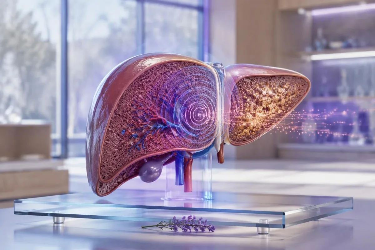
FibroScan-ի արդյունքները գնահատում են լյարդի կարծրությունը կՊա-ով և լյարդի ճարպը CAP գնահատականի միջոցով. դրանք միայնակ չեն ախտորոշում ֆիբրոզ կամ ցիռոզ։ Սեպտեմբերի 28, 2026-ի դրությամբ, ամենաօգտակար զեկույցը համատեղում է թիվը, սկանավորման որակը, պահքի կարգավիճակը, լյարդի արյան թեստերը և այն պատճառը, որի համար սկանավորվել եք։.


- Լյարդի կարծրության չափում 8 kPa-ից ցածրը սովորաբար անհավանական է դարձնում առաջադեմ ֆիբրոզը մետաբոլիկ լյարդի հիվանդության դեպքում, երբ սկանը հուսալի է։.
- FibroScan kPa գնահատական 8-12 kPa-ն անորոշ գոտի է, որը սովորաբար պահանջում է արյան թեստի համատեքստ կամ կրկնակի գնահատում։.
- Ավելի բարձր կարծրություն 12-15 kPa-ից բարձրը մտահոգություն է առաջացնում առաջադեմ ֆիբրոզի կամ ցիռոզի վերաբերյալ, բայց դրան կարող են նպաստել նաև այտուցը, հեպատիտը, խոլեստազը և կերակուրը։.
- CAP գնահատականի իմաստը վերաբերում է լյարդի ճարպին, ոչ թե սպիացած հյուսվածքին. մոտավորապես 238, 260 և 290 dB/m-ը սովորաբար օգտագործվող հետազոտական շեմեր են՝ ստեատոզի աճի համար։.
- Ծոմ պահել առնվազն 3 ժամ պահքը մինչև անցողիկ էլաստոգրաֆիան նվազեցնում է սննդի հետ կապված լյարդի կարծրության կեղծ բարձրացումը։.
- Հուսալի սկաններ սովորաբար ներառում են առնվազն 10 վավերական ընթերցումներ և IQR/միջին հարաբերակցություն 30% կամ ցածր։.
- Անհապաղ վերանայում հարկավոր է դեղնախտի, շփոթության, փսխման արյան, սև աթոռի կամ որովայնի այտուցվածության դեպքում, անկախ FibroScan թվից։.
- Մասնագետի հետագա հետևում ավելի հարմար է, քան ինքնաախտորոշումը, երբ կոշտությունը 8 կՊա կամ ավելի բարձր է, թրոմբոցիտները ցածր են, կամ լյարդի ֆերմենտները մնում են աննորմալ։.
Ինչ են չափում FibroScan զեկույցի երկու հիմնական թվերը
FibroScan-ի արդյունքները սովորաբար ցույց են տալիս երկու տարբեր չափում՝ լյարդի կոշտություն կիլոպասկալներով (կՊա) և ճարպի թուլացում դեցիբել մետրով (դԲ/մ):. Ավելի բարձր կՊա արժեքը կարող է արտացոլել ֆիբրոզ, բայց այն նաև կարող է ժամանակավորապես բարձրանալ վերջին ուտելուց, ակտիվ հեպատիտից, լեղու խցանումից կամ լյարդի շուրջ հեղուկի ճնշումից։.
Լյարդի կարծրության չափում օգտագործում է թրթռման վերահսկվող անցողիկ էլաստոգրաֆիա. փոքր մեխանիկական ազդակն անցնում է լյարդի միջով, և սարքը գնահատում է, թե որքան արագ է այդ ալիքը շարժվում։ Ավելի կոշտ հյուսվածքն ավելի արագ է փոխանցում ալիքը։ Իմ կլինիկայում տարածված սխալն է 9.5 կՊա արդյունքը բուժելը որպես ախտորոշում, այլ ոչ թե հավանականության ազդանշան, որը պետք է ստուգվի լյարդի հիվանդության պատճառի հետ։.
CAP, կամ վերահսկվող աթենուացիոն պարամետրը, գնահատում է, թե որքան է ուլտրաձայնային ազդանշանը թուլանում, երբ անցնում է լյարդի միջով։ CAP-ը հաղորդվում է դԲ/մ, սովորաբար մոտ 100-400 դԲ/մ սանդղակով, և հիմնականում արտացոլում է ստեատոզը։ Բարձր CAP գնահատականը չի ապացուցում ստեատոհեպատիտ, իսկ ցածր CAP գնահատականը չի բացառում ալկոհոլի, վիրուսային հեպատիտի, աուտոիմունային հիվանդության կամ երկաթի ավելցուկի պատճառով ֆիբրոզը։.
Կանտեստին մի է AI արյան անալիզատոր որը լյարդի ֆերմենտները, թրոմբոցիտները, գլյուկոզան, լիպիդները և երկաթի մարկերները տեղադրում է պատկերային արդյունքների կողքին, այլ ոչ թե առանձին-առանձին կարդալով։ Այդ տարբերությունը կարևոր է. 7.2 կՊա արդյունքը նորմալ թրոմբոցիտներով և նորմալ բիլիռուբինով նշանակում է բոլորովին այլ բան, քան 7.2 կՊա՝ ընկնող թրոմբոցիտներով և բարձրացած INR-ով։ Սկսեք FIB-4 հաշվարկը երբ ունեք անհրաժեշտ լաբորատոր արժեքները։.
Ինչու՞ մեկ թիվը չի կարող անվանել հիվանդությունը
FibroScan-ը չի որոշում կոշտության պատճառը։ Սրտի անբավարարությամբ, ալկոհոլային սուր հեպատիտով կամ խցանված լեղուղով մարդը կարող է ունենալ կՊա արժեք այն տիրույթում, որը սովորաբար կապված է զգալի ֆիբրոզի հետ։ Հետևաբար, կլինիկական հարցը ոչ թե “Ինչ փուլում եմ ես” է, այլ “Արդյոք այս արդյունքը հավաստի է, կայուն է և համապատասխանում է իմ լյարդի ավելի լայն գնահատմանը”։”
Ինչպես կարդալ FibroScan kPa գնահատականը՝ առանց ֆիբրոզի գերագնահատման
FibroScan kPa գնահատականը չափում է դիմադրությունը կտրող ալիքին, այլ ոչ թե անմիջապես սպի հյուսվածքը։. Շատ մեծահասակների մոտ, ովքեր ունեն մետաբոլիկ դիսֆունկցիա-ասոցիացված ստեատոտիկ լյարդային հիվանդություն, հուսալի արժեքը 8 կՊա-ից ցածր ունի առաջադեմ ֆիբրոզի լավ բացասական կանխատեսողական արժեք։.
ԿՊԱ սանդղակը լինիկական իմաստով գծային չէ։ 5-ից 7 կՊա փոփոխությունը կարող է արտացոլել սովորական կենսաբանական կամ տեխնիկական փոփոխություն, մինչդեռ 9-ից 15 կՊա-ի կայուն տեղաշարժն ավելի անհանգստացնող է՝ պայմանով, որ կիրառվում են նույն զոնդը, նախապատրաստումը և հիվանդության համատեքստը։ EASL ուղեցույցը 8 կՊա-ից ցածր լյարդի կոշտությունը համարում է օգտակար՝ մի քանի սովորական իրավիճակներում (EASL, 2021) կոմպենսացված առաջադեմ քրոնիկ լյարդային հիվանդությունները բացառելու համար։.
Տարբեր պայմաններ պահանջում են տարբեր կտրման կետեր։ Չբուժված քրոնիկ հեպատիտ C-ում 7-8 կՊա-ի շրջակայքում գտնվող արժեքները հաճախ օգտագործվել են որպես զգալի ֆիբրոզի հնարավոր շեմեր, մինչդեռ ցիռոզի շեմերը սովորաբար ընկնում են 12-14 կՊա-ի շրջանակներում։ Մետաբոլիկ լյարդային հիվանդության դեպքում, գիրությունը և XL զոնդը փոխում են կատարումը, ուստի հեպատիտի ուսումնասիրությունից կտրման կետի տեղափոխումը կարող է մոլորեցնել։.
Ես վերանայել եմ մոդելներ՝ բարակ հիվանդների մոտ 6.5 կՊա ընթերցումով և զգալի մետաբոլիկ ռիսկով, ապա ավելի մեծ հիվանդների մոտ 10 կՊա ընթերցումներով, որոնք ստացվել են ցածր որակի M-զոնդով փորձերի միջոցով։ Ոչ մեկը չպետք է պատահականորեն պիտակավորվի։ Ավելի լայն կլինիկական համատեքստի համար դիտեք մեր MASLD թեստավորման ուղեցույցը.
Ինչու՞ միավորը կՊա է
Կիլոպասկալը ճնշման միավոր է։ FibroScan-ը փոխակերպում է ալիքի տարածման տվյալները հյուսվածքի կոշտության գնահատման, ինչի պատճառով արդյունքները հաղորդվում են կՊա-ով, այլ ոչ թե ֆիբրոզի տոկոսով։ 15 կՊա լյարդի կոշտության չափումը 7.5 կՊա-ից “երկու անգամ ավելի սպի” չէ։.
Լյարդի կարծրության արդյունքների համար պրակտիկ ֆիբրոզ-ռիսկային գոտիներ
Շատ մեծահասակների համար, ովքեր կասկածվում են մետաբոլիկ լյարդային հիվանդության մասին, 8 կՊա-ից պակասը ցածր ռիսկի խումբ է, 8-12 կՊա-ն անորոշ է, իսկ 12-15 կՊա-ից բարձրը առաջադեմ ֆիբրոզի բարձր ռիսկի խումբ է։. Սրանք տրիաժ խմբեր են, ոչ թե համընդհանուր ֆիբրոզի փուլեր կամ մասնագետի գնահատման փոխարինող միջոց։.
EASL's 2021 non-invasive test guidance uses 8 կՊա-ից պակաս to help exclude compensated advanced chronic liver disease and 12-15 kPa or higher to help rule it in, depending on the underlying disease and test quality (EASL, 2021). Values around 8-12 kPa are where I most often avoid reassuring or alarming language until I see platelets, AST, ALT, bilirubin, albumin, and the scan reliability metrics.
A reliable reading of 20-25 kPa or more can raise concern for clinically significant portal hypertension, especially when platelets are below 150 × 10^9/L. It still does not diagnose varices or replace clinical examination, ultrasound, or endoscopy. The combination of a high kPa score and thrombocytopenia is more meaningful than either finding alone.
A 52-year-old endurance runner I saw had a stiffness result of 11.8 kPa after a viral illness, AST 89 IU/L, and ALT 104 IU/L. A repeat scan after recovery and appropriate fasting was 7.1 kPa. If liver enzymes remain high, use our guide to սահմանային ALT-ի հետագա ստուգում rather than assuming that one scan settled the question.
CAP գնահատականի իմաստը. ինչ է ասում dB/m արդյունքը լյարդի ճարպի մասին
A CAP score estimates liver fat, with higher dB/m values generally indicating more steatosis. It does not measure fibrosis, liver-cell injury, or whether fat is causing progressive disease.
Research thresholds often place mild steatosis near 238 dB/m, moderate steatosis near 260 dB/m, and more marked steatosis near 290 dB/m. Karlas and colleagues' individual-patient meta-analysis found useful CAP cutoffs, but also showed substantial overlap between categories (Karlas et al., 2017). A score of 275 dB/m is therefore an estimate, not a precise liver-fat percentage.
CAP can run higher with greater body mass, diabetes, and use of the XL probe; probe-specific reference ranges are not perfectly interchangeable. The reading also has less clarity at the individual level than many reports imply. I tell patients that CAP is best used to monitor direction over time under comparable conditions, not to chase a single “ideal” dB/m target.
Fatty liver often travels with triglyceride elevation, glucose dysregulation, and high fasting insulin. Our review of fasting insulin signals explains why insulin resistance can matter even when an HbA1c is not yet in the diabetes range.
CAP is not a severity score for liver damage
A CAP score of 320 dB/m may reflect substantial fat while stiffness remains 5.5 kPa, which is reassuring about current advanced fibrosis risk but not a free pass. Conversely, a CAP score below 238 dB/m can coexist with advanced fibrosis from hepatitis C, alcohol exposure, autoimmune hepatitis, or previous steatotic injury.
Ինչու է պահքը փոխում FibroScan-ի արդյունքները և որքան ժամանակ պետք է պահք պահել
Fasting for at least 3 hours before FibroScan is the usual practical standard because eating can temporarily increase liver stiffness. Water is normally acceptable, but follow the testing centre's instructions if medication timing or diabetes management is involved.
After a meal, blood flow through the portal circulation increases and can push stiffness readings upward for roughly 1.5-3 hours. The effect may be clinically relevant in people near an 8 or 12 kPa decision boundary. This is one reason a non-fasting 9.0 kPa result should not automatically trigger a fibrosis label.
Avoiding a heavy meal is not the same as fasting. Most centres ask for no food or caloric drinks for 3 ժամ, and some use longer intervals. I also prefer patients avoid vigorous exercise on the day of the scan when possible; the evidence is less settled than for food, but it removes one avoidable source of physiological noise.
If your scan and routine blood draw are planned together, preparation differs between tests. Our արյան անալիզի ծոմապահության ուղեցույցը covers supplements, coffee, and medication questions that can affect a same-day laboratory panel.
Do not stop prescribed medicines without advice
Diabetes medicines, blood-pressure medicines, and essential therapies should not be skipped simply to prepare for elastography. A person using insulin or a sulfonylurea may need an individual fasting plan to avoid hypoglycaemia. Call the scanning unit beforehand rather than making a last-minute medication change.
Ինչպես իմանալ՝ արդյոք FibroScan-ի ընթերցումը հուսալի է
A credible FibroScan report usually records at least 10 valid measurements and an interquartile-range-to-median ratio, often called IQR/Med. An IQR/Med of 30% or less is commonly considered acceptable when the median stiffness is in the clinically important range.
The median is the central stiffness value from repeated acquisitions; the IQR shows how widely those individual readings scattered. A median of 10.0 kPa with an IQR of 1.5 kPa is generally more persuasive than the same median with an IQR of 5 kPa. In practical terms, a noisy scan is not rescued by a dramatic-looking final number.
The M probe is commonly used in standard body habitus, while the XL probe improves feasibility when the skin-to-liver distance is greater. Do not directly compare an old M-probe value with a new XL-probe value as though they were identical instruments. The report should state the probe type, fasting status, and whether the operator obtained an adequate number of valid shots.
Կանտեստին մի է ԱԻ լաբորատոր թեստի մեկնաբանման ծառայությունում that can organise the relevant liver-panel and platelet context, but it does not replace the technical quality statement from the scan provider. Our կլինիկական վավերացման մոտեցումը treats low-quality source data as a reason for caution, not a reason to manufacture certainty.
The often-missed reliability clue
A report can look normal at first glance while being technically limited. Ask for the median kPa, CAP, IQR/Med, number of valid measurements, success rate if supplied, and probe type. Those five details make a repeat result far easier for a hepatology team to interpret.
Վիճակներ, որոնք բարձրացնում են kPa-ն՝ առանց լյարդի մշտական սպիացման
Acute liver injury, bile obstruction, venous congestion, recent meals, and heavy alcohol exposure can raise a FibroScan kPa score without representing fixed fibrosis. These reversible causes are particularly relevant when the stiffness result and the rest of the clinical picture disagree.
ALT or AST values more than five times the laboratory upper limit of normal make stiffness harder to interpret because active hepatocyte injury can increase tissue rigidity. For example, an ALT of 450 IU/L during acute viral hepatitis is a poor moment to stage chronic fibrosis by elastography. Repeat assessment after the acute process settles is usually more informative.
Cholestasis can elevate stiffness through increased biliary pressure. A person with jaundice, dark urine, pale stools, bilirubin above 50 µmol/L, or a rising alkaline phosphatase needs clinical assessment for obstruction rather than a casual fibrosis discussion. Our guide to cholestatic liver patterns explains the laboratory clues.
Right-sided heart failure and severe fluid overload can also elevate liver stiffness through congestion. I have seen a high kPa value fall substantially after cardiac treatment, which is why breathlessness, ankle swelling, raised jugular venous pressure, and medication changes belong in the history. A FibroScan is measuring a physical property of the liver at that moment.
Alcohol changes the interpretation
Recent sustained alcohol use can elevate GGT, AST, and liver stiffness through steatohepatitis and inflammation. A period of abstinence may improve values, but a lower repeat score does not erase prior risk. See our explanation of biomarker trends after alcohol cessation for realistic timing.
Ինչու են ֆիբրոզի կտրվածքները փոխվում լյարդի հիվանդության պատճառով
FibroScan cutoffs vary because hepatitis B, hepatitis C, alcohol-related liver disease, MASLD, cholestatic disease, and congestive hepatopathy alter stiffness in different ways. A number that is concerning in one setting can be less specific in another.
In chronic hepatitis B, viral activity can raise ALT and stiffness, so timing relative to a flare matters. In chronic hepatitis C, published cirrhosis cutoffs often cluster around 12-14 kPa, but cure with antiviral therapy can lower stiffness through reduced inflammation before established scar tissue has fully regressed. The հեպատիտ B ախտանիշների ուղեցույցը explains why normal appearance does not exclude infection.
In MASLD, stiffness is interpreted alongside obesity, type 2 diabetes, hypertension, triglycerides, and FIB-4. The 2023 AASLD guidance recommends an initial FIB-4-based risk assessment in appropriate adults and secondary assessment with elastography or ELF when risk is not low (Rinella et al., 2023). This staged approach reduces unnecessary referral while missing fewer people with advanced fibrosis.
Cholestatic conditions and infiltrative disorders can produce stiffness values that overestimate fibrosis. If alkaline phosphatase is disproportionately high, autoimmune markers or imaging may matter more than a generic kPa cutoff. Patients with relevant exposure or symptoms should also understand hepatitis C testing rather than relying on elastography alone.
Pregnancy and paediatric scans need separate expertise
Pregnancy changes blood volume, liver physiology, and the differential diagnosis of abnormal liver tests; adult fibrosis bands are not a self-management tool in this situation. Children also require age-appropriate probes and specialist interpretation. A high result in either group deserves clinician-led evaluation.
Արյան թեստեր, որոնք ավելի օգտակար են դարձնում FibroScan-ի արդյունքները
Platelets, AST, ALT, bilirubin, albumin, INR, glucose, lipids, ferritin, and viral hepatitis tests can materially change the meaning of a FibroScan result. A kPa value becomes safer to interpret when it agrees with these independent markers.
თრომბოციტები ქვემოთ 150 × 10^9/L, albumin below the local reference interval, bilirubin elevation, and a prolonged INR can suggest more advanced liver dysfunction or portal hypertension when paired with high stiffness. Normal ALT does not rule out advanced fibrosis; I have seen quiet enzyme panels in patients whose platelets and imaging told the more important story.
Ferritin is an acute-phase protein and can rise with metabolic inflammation, but a ferritin above 1,000 µg/L or persistently high transferrin saturation deserves a structured iron-overload assessment. This is particularly useful when CAP is low but stiffness is unexpectedly elevated. Review iron studies in context before assuming raised ferritin means iron overload.
Կանտեստին մի է AI բիոմարկերների մեկնաբանման հարթակ that reads liver results as patterns across time, including whether platelet count, GGT, ALT, and triglycerides are moving together. The liver panel reference guide can help you bring the correct values to a GP or hepatology appointment.
A useful discrepancy pattern
High stiffness with normal CAP should prompt a search beyond fatty liver, including viral hepatitis, alcohol-related injury, congestion, cholestasis, medication injury, autoimmune disease, and iron overload. High CAP with low stiffness usually shifts the immediate focus toward cardiometabolic risk reduction and periodic fibrosis-risk reassessment.
Երբ արդյունքի ոչ նորմալ FibroScan-ը պահանջում է կրկնակի թեստավորում կամ մասնագետի ուղեգրում
A reliable FibroScan result of 8 kPa or more commonly warrants clinician review when liver tests are abnormal, metabolic risk is present, or there is known liver disease. A value above 12-15 kPa generally merits hepatology assessment rather than watchful waiting alone.
Repeat testing is reasonable when fasting was inadequate, IQR/Med was high, ALT was acutely elevated, alcohol intake changed recently, or the result conflicts with the clinical picture. For low-risk MASLD, repeat non-invasive fibrosis assessment is often considered every 1-3 տարի, with shorter intervals for type 2 diabetes or several metabolic risk factors. The exact schedule is individual.
Refer sooner if kPa is above 12-15, FIB-4 is elevated, platelets fall, bilirubin rises, or ultrasound suggests nodularity, splenomegaly, or ascites. A specialist may choose ELF testing, MR elastography, targeted imaging, or occasionally tissue examination if the answer will change management. Our ELF score guide compares one common second-line option.
Kantesti AI can flag a mismatch—such as 14 kPa with normal enzymes but platelets of 118 × 10^9/L—as a follow-up trigger rather than a diagnosis. In my experience, that is the useful role for automated interpretation: preparing better questions for the clinician, not replacing the consultation.
Ինչ վերցնել նշանակման համար
Bring the complete FibroScan report, not only the kPa and CAP figures. Include the scan date, fasting duration, probe type, IQR/Med, recent illness, alcohol history, medication and supplement list, and liver tests from the previous 12 months. This often avoids an unnecessary repeat.
Ինչ են ստուգում մասնագետները բարձր լյարդի կարծրության ընթերցումից հետո
After a high liver stiffness measurement, specialists usually confirm the result, identify the cause, and assess for complications rather than immediately assigning a fibrosis stage. The work-up often includes repeat laboratory testing, abdominal ultrasound, viral serology, medication review, and fibrosis-risk calculations.
A targeted work-up may include hepatitis B surface antigen, hepatitis C antibody with RNA testing if positive, iron saturation, ferritin, autoimmune markers, immunoglobulins, alpha-1 antitrypsin testing, and coeliac testing when indicated. Not everyone needs every test. The history—family disease, alcohol, metabolic risk, medicines, and ethnicity—guides sensible selection.
If cirrhosis is suspected, clinicians may assess platelet trends, spleen size, portal-vein features, kidney function, and whether liver-cancer surveillance is indicated. Ultrasound surveillance is commonly performed at 6-month intervals for people with established cirrhosis who are appropriate candidates, but a high FibroScan alone does not automatically establish that programme.
Unexpectedly high stiffness with emphysema, family liver history, or unexplained abnormal liver tests can make alpha-1 antitrypsin testing relevant. Kantesti's AI տեխնոլոգիայի ուղեցույց explains how our system presents possible follow-up themes without claiming to determine a diagnosis.
Why biopsy is now less common, not obsolete
Tissue examination is no longer required for every patient because combined blood and imaging tests classify many people well enough for care decisions. It remains useful when non-invasive tests disagree, an alternative diagnosis is plausible, or treatment eligibility depends on histological detail.
Ինչը կարող է ժամանակի ընթացքում բարելավել CAP-ը, լյարդի ֆերմենտները և ֆիբրոզի ռիսկը
Weight loss, regular physical activity, diabetes control, and avoiding alcohol when liver disease is present can reduce liver fat and improve liver risk markers. A falling CAP or ALT is encouraging, but it does not by itself prove that fibrosis has reversed.
For many people with MASLD, sustained loss of 5% մարմնի քաշի կորուստը բարելավում է լյարդի ճարպը, մինչդեռ 7-10% is more often associated with improvement in steatohepatitis and fibrosis-related outcomes. Response varies: a person starting at 100 kg may see metabolic benefit from 5 kg lost, but their individual treatment plan should account for nutrition, medicines, and muscle mass.
Ձգտեք առնվազն շաբաթական 150 րոպե of moderate aerobic activity plus resistance work where safe. Exercise can reduce liver fat even without major weight loss. Avoid “liver detox” supplements; concentrated green-tea extract, anabolic products, and multi-ingredient weight-loss remedies are recurrent causes of drug-induced liver injury.
Food choices work best when they are specific and sustainable: fibre-rich plants, minimally processed protein, unsweetened drinks, and fewer refined carbohydrates are reasonable starting points. Our evidence-based liver-supportive food guide separates practical dietary measures from detox marketing.
Do not use a scan to punish yourself
CAP and kPa are clinical measurements, not report cards. I have had patients dramatically restrict food before a scan in an effort to improve the number; that only makes serial comparison less useful. Use the same preparation each time and focus on changes that remain workable after the appointment.
Ախտանշաններ, որոնք պահանջում են անհապաղ խնամք՝ անկախ ձեր FibroScan-ի թվից
Jaundice, confusion, vomiting blood, black tarry stools, new abdominal swelling, severe drowsiness, or fever with upper abdominal pain require urgent medical assessment regardless of FibroScan results. Elastography does not assess acute liver failure, gastrointestinal bleeding, infection, or bile-duct obstruction safely enough to delay care.
New confusion, sleep-wake reversal, or marked drowsiness in someone with known liver disease can signal hepatic encephalopathy or another serious condition. Vomiting blood or passing black stools can indicate upper gastrointestinal bleeding. Call emergency services or seek emergency care rather than waiting for a repeat scan or online interpretation.
Yellow eyes with dark urine, pale stools, fever, and right-upper abdominal pain may indicate cholestasis, gallbladder disease, or acute hepatitis. Bilirubin can rise quickly in these settings, while kPa may be temporarily inflated by biliary pressure. Our guide to երբ բիլիռուբինին անհետաձգելի օգնություն է հարկավոր ծածկում է նախազգուշացման նախշը։.
As Chief Medical Officer, Dr. Thomas Klein advises against using any result app as emergency triage. Kantesti can preserve longitudinal records and help list relevant abnormalities, but symptoms and deterioration always outrank a reassuring older number.
Pregnancy is a special case
Itching of the palms or soles, jaundice, headache with visual changes, severe nausea, or upper abdominal pain during pregnancy needs same-day maternity assessment. Pregnancy-related liver disorders can evolve quickly, and standard FibroScan thresholds are not designed to exclude them.
Ինչպես անվտանգ հետևել FibroScan-ի արդյունքներին ժամանակի ընթացքում
The best comparison is a repeat FibroScan done with the same fasting conditions, similar probe selection, and the same supporting blood tests. One isolated kPa result is less informative than a trend that remains consistent after technical and clinical confounders are checked.
Record the date, kPa, CAP, probe, IQR/Med, fasting interval, weight, alcohol exposure, recent illness, and ALT value each time. A shift from 6.4 to 7.0 kPa may be ordinary variation, while a confirmed move from 7.0 to 13.5 kPa alongside falling platelets needs prompt review. Trends only mean something when the conditions are comparable.
Kantesti AI supports longitudinal review of uploaded laboratory reports and can identify changes in enzyme, platelet, glucose, and lipid patterns across visits. Kantesti is used by people in more than 127 countries, but local laboratory ranges, access to elastography, and referral pathways still matter. Dr. Thomas Klein's practical rule is simple: preserve the original report and ask what changed physiologically before assuming fibrosis progressed.
Our doctors and clinical standards are described through the Բժշկական խորհրդատվական խորհուրդ. For readers who want supplementary lab-interpretation publications, the two DOI records below concern iron and coagulation testing; they do ոչ validate FibroScan or replace liver-specific guidelines. They are included for transparent research indexing only.
A final practical question for your clinician
Ask: “Does my kPa result agree with my platelet count, liver function, metabolic risk, ultrasound, and scan quality?” That question usually produces a more useful plan than asking for a fibrosis stage from a single number. Persistently abnormal results deserve follow-up, while a technically flawed scan deserves repetition.
Հաճախակի տրվող հարցեր
Ուղիղ ֆիբրոսկան kPa միավորը որքա՞ն է։
Մեծահասակների մոտ, որոնց մոտ կասկածվում է նյութափոխանակության հետ կապված լյարդի հիվանդություն, FibroScan-ի կողմից լյարդի կարծրության չափումը 8 կՊա-ից ցածր համարվում է ցածր ռիսկի արդյունք՝ առաջադեմ ֆիբրոզի համար, երբ սկանավորումը տեխնիկապես հուսալի է։ Շատ առողջ մեծահասակների մոտ ցուցիչները տատանվում են 2-7 կՊա-ի սահմաններում, թեև չկա մեկ համընդհանուր նորմալ տիրույթ, քանի որ տարիքը, մարմնի չափը, զոնդի տեսակը և լյարդի վիճակը ազդում են մեկնաբանության վրա։ 8 կՊա-ից ցածր արժեքը չի բացառում լյարդի ցանկացած վիճակ, հատկապես սուր հեպատիտը կամ ոչ-ֆիբրոտիկ հիվանդությունը։ Հաշվետվության IQR/Med-ը, ծոմապահության կարգավիճակը և արյան ստուգումները պետք է վերանայվեն թվի հետ մեկտեղ։.
7 kPa FibroScan-ին վատ է։
FibroScan-ի 7 կՊա արդյունքը հաճախ հուսադրող է, քանի որ այն ցածր է MASLD-ի բազմաթիվ ուղիներում առաջադեմ ֆիբրոզի ռիսկի համար սովորաբար օգտագործվող 8 կՊա շեմից։ Այն ինքնաբերաբար նորմալ չէ բոլոր դեպքերում. չբուժված վիրուսային հեպատիտը, լյարդի թեստերի աննորմալ արդյունքները կամ թրոմբոցիտների թվի նվազումը կարող են փոխել դրա նշանակությունը։ 7 կՊա արդյունքը, որը ստացվել է ուտելուց հետո կամ սուր հիվանդության ժամանակ, կարող է ավելի քիչ մեկնաբանելի լինել։ Հիվանդներից շատերը պետք է քննարկեն միավորը այն բժշկի հետ, ով գիտի, թե ինչու է կատարվել սկանավորումը։.
Ի՞նչ է նշանակում ճարպային լյարդի CAP գնահատականը։
CAP-ի մոտ 238 դԲ/մ կամ ավելի բարձր միավորը կարող է ենթադրել առնվազն թեթև լյարդային ճարպ, մինչդեռ 260 դԲ/մ և 290 դԲ/մ-ի շուրջ հետազոտության շեմերը հաճախ օգտագործվում են միջին և ավելի արտահայտված ստեատոզի համար։ CAP-ը լյարդային ճարպի տոկոսի ուղղակի չափման փոխարեն էխոգրաֆիկ ուղղակի թուլացման գնահատական է։ Մարմնի չափը, դիաբետը, զոնդի տեսակը և տեխնիկական գործոնները կարող են տեղափոխել արդյունքը։ CAP-ի բարձր միավորը չի ասում, թե արդյոք առկա է ֆիբրոզ, ուստի kPa միավորը և լյարդի արյան թեստերը դեռևս կարևոր են։.
Կարո՞ղ է արդյոք FibroScan-ից առաջ ուտելը հանգեցնել kPa-ի բարձր միավորի:
Այո, FibroScan-ից առաջ ուտելը կարող է ժամանակավորապես բարձրացնել լյարդի կոշտությունը, քանի որ սնունդը մեծացնում է պորտալ արյան հոսքը և փոխում լյարդի հեմոդինամիկան։ Որոշ կենտրոններ խնդրում են հիվանդներին խուսափել սնունդից և կալորիական ըմպելիքներից առնվազն 3 ժամ առաջ քննությունից։ Այս ազդեցությունը կարող է բավականաչափ նշանակալից լինել, երբ արդյունքը մոտ է 8 կՊա կամ 12 կՊա որոշման սահմաններին։ Սովորաբար ջուրը թույլատրվում է, բայց գլյուկոզան իջեցնող դեղորայք ընդունող մարդիկ պետք է հաստատեն իրենց նախապատրաստական պլանը փորձաքննական կենտրոնի հետ։.
12 kPa FibroScan միավորը ցիրոզ ախտորոշում է նշանակում։
12 kPa FibroScan միավորը չի ապացուցում ցիռոզ, բայց այն բավականաչափ բարձր է, որպեսզի արդարացնի կառուցված կլինիկական հետևումը լյարդի հիվանդությունների շատ պայմաններում։ Մետաբոլիկ լյարդի հիվանդության դեպքում 12-15 kPa-ն հաճախ դիտարկվում է որպես առաջադեմ ֆիբրոզի ավելի բարձր ռիսկային տիրույթ. վիրուսային հեպատիտում ցիռոզի հրապարակված շեմերը կարող են նման լինել, բայց տարբերվում են ըստ ուսումնասիրության։ Վերջին կերակուրները, լյարդի սուր բորբոքումը, լեղուղիների խցանումը և սրտի հետ կապված այտուցը կարող են բարձրացնել կարծրությունը առանց ցիռոզի։ Մասնագետը կարող է հաստատել ռիսկը՝ օգտագործելով թիթեղիկներ, FIB-4, ELF, պատկերացում կամ MR էլաստոգրաֆիա։.
Որքան հաճախ պետք է կրկնել FibroScan-ը:
FibroScan-ի կրկնության ժամկետը կախված է լյարդի հիվանդության պատճառից և առաջին արդյունքից, սակայն ցածր ռիսկի մետաբոլիկ լյարդի հիվանդությունը հաճախ վերագնահատվում է 1-3 տարին մեկ։ 2-րդ տիպի դիաբետ, մի քանի մետաբոլիկ ռիսկի գործոններ, լյարդի ֆերմենտների աննորմալություն կամ անորոշ կարծրություն ունեցող մարդկանց կարիք կարող է լինել ավելի վաղ վերագնահատման։ Տեխնիկապես սահմանափակ սկանավորումը, ոչ ստամոքսի վրա սկանավորումը կամ սուր հիվանդության ժամանակ կատարված սկանավորումը հաճախ ավելի շուտ է կրկնվում, երբ խանգարող գործոնը վերանա։ Նույն պահքի և զոնդի պայմաններում կրկնությունը միտումը ավելի իմաստալից է դարձնում։.
Ստացեք AI-ով աշխատող արյան անալիզի վերլուծություն այսօր
Միացեք ավելի քան 2 միլիոն օգտատերերի ամբողջ աշխարհում, ովքեր վստահում են Kantesti-ին՝ լաբորատոր թեստերի ակնթարթային, ճշգրիտ վերլուծության համար։ Վերբեռնեք ձեր արյան անալիզի արդյունքները և ստացեք 15,000+ բիոմարկերների համապարփակ մեկնաբանություն վայրկյանների ընթացքում։.
📚 Հղված հետազոտական հրապարակումներ
Klein, T., Mitchell, S., & Weber, H. (2026). Kantesti LTD. (2026). Iron Studies Guide: TIBC, Iron Saturation & Binding Capacity. Zenodo. https://doi.org/10.5281/zenodo.18248745.։ Kantesti AI Medical Research.
Klein, T., Mitchell, S., & Weber, H. (2026). Kantesti LTD. (2026). aPTT Normal Range: D-Dimer, Protein C Blood Clotting Guide. Zenodo. https://doi.org/10.5281/zenodo.18262555.։ Kantesti AI Medical Research.
📖 Արտաքին բժշկական հղումներ
⚕️ Բժշկական հրաժարում
Այս հոդվածը միայն կրթական նպատակների համար է և չի հանդիսանում բժշկական խորհրդատվություն։ Ախտորոշման և բուժման որոշումների համար միշտ խորհրդակցեք որակավորված բուժաշխատողի հետ։.
E-E-A-T վստահության ազդանշաններ
Փորձառություն
Բժշկի կողմից ղեկավարվող կլինիկական վերանայում՝ լաբորատոր մեկնաբանության աշխատանքային հոսքերի համար։.
Մասնագիտություն
Լաբորատոր բժշկության կենտրոնացում՝ այն բանի վրա, թե ինչպես են բիոմարկերները դրսևորվում կլինիկական համատեքստում։.
Հեղինակություն
Գրված է դոկտոր Թոմաս Քլայնի կողմից՝ դոկտոր Սառա Միթչելի և պրոֆեսոր դոկտոր Հանս Վեբերի կողմից վերանայմամբ։.
Հուսալիություն
Ապացույցների վրա հիմնված մեկնաբանություն՝ հստակ հետագա քայլերի ուղիներով՝ ահազանգերը նվազեցնելու համար։.