in lege mean corpuscular volume wjert meastentiids feroarsake troch izer-beheinde produksje fan reade bloedsellen of in erflike hemoglobine-eigenskip. De fluchste feilige triearring komt fan it lêzen fan MCV tegearre mei RBC-telling, RDW, ferritine, transferrine-saturaasje, en it klinyske ferhaal.



- Lege MCV betsjut dat reade sellen lytser binne as ferwachte; de measte laboratoaria foar folwoeksenen markearje mikrozytose ûnder 80 fL.
- Ferritine ûnder 15 ng/mL is tige spesifyk foar útputte izerfoarrieden by in fierders sûne folwoeksene, wylst wearden ûnder 30 ng/mL yn 'e praktyk faak izertekoart stypje.
- RBC-telling boppe 5,0 ×10¹²/L mei MCV ûnder 75 fL en in normale RDW wiist faak op thalassemia-eigenskip ynstee fan ienfâldich izertekoart.
- RDW boppe 14.5% stipe ûntwikkeljend izertekoart, om't nije sellen lytser wurde foardat alle sirkulearjende sellen beynfloede binne.
- Transferrine-saturaasje ûnder 20% jout beheinde beskikberens fan sirkulearjend izer oan; ûnder 15% is mear oertsjûgjend as ferritine ûndúdlik is.
- Mentzerindex ûnder 13 foardieliget thalassemia-eigenskip, wylst in yndeks boppe 13 foardieliget izertekoart, mar it is in screening-oanwizing ynstee fan in diagnoaze.
- HbA2 boppe 3.5% op hemoglobine-elektroforese stipe beta-thalassemia-eigenskip nei't izertekoart korrizjearre of útsletten is.
- Nij izertekoart by manlju of postmenopausale froulju hat in op oarsaken rjochte evaluaasje nedich, faak ynklusyf gastrointestinale beoardieling ynstee fan allinich izerenabletten.
Wat betsjut in lege MCV op in folsleine bloedtelling
A leech MCV betsjut dat de reade bloedsellen yn trochsneed lyts binne, meastentiids ûnder 80 fL by folwoeksenen. De twa wichtichste oarsaken fan lege MCV binne izertekoart en talassemia trait, en de praktyske ferdieling begjint mei ferritine, RBC-telling, RDW, en oft der eins bloedearmoed is.

MCV is in trochsneed, dus it kin mar mild leech lykje op 78 fL sels as twa ferskillende selpopulaasjes oanwêzich binne. In tanimmende RDW lit dy mingeling earder sjen as allinich MCV, dêrom is in CBC-yndekskomparaasje nuttiger as it efterfolgjen fan ien markearre resultaat.
In normale hemoglobine slút in betsjuttend probleem net út. Ik sjoch regelmjittich ferritine fan 8 oant 18 ng/mL mei hemoglobine noch boppe 120 g/L, benammen by minsken mei swiere menstruaasjeferlies, úthâldingsfermogen training, restriktive diëten, of faak bloedjaan.
Kantesti is in AI bloedtestanalysator dat MCV lêst tegearre mei hemoglobine, RBC-telling, RDW, ferritine, en foarige resultaten ynstee fan in oarsaak ta te jaan fan ien reade flagge. Fan 9 septimber 2026 ôf bliuwt dy patroan-basearre oanpak feiliger dan selsbehanneling fan elke lege MCV as izertekoart.
Mikrosytose is in fynst, gjin diagnoaze
Mikrosytose beskriuwt selgrutte; bloedearmoed beskriuwt ûnfoldwaande soerstofdragerskapasiteit. In persoan kin mikrosytose hawwe sûnder bloedearmoed, bloedearmoed sûnder mikrosytose, of beide - elk patroan hat in oare wurking.
Brûk RBC-telling en RDW foardat jo izertekoart oannimme
In RBC-telling dy't relatyf heech is nettsjinsteande in lege MCV komt oerien mei talassemia trait, wylst in lege of gewoane RBC-telling mei hege RDW oerienkomt mei izertekoart. Dit is it nuttichste earste-passażje CBC-patroan as ferritine noch net werom is.
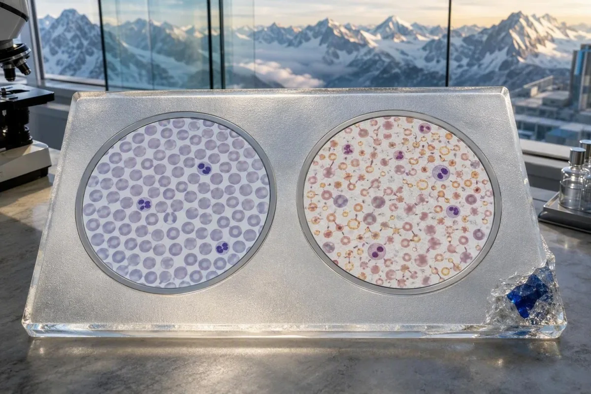
By ûnkomplisearre izertekoart mist it bienmurch substraat en produseart faak minder sellen: hemoglobine falt, RBC-telling kin ûnder 4.5 ×10¹²/L, lizze, en RDW stiet faak boppe 14.5%. By talassemia trait wurdt de output fan it bienmurch behâlden, dus in RBC-telling fan 5.2 oant 6.2 ×10¹²/L is net ûngewoan nettsjinsteande in MCV yn de lege 60s of 70s.
De Mentzer-yndeks ferdielt MCV troch RBC-telling. Bygelyks, 68 fL ÷ 5.8 = 11.7, in resultaat dat foar thalassemia-eigenskip falt; 74 fL ÷ 4.3 = 17.2 neigje nei izertekoart, hoewol't mingde sykte de regel ferslaan kin.
In lege MCV mei normale hemoglobine en in stabile hege RBC-telling oer ferskate jierren is faak erflik ynstee fan fiedingsstoffen. Ferlykje it gehiel folsleine bloedtelling mei eardere tests foardat jo beslute dat in resinte libbensstylferoaring it feroarsake hat.
Wêrom RDW faak it iere izertekoart skiedt fan eigenskip
Hege RDW mei lege MCV wjerspege meastentiids izertekoart, om't âlde sellen fan normale grutte neist nijere lytsere sellen sirkulearje. RDW kin normaal bliuwe yn thalassemia-eigenskip, om't lytse sellen konstant oer tiid produsearre wurde.

RDW-CV hat faak in referinsjeinterval om 11.5% oant 14.5%, hoewol't it eigen interval fan it laboratoarium wint. In wearde fan 16.8% mei MCV 76 fL hat mear diagnostyske wearde as ien fan beide resultaten allinich, benammen as de foarige MCV seis moanne earder 87 fL wie.
RDW is net spesifyk: resinte transfúsje, B12-tekoart, folaat-tekoart, hemolyse en herstel nei izerterapy kinne it allegear ferheegje. In ferrassende stiging nei behanneling kin gewoan nije izerrike sellen yn de sirkulaasje fertsjintwurdigje; ús gids foar RDW nei izerterapy ferklearret dizze tsjinoerstelde faze.
Nei myn klinyske ûnderfining moat in normale RDW de wurking foar izertekoart fertrage, net stopje. Lang duorjend tekoart kin úteinlik de measte sellen unifoarm lyts meitsje, wylst alfa-thalassemia-eigenskip plus izerferlies in heul hege RDW produsearje kin.
Trend wint fan in inkelde RDW
In ferskowing fan RDW 12.8% nei 15.6% is betsjuttend, sels as beide wearden de referinsjelimiten fan ferskate laboratoaria passe. De rjochting fan feroaring ferskynt faak foardat in pasjint wurch of bûten sykheljen fielt.
Ferritine is de bêste earste opfolgingstest foar lege MCV
Ferritine is de earste test nei in lege MCV by de measte folwoeksenen omdat it opslein izer skat. Ferritine ûnder 15 ng/mL jout sterk izertekoart oan, en in protte klinisy ûndersykje wearden ûnder 30 ng/mL as symptomen of mikrosytose oanwêzich binne.

Ferritine is in akute-faze reaktant, dus ûntstekking kin útputte foarrieden ferbergje. Camaschella's resinsje fan izertekoart-anemy beskriuwt wêrom't ferritine stige mei ûntstekking sinalen; in ferritine fan 55 ng/mL slút net betrouber izertekoart út as de CRP ferhege is (Camaschella, 2015).
Kantesti AI is in AI bloedtest-útslachplatfoarm dat markearret de spesifike mismatch fan lege MCV, lege transferrinsaturaasje, en skynber normale ferritine as de CRP of levermarkers in akute-faze effekt suggerearje. Dit is ien fan dy gebieten wêr't kontekst mear is as in fet laboratoariumflagge.
Foar in nuttige folgjende tekening, mije it ynterpretearjen fan serumin izer allinnich, om't it oer de dei en nei supplementen feroaret. Koppel ferritine mei transferrinsaturaasje, CRP, en de hantlieding foar izerstúdzjes wannear't it patroan ûndúdlik is.
It typyske patroan fan izertekoart efter mikrozytose
Izertekoart produseart meastentiids lege ferritine, lege transferrinsaturaasje, stige RDW, en úteinlik lege hemoglobine. De ierste oanwizing kin ferritine delgong moannen foar MCV ûnder 80 fL falt.
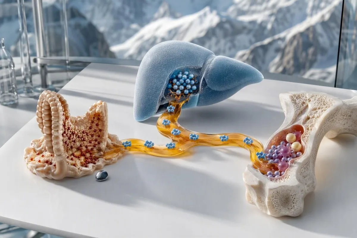
In praktysk patroan is ferritine 12 ng/mL, transferrinsaturaasje 9%, MCV 74 fL, en RDW 17%. Serumin izer allinnich is minder betrouber, om't in resinte miel, izer tablet, of moarn-tsjin-middei kolleksje it gâns feroarje kin.
Menstruale ferlies is gewoan, mar it moat gjin refleksferklearring wurde. In 38-jierrige mei perioaden beskreaun as normaal, ferritine 6 ng/mL, en oanhâldende mikrozytose hat dieet-, donaasje-, medisinen-, celiac- en gastrointestinale fragen nedich ynstee fan geruststelling; sjoch leech ferritine sûnder swiere menstruaasje.
Orale elemintêre izerdoses fan 40 oant 65 mg ien kear deis of elke oare dei wurde faak better tolerearre as âldere meardere-dei-dosis skema's. Reaksje moat wurde kontrolearre mei in klinikus, om't hemoglobine meastal stige mei rûchwei 10 g/L oer 2 oant 4 wiken as de opname en de diagnoaze goed binne.
Wannear't ynfeksje ferritine better lit lykje as it is
ûntstekking kin lege MCV feroarsaakje mei normale of ferhege ferritine troch izer fuort te sluten fan it bienmurch. Lege transferrin-saturaasje, ferhege CRP, chronike nier sykte, auto-immune sykte, of aktive ynfeksje kinne wize op dit funksjonele izer-beheining patroan.
Transferrin-saturaasje is lyk oan seerum izer dield troch totale izer-bindende kapasiteit, fermannichfâldige mei 100. In saturaasje ûnder 20% suggerearret beheinde izer beskikberens, mar wearden ûnder 15% binne mear oertsjûgjend as ferritine tusken 30 en 100 ng/mL.
ûntstekking ferleget transferrin faak, wylst it ferritine ferheget, wat in misliedende kombinaasje produseart. De transferrin en ûntstekking patroan is fan belang, om't in lege TIBC net betsjut dat de izer reserves folop binne.
Oplosbere transferrin receptor kin helpe as resultaten it net iens binne, om't it minder beynfloede wurdt troch akute ûntstekking, hoewol't beskikberens en assay-fariaasjes ferskille. Ik soe in normale ferritine fan 70 ng/mL net brûke as reden om izer beheining ôf te wizen by immen mei CRP 35 mg/L en transferrine-saturaasje 11%.
Anemia fan chronike sykte en oare ferwurven oarsaken fan lege MCV
Chronike ûntstekking sykte, chronike nier sykte, lead exposure, en sideroblastyske prosessen kinne lege MCV feroarsaakje, mar se binne minder algemien as izertekoart of talassemia trait. Har izer-stúdzje patroanen en klinyske setting foarkomme ûnnedige izer behanneling.

Anemia fan chronike ûntstekking is faak earst normocytysk en wurdt by in minderheid fan gefallen mikrocytysk. Ferritine is meastentiids normaal of heech, seerum izer leech, en TIBC leech of normaal—gâns oars as klassike izertekoart, dêr't TIBC faak ferhege is.
Chronike nier sykte kin fermindere erytropoietine produksje kombinearje mei izer-beheinde erytropoese. As eGFR ûnder 60 mL/min/1.73 m² is foar op syn minst 3 moannen, moat izer ynterpretaasje koördinearre wurde mei it nierteam; nim de CKD-staginggids.
Lead toksisiteit en sideroblastyske anemia fertsjinje rjochte testen allinich by exposure, medisinen, alkoholgebrûk, neurologyske symptomen, basofiliske stippeling, of in ûngewoane smear dy't dêrhinne wize. In bloedfilm hantearre troch in hematology laboratoarium is mear ynformatyf as it bestellen fan in breed paniel sûnder in klinyske lead.
It CBC-patroan dat suggerearret thalassemia-eigenskip
Talassemia trait feroarsaket typysk in ûnproportioneel lege MCV mei in normale of hege RBC-telling en mylde of ôfwêzige bloedearmoede. Ferritine is meastentiids normaal, útsein as izertekoart tegearre bestiet, en RDW is faak normaal of mar licht ferhege.
In klassike beta-talassemia trait paniel kin hemoglobine sjen 118 g/L, MCV 66 fL, RBC 5.7 ×10¹²/L, RDW 13.2%, en ferritine 48 ng/mL. De lege MCV kin skrikje, mar it stabile hege RBC-telle is de oanwizing dy't in protte lêzers misse.
Trait is gjin sykte om te korrizjearjen mei izer, útsein as izertekoart bewiisd is. Werhelle izerkuren by immen mei ferritine 80 ng/mL en in libbenslange MCV fan sa'n 68 fL ferbetterje de telling selden en kinne genetyske rieplachting of in juste ferklearring fertrage.
Famylje-oarsprong kin de wierskynlikheid ynformearje, mar it ferfangt nea testen; talassemia traits komme wrâldwiid foar. In skynber hege RBC-telling kin ek wjerspegelje dehydraasje, dus ferlykje mei hematokryt, albumine, en de kontekst yn ús review fan hege RBC-telling.
Alpha en beta trait binne net identyk
Beta-talassemia trait ferheget faak HbA2, wylst alpha-talassemia trait normale elektroforeze hawwe kin. Normale elektroforeze slút dêrom alpha-talassemia net út as it CBC-patroan en famyljeskiednis oertsjûgjend binne.
Wannear't hemoglobine-elektroforese en genetyske testen helpe
Hemoglobine elektroforeze is it meast nuttich as ferritine normaal is en de CBC talassemia trait suggerearret. HbA2 boppe likernôch 3.5% stipet beta-talassemia trait, wylst alpha-talassemia molekulêre testen nedich hawwe kin nei spesjalistyske beoardieling.
Korrigearje izertekoart foardat jo fertrouwe op in râne HbA2-resultaat, om't izertekoart HbA2 genôch ferleegje kin om beta-talassemia trait te maskeren. In ferstannige folchoarder is earst izerstúdzjes, behanneling as útput, dan werhelje CBC en hemoglobine analyze nei't it tekoart oanpakt is.
Galanello en Origa beskriuwe beta-talassemia as in globinproduksjedistúrbanje mei dragerstaten dy't meastentiids mild binne, mar wichtich foar reproduktive rieplachting (Galanello en Origa, 2010). As beide biologyske âlden klinysk relevante hemoglobinefarianten drage, kin elke swangerskip in 25% kâns hawwe op in troffen bern ôfhinklik fan de belutsen farianten.
Nim net oan dat elk lyts-sel patroan talassemia is. Hemoglobinefarianten kinne tegearre foarkomme, en in screeninggids foar sikkel-sel ferklearret wêrom't definitive ynterpretaasje heart by it laboratoariumrapport en in klinikus dy't bekend is mei hemoglobineopaty-testen.
Mingde izertekoart en thalassemia: it patroan dat de measte betize
Izertekoart kin tegearre foarkomme mei talassemia trait, wat resultearret yn lege ferritine plus in heger dan ferwachte RBC-telling. Dit mingde patroan is sa gewoan dat in lege MCV nea as trait bestimpele wurde moat oant de izerreserves kontrolearre binne.
Tink oan in persoan mei MCV 64 fL, RBC 5,4 ×10¹²/L, RDW 18.1%, ferritine 9 ng/mL, hawwe, en hemoglobine 104 g/L. De RBC-telling suggerearret oanlis; de tige lege ferritine en hege RDW bewiizje klinysk relevante izertekoart ek.
Nei izerferfanging kinne hemoglobine en RDW ferbetterje, wylst MCV leech bliuwt, wat de ûnderlizzende oanlis iepenbieret. Dat restearjende MCV is gjin behannelingsmislearring as ferritine is oanfolle en de klinyske oarsaak fan izerferlies is oanpakt.
Kantesti is in AI-oandreaune ark foar analyse fan bloedtests dy't foar- en neibehandeling CBC-patroanen fergeliket, sadat in oanhâldende erflike mikrosytose net betize wurdt mei oanhâldend tekoart. Us longitudinale labgids leit út wêrom't twa goed-timed resultaten mear kinne beantwurdzje dan ien oversized paniel.
Minder foarkommende fiedings-, medisyn- en bien-oarsaken fan lege MCV
Kopertekoart, tefolle sinkeksposysje, alkohol-relatearre sideroblastyske feroaring, bepaalde medisinen, en selden mergstoornissen kinne mikrosytose of in mingd read-sellenbyld feroarsaakje. Dizze oarsaken wurde plausibel wannear't ferritine net leech is en it gewoane izer-tsjinoer-oanlis patroan net past.

Kopertekoart feroarsaket faker neutropenia en bloedearmoed dy't normocytysk of makrosytysk is, mar mikrosytose kin foarkomme. It is benammen relevant nei bariatryske sjirurgy, slimme malabsorpsje, of langduorjend heech-dose sinkgebrûk; sinkyntak boppe 40 mg/dei foar moannen kin koperabsorpsje hinderje.
Sideroblastyske bloedearmoed hat faak heech serume izer en ferritine ynstee fan útputte foarrieden, dus mear izer jaan kin ferkeard wêze. Medikaasje-evaluaasje is wichtich: isoniazid, linezolid, chloramphenicol, en swiere alkoholútskieding binne ûnder de erkende klinyske oanwizings, net routineferklearrings.
Wannear't lege MCV foarkomt mei lege neutrofilen, ûnferklearbere neurologyske symptomen, of sinksupplemintaasje, freegje nei kopertekoart-wizers en belûk in klinikus. Dit binne gjin diagnoazen om allinich fan in online resultaat te meitsjen.
It finen fan de boarne fan bewezen izertekoart is fan belang
Bewiisd izertekoart fereasket in op de oarsaken rjochte anamnese, net allinich ferfangende terapy. Swiere menstruale ferlies, swangerskip, dieet, bloed donaasjon, medisinen, malabsorpsje, en gastrointestinale bloedferlies binne allegear mienskiplike rûtes nei útputte izerfoarrieden.

De British Society of Gastroenterology riedt gastrointestinale ûndersyk oan foar ûnferklearre izertekoart-bloedearmoed by manlju en postmenopausale froulju, om't wichtige gastrointestinale patology oanwêzich wêze kin (Snook et al., 2021). Dit betsjut net dat kanker de gewoane ferklearring is; it betsjut dat okkulte ferlies te wichtich is om te negearjen.
Swarte krukken, sichtber rektale bloeding, ûnferklearbere gewichtsverlies, oanhâldende buikpine, nije feroaring yn darmgewoante, flauwvallen, of hemoglobine ûnder 80 g/L fertsjinje prompt klinyske beoardieling. In posityf stoelscreening moat tiidige opfolging krije; lês ús gids foar in posityf FIT-resultaat.
Foar menstruearjende folwoeksenen, kwantifisearje bloedferlies ynstee fan de útdrukking “swiere menstruaasje” te akseptearjen. Beskerming elke 1 oant 2 oeren, trochklots grutter dan sa'n 2,5 sm, of bloeden langer dan 7 dagen makket izerferlies mear plausibel en fertsjinnet in gynekologyske diskusje.
Wat moat feroarje nei izerbehanneling en wannear't opnij testen
Mei effektyf mûnlik izerterapy begjinne retikulocyten te tanimmen binnen sa'n 7 oant 10 dagen en ferheget hemoglobine meastentiids mei sa'n 10 g/L binnen 2 oant 4 wiken. MCV normalisearret stadiger om't âldere mikrosytyske sellen oant 120 dagen yn sirkulaasje bliuwe.

In praktyske kontrôle op 2 oant 4 wiken siket nei hemoglobine antwurd, neilibjen, bywurkingen, en trochgeand ferlies; ferritin wurdt faak opnij kontrolearre nei hemoglobine herstel of sa'n 8 oant 12 wiken letter, ôfhinklik fan it plan fan de klinikus. It nimmen fan izer mei tee, kofje, kalsium, of medisinen dy't soerûnderdrukke kin de opname ferminderje.
It ûntbrekken fan in betsjuttende hemoglobine stiging moat fragen oproppe foardat de dosis ferhege wurdt: Wie de diagnoaze korrekt? Is der celiac sykte, trochgeand ferlies, min neilibjen, ûntstekking, of in besteande eigenskip? De celiac testing tariedingsgids is relevant as malabsorpsje fertocht wurdt.
Thomas Klein, MD, sjocht hjir in weromkommende flater: stopjen fan izer as hemoglobine earst normaal is, mar foardat de foarrie wer opboud is. Oan de oare kant is trochgean mei ûnbepaalde tiid sûnder in werhellege ferritin en in boarnebeoardieling ek gjin goede medisinen.
Wannear't lege MCV urgint ynstee fan routine soarch nedich hat
Leech MCV sels is selden in needgefallen, mar swiere bloedearmoede, aktyf bloedferlies, boarstpine, sykheljen yn rêst, flaufallen, of swangerskip feroarje de urgency. Symptomen en hemoglobine nivo binne wichtiger as oft MCV 72 fL of 78 fL is.
Sykje driuwende evaluaasje foar boarstpine, nije betizing, ynstoarten, swarte teare stoelgong, braakjen fan materiaal dat op kofjegrûn liket, swiere sykheljen, of in rap sakkend hemoglobine. In hemoglobine ûnder 70 g/L is faak in drempel foar driuwende beoardieling by stabile folwoeksenen, hoewol transfúzjebeslissingen ôfhingje fan symptomen, bloeden, hert sykte, en lokale protokollen.
Swangerskip fertsjinnet earder beoardieling, om't de izerfraach substansjeel tanimt, en ûnbehandele tekoart is assosjearre mei memmeta moedichheid en negative befallingsoftkomsten. Ferritin ûnder 30 ng/mL yn swangerskip wurdt faak brûkt as in behanneling drempel yn in protte ferloskundige paadwizers; sjoch swangerskip ferritin berikken.
Kantesti AI ynterpretearret mikrosytose resultaten as in opfolgingssinjaal, net as ferfanging foar akute soarch of diagnoaze. As in ferslach slimme bloedearmoede of soarchlike symptomen befettet, wurde ús klinyske noarmen beoardiele tsjin de prinsipes beskreaun yn medyske falidaasje.
In praktysk plan foar opfolging fan lege MCV om mei jo dokter te nimmen
De feilichste leech MCV opfolging is CBC resinsje, ferritin, transferrin saturaasje, CRP as ûntstekking mooglik is, en hemoglobine testen as izerfoarrieden net dúdlik leech binne. It werheljen fan MCV allinich beäntwurdet selden de eigentlike fraach.

Bring eardere folsleine bloedtellingen, izer supplements en doses, dieetwizigingen, donaasjeskiednis, menstruaasje- of gastrointestinale symptomen, famyljeanemyeskiednis, etnysk komôf allinich as jo it diele wolle, en swangerskipsplannen. Ien eardere MCV fan 69 fL út de adolesinsje kin fuortendaliks de kâns op in erflike eigenskip fergrutsje.
Thomas Klein, MD, riedt oan om de krekte laboratoarium-ienheden en referinsje-intervallen op te skriuwen foar in ôfspraak. Kantesti AI kin de folsleine bloedtelling en izerpatroan organisearje út in PDF of foto, mar in dokter beslút noch altyd oft de folgjende stap izerbehanneling, ûndersyk fan krukken, elektroforese, genetyske begelieding, of observaasje is.
Foar lêzers dy't begripe wolle hoe't ark foar resultaat-kontekst boud en medysk beoardiele wurde, beskriuwe de AI-technologygids en ús Medyske Advysried de grinzen fan ynterpretaasje. It doel is in dúdlike fraach foar jo dokter, net in selsdiagnoaze.
Faak stelde fragen
Wat is de meast foarkommende oarsaak fan lege MCV?
Izertekoart is de meast foarkommende oarsaak fan lege MCV wrâldwiid, benammen as ferriitin ûnder 15 oant 30 ng/mL is en RDW boppe it laboratoariumreferinsjebestân is. Thalassemytreit is in oare faak foarkommende oarsaak, benammen as de RBC-telling heech bliuwt op mear as 5,0 ×10¹²/L nettsjinsteande in MCV ûnder 80 fL. Ferriitin, transferrin saturaasje, RBC-telling, en RDW ûnderskiede dizze patroanen betrouberder as MCV allinich. In klinikus moat ûndersykje wêrom't izer leech is ynstee fan gewoan oan te nimmen dat dieet ferantwurdlik is.
Kin lege MCV normaal wêze as hemoglobine normaal is?
Lege MCV kin foarkomme by in normale hemoglobine, benammen by iere izertekoart en thalassemie-eigenskip. In folwoeksene mei hemoglobine boppe de 120 g/L kin noch altyd ferritine hawwe ûnder de 15 ng/mL of in erflike eigenskip dy't in stabile MCV fan 65 oant 78 fL feroarsaket. Normale hemoglobine betsjut dat de soerstofdragende kapasiteit op dat stuit beholden bliuwt; it identifisearret de oarsaak fan lytse reade bloedsellen net. Ferritine en it RBC-telle/RDW-patroan bliuwe geskikte opfolgingstests.
Hoksa RBC-telling jout thalassemie-eigenskip oan?
In RBC-telling boppe sa'n 5,0 ×10¹²/L kombinearre mei lege MCV, normale ferritine, en in normale of mild ferhege RDW stipet talassemia-trek. Bygelyks, in MCV fan 67 fL mei in RBC-telling fan 5,8 ×10¹²/L is mear typysk foar in trek dan ienfâldich izertekoart. Dit is net diagnostysk, om't útdroeging, mingd izertekoart, en laboratoariumfariaasje de telling oanpasse kinne. Hemoglobine-elektroforeze en, yn guon gefallen, genetyske testen befêstigje de oarsaak.
betsjut in lege MCV altyd izertekoart?
In lege MCV betsjut net altyd izertekoart. Talassemytrek, groanyske ûntstekking, groanyske niersykte, leadbelesting, sideroblastyske steuringen, en sa no en dan koperrelatearre problemen kinne ek mikrozytose feroarsaakje. Ferritin ûnder de 15 ng/mL stipet sterk izertekoart by in folwoeksen persoan, wylst in normale ferritin mei in RBC-telling boppe de 5,0 ×10¹²/L de oandacht ferskoot nei talassemytrek. Mingd izertekoart en talassemy kinne tegearre foarkomme, dus ien normaal lijkende oanwizing moat de beoardieling net beëinigje.
Hokker ferritinnivo slút izertekoart út?
Gjin inkele ferritine wearde slút izertekoart yn elke klinyske kontekst út, om't ferritine stjget by ûntstekking, leversykte, ynfeksje en metaboalsk sykte. Ferritine boppe 100 ng/mL makket algemien absolút izertekoart minder wierskynlik by in sûwassen folwoeksene, mar in wearde fan 50 oant 100 ng/mL kin gearrinne mei izerbeheining as de CRP ferhege is en de transferrinsaturaasje ûnder 20% is. Ferritine ûnder 15 ng/mL is tige spesifyk foar útputte foarrieden, en in protte klinisy behannelje in wearde ûnder 30 ng/mL as wierskynlik tekoart by lege MCV. Transferrinsaturaasje en CRP ferdúdlikje grinsoerskridende resultaten.
Hoe fluch moat MCV ferbetterje nei it nimmen fan izer?
MCV ferbetteret faak stadichoan nei izerbehanneling omdat besteande lytsere reade bloedsellen oant 120 dagen sirkulearje. Retikulocytproduksje feroaret meastentiids binnen 7 oant 10 dagen, en hemoglobine stjit meastentiids mei sawat 10 g/L omheech oer 2 oant 4 wiken as de behanneling wurdt opnommen en it trochgeande ferlies wurdt kontrolearre. In oanhâldend lege MCV nei't de ferritine en hemoglobine hersteld binne, kin de talassemy-eigenskip iepenbierje ynstee fan behannelingsmislearring. Herheljen fan tests moat pland wurde mei de behanneljende dokter dy't de ûnderlizzende oarsaak behannelet.
Krij hjoed noch AI-oandreaune bloedtest-analyse
Doch mei oan mear as 2 miljoen brûkers wrâldwiid dy’t Kantesti fertrouwe foar direkte, krekte analyse fan laboratoariumtests. Upload jo bloedtest resultaten en ûntfange wiidweidige ynterpretaasje fan 15,000+-biomarkers yn sekonden.
📚 Ferwiisde ûndersykspublikaasjes
Klein, T., Mitchell, S., & Weber, H. (2026). Gids foar frouljus sûnens: Ovulaasje, menopauze en hormonale symptomen. Kantesti AI Medysk Undersyk.
Klein, T., Mitchell, S., & Weber, H. (2026). Meartalige AI-stipe Klinyske Beslútfoarming foar Iere Hantavirus Trije: Untwerp, Yngenieursvalidaasje, en Echte Ynset oer 50.000 ynterpretearre Bloedtestrapporten. Kantesti AI Medysk Undersyk.
📖 Eksterne medyske referinsjes



⚕️ Medyske disclaimer
Dit artikel is allinnich foar ûnderwiisdoelen en foarmet gjin medysk advys. Rieplachtsje altyd in kwalifisearre soarchferliener foar besluten oer diagnoaze en behanneling.
E-E-A-T fertrouwensignalen
Ûnderfining
Dokter-oandreaune klinyske resinsje fan lab-ynterpretaasje-wurkprosessen.
Ekspertize
Fokus fan laboratoariummedisyne op hoe’t biomarkers har gedrage yn in klinyske kontekst.
Autoriteit
Skreaun troch dr. Thomas Klein mei resinsje troch dr. Sarah Mitchell en prof. dr. Hans Weber.
Betrouberens
Bewiis-basearre ynterpretaasje mei dúdlike ferfolchpaadkes om alarm te ferminderjen.