
High anti MAG antibodies can help explain numb feet when symptoms, nerve conduction findings and an IgM-producing clone fit together. A low-positive result—or even a very high result without that pattern—is not a diagnosis.

Wichtichste punten
- Anti MAG antibodies support a specific immune-mediated peripheral neuropathy when distal sensory symptoms, demyelinating nerve conduction findings and usually monoclonal IgM occur together.
- Antibody thresholds depend on the assay: below approximately 1,000 BTU is commonly negative on one commercial ELISA, while results above 7,000 BTU provide stronger diagnostic support.
- Low-positive results around 1,000–7,000 BTU can overlap with other neuropathies; this range is not a reliable stand-alone diagnosis or a universal laboratory definition.
- Typical symptoms develop over months to years: symmetric foot numbness, reduced vibration sense, balance difficulty and sometimes hand tremor, with weakness often appearing later.
- Nerve conduction studies assess the pattern of myelin dysfunction; advanced anti MAG neuropathy can also cause axonal loss, making interpretation less straightforward.
- Monoklonaal IgM requires serum protein electrophoresis and immunofixation assessment; a normal total IgM concentration does not exclude a small monoclonal band.
- Alternative causes include diabetes, vitamin B12 deficiency, alcohol exposure, medication effects and nerve compression; a positive antibody does not remove these possibilities.
- Behannelbesluten depend on progression and disability, not antibody height alone; a 54-participant rituximab trial did not meet its primary efficacy endpoint.
- Dringende symptomen such as rapidly increasing weakness, breathing difficulty or new bladder dysfunction require immediate assessment rather than waiting for another antibody result.
When do high anti MAG antibodies explain numb feet?
High anti MAG antibodies support anti MAG neuropathy when the examination and nerve conduction studies show a compatible distal, usually demyelinating neuropathy. A monoclonal IgM finding strengthens that explanation, but neither the antibody result nor an IgM band can establish the cause of numb feet independently.

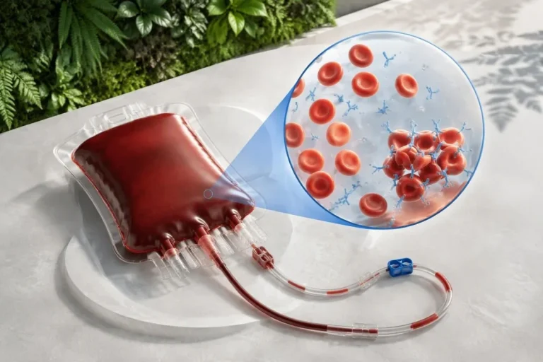
Anti-MAG antibodies usually belong to the IgM antibody class and recognize myelin-associated glycoprotein, a protein associated with the insulating sheath around peripheral nerves. MAG is not a vitamin or a routine inflammation marker; antibody positivity answers a different question from glucose, B12 or C-reactive protein testing.
Diagnosis rests on 3 connected domains: the symptom pattern, electrophysiology and laboratory evidence. Strong agreement between those domains raises confidence; disagreement should prompt a search for another explanation, a coexisting disorder or an assay limitation rather than an automatic immune-neuropathy label.
For a hypothetical 68-year-old with slowly worsening balance, reduced toe vibration and distinctly prolonged distal nerve latencies, a result above 7,000 BTU has more diagnostic weight than it would in someone with a single numb toe after tight footwear. Our guide to ynterpretaasje fan in posityf antistofresultaat explains this distinction between detection and diagnosis.
Kantesti is an AI blood test analyzer that can help organize antibody results alongside relevant laboratory findings, but an examination and nerve conduction study remain outside a blood-test-only assessment. Information about ús organisaasje explains who provides the service; no AI interpretation should substitute for specialist confirmation of this uncommon condition.
Which anti MAG neuropathy symptoms fit the condition?
Anti MAG neuropathy symptoms usually begin with slowly progressive, symmetric sensory changes in the feet, particularly numbness, reduced vibration sense and unsteadiness. The pattern generally develops over months or years, not within a few hours, and weakness may be less prominent than impaired sensation early on.

Loss of position sense can make walking harder in dim light because vision compensates for unreliable sensory information from the feet. A person may describe feeling as though they are walking on cushions or being less certain where their feet land, even when muscle strength seems relatively preserved.
Tremor is a recognized accompanying feature, especially in the hands, but tremor alone does not identify anti-MAG disease. A hypothetical person with foot numbness over 3 years and a new hand tremor has a different evaluation pathway from someone whose only problem is a longstanding familial tremor.
The distribution helps separate generalized neuropathy from focal compression. Symptoms restricted to selected fingers, especially at night, can suggest carpal tunnel nerve compression, whereas length-dependent neuropathy usually affects the feet before substantial hand involvement; both conditions can exist in the same person.
Pain does not rule out anti-MAG neuropathy, but intense burning pain with normal routine nerve conduction results should also raise consideration of small-fiber neuropathy. Record 3 practical observations before review: whether symptoms are symmetric, how balance has changed and whether weakness is genuinely increasing rather than simply feeling fatigued.
How should anti MAG antibody levels be interpreted?
Anti MAG antibody levels must be interpreted using the laboratory's assay and units. For one commonly used ELISA reported in Bühlmann titer units, approximately 1,000 BTU is the manufacturer's positivity threshold, while levels above 7,000 BTU provide stronger support for anti-MAG neuropathy in the appropriate clinical setting.

Liberatore et al. (2020), in the Journal of Neuroimmunology, evaluated the diagnostic performance of a commercial anti-MAG ELISA and described a diagnostic gray zone between 1,500 and 7,000 BTU. Their findings explain why a laboratory can correctly flag a positive result that still does not provide sufficient specificity for a clinical diagnosis.
The 2021 EAN/PNS CIDP guideline identifies high anti-MAG titers above 7,000 BTU as strongly suggesting a diagnosis other than CIDP when the relevant assay is used (Van den Bergh et al., 2021). That threshold helps distinguish disease categories; it is not an emergency-treatment threshold or a measure of how much nerve function remains.
Results expressed as a dilution titer, an index or another laboratory's arbitrary units cannot be converted reliably into BTU using a simple formula. The distinction between kwalitative en kwantitative resultaten matters here: the words positive and high may conceal very different analytical methods.
A hypothetical report reading greater than 70,000 BTU may reflect the assay's reporting ceiling rather than an exact concentration. Ask whether dilution testing was performed before comparing that result with a later numerical value; a change from greater than 70,000 to 60,000 does not establish a precisely measurable percentage reduction.
| Nivo | Berik | Wat it betsjut |
|---|---|---|
| Usually negative on this ELISA | Below approximately 1,000 BTU | Does not support anti-MAG neuropathy on this assay; other causes of neuropathy still require assessment. |
| Low-positive near the assay cutoff | Approximately 1,000–1,499 BTU | Limited diagnostic specificity; assess why testing was ordered and whether the nerve pattern fits. |
| Overlapping diagnostic range | Approximately 1,500–7,000 BTU | Compatible results can occur in different neuropathies; clinical and electrophysiological correlation is necessary. |
| Stronger diagnostic support | Above 7,000 BTU | Supports anti-MAG neuropathy when the clinical pattern fits; does not independently indicate severity or urgency. |
Why can a low-positive anti-MAG result be misleading?
A low-positive anti-MAG result may be incidental, analytically nonspecific or associated with a different neuropathy. Its meaning depends heavily on the probability of anti-MAG disease before testing, so broad antibody screening in someone with nonspecific tingling creates more interpretive problems than targeted testing after a neurological assessment.

A test's specificity does not guarantee that every positive result represents disease. Consider a purely hypothetical screening test with 95% sensitivity and 95% specificity in 1,000 people, only 10 of whom have the target condition: it would produce roughly 10 true positives and 50 false positives.
Those example percentages are not measured performance estimates for anti-MAG testing; they demonstrate why low disease prevalence changes the meaning of a positive result. At Kantesti, we explain a flagged antibody as a finding to reconcile with other evidence, not proof that it caused the person's symptoms.
A similar interpretive problem occurs with low-positive MOG antibody findings, although MOG and MAG identify different proteins and disorders. Shared naming does not make the tests interchangeable, and a positive result for one cannot confirm a condition associated with the other.
If a hypothetical result of 1,200 BTU conflicts with an entirely axonal, diabetes-compatible neuropathy, clinicians may review the original method, repeat testing or seek specialist laboratory advice. Repeating an antibody panel every few weeks without a specific question is less useful than confirming whether the phenotype actually supports anti-MAG disease.
What do nerve conduction studies add to diagnosis?
Nerve conduction studies establish whether numb feet reflect large-fiber neuropathy and whether the electrical pattern suggests myelin dysfunction. Anti-MAG neuropathy commonly shows disproportionately slowed conduction in distal nerve segments, prolonged distal motor latencies and reduced or absent sensory responses; the whole pattern matters more than one abnormal value.
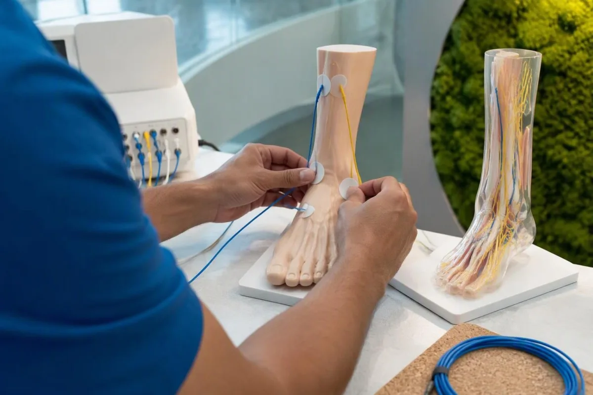
De distal motor latency measures how long a stimulated signal takes to reach a muscle from a distal stimulation site. A hypothetical latency of 7.2 milliseconds against that laboratory's upper limit of 4.5 milliseconds suggests slowing, but it cannot establish anti-MAG neuropathy without reviewing other nerves, distances and technical conditions.
Cold limbs can falsely prolong latency and slow conduction, so temperature control is part of a reliable examination. Common technical targets are at least approximately 32°C for the hands and 30°C for the feet, although laboratories follow their own protocols and should document whether warming was needed.
Severe anti-MAG neuropathy can involve secondary axonal loss, leaving responses too small or absent for standard demyelination criteria to be demonstrated cleanly. An advanced study that appears partly axonal therefore does not automatically exclude the condition; earlier recordings and the distribution of surviving responses may become especially useful.
A neurofilament light chain result cannot replace nerve conduction testing because it does not localize or classify the neuropathy in the same way. Ask for the specialist's written interpretation—not just a page of numbers—and whether the findings are distal-predominant, generalized, focal or technically limited.
How does anti-MAG neuropathy differ from CIDP or MS?
Anti-MAG neuropathy is generally distinguished from CIDP and is not the same disorder as multiple sclerosis. Anti-MAG disease predominantly affects peripheral nerves; MS affects the central nervous system, while CIDP is another immune-mediated peripheral neuropathy with different diagnostic criteria and often different treatment responsiveness.
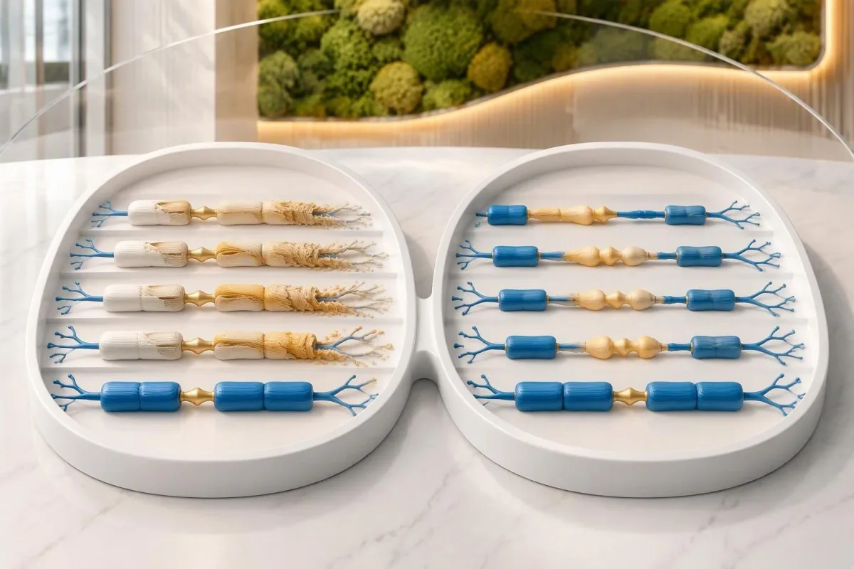
The typical anti-MAG phenotype is sometimes described as distal acquired demyelinating symmetric neuropathy, or DADS. By contrast, typical CIDP commonly causes both proximal and distal weakness, although distal CIDP variants exist; this overlap is one reason an IgM-associated distal phenotype needs more than a diagnostic label from electrophysiology alone.
The EAN/PNS guideline recommends considering anti-MAG testing when a person with an IgM paraprotein meets CIDP criteria, especially in a distal presentation (Van den Bergh et al., 2021). Its greater-than-7,000-BTU discussion concerns disease differentiation, not the assumption that any positive anti-MAG result invalidates every possible CIDP diagnosis.
A spinal fluid protein elevation is not specific enough to settle the distinction by itself. Likewise, oligoklonale bannen yn it harsensfocht answer a different neurological question; neither a CSF finding nor an MRI abnormality should be treated as confirmation that an anti-MAG result explains distal numbness.
Treatment response also requires care in interpretation: limited benefit from IVIG may fit anti-MAG disease, but response or nonresponse alone cannot classify a neuropathy. For a hypothetical person whose diagnosis changes after 2 specialist reviews, the useful question is which examination, antibody and nerve-study findings justified the reclassification.
Why does monoclonal IgM matter in anti-MAG disease?
Monoclonal IgM matters because an antibody-producing cell clone can generate the anti-MAG antibody associated with nerve dysfunction. Many affected people have an IgM monoclonal gammopathy, but an IgM band is not automatically a cancer diagnosis, and an incidental clone does not automatically explain a neuropathy.

Serum protein electrophoresis estimates a monoclonal protein when it is detectable, while immunofixation identifies its immunoglobulin type and light-chain restriction. A small band may appear on immunofixation testing even when electrophoresis does not show a clearly quantifiable M-spike, so the two tests are complementary rather than redundant.
A common adult total IgM reference interval is approximately 0.4–2.3 g/L, with laboratory-specific variation. A normal total IgM does not exclude a small monoclonal IgM band, while a high total IgM may be polyclonal; our útlis oer it immunoglobulinepaniel separates antibody quantity from clonality.
Hematology assessment determines whether the clone represents an IgM monoclonal gammopathy or a condition such as Waldenström macroglobulinemia, using the broader clinical and laboratory picture. Marrow assessment or imaging may be appropriate, but neither is automatically required solely because one antibody test is positive; anemia, organ enlargement and other features influence the decision.
A hypothetical monoclonal protein of 0.5 g/L can still warrant attention if its antibody has a credible nerve-directed effect. Conversely, a larger IgM concentration can coexist with unrelated diabetic neuropathy; clone size, antibody specificity and neurological disability answer different questions and should not be collapsed into one severity score.
Which blood tests usually accompany anti-MAG evaluation?
The accompanying workup usually assesses monoclonal proteins and common treatable causes of neuropathy. Depending on the presentation, clinicians consider serum protein electrophoresis, immunofixation, quantitative immunoglobulins, free light chains, glucose or HbA1c, vitamin B12, a complete blood count, kidney function and thyroid testing rather than repeating anti-MAG antibodies alone.
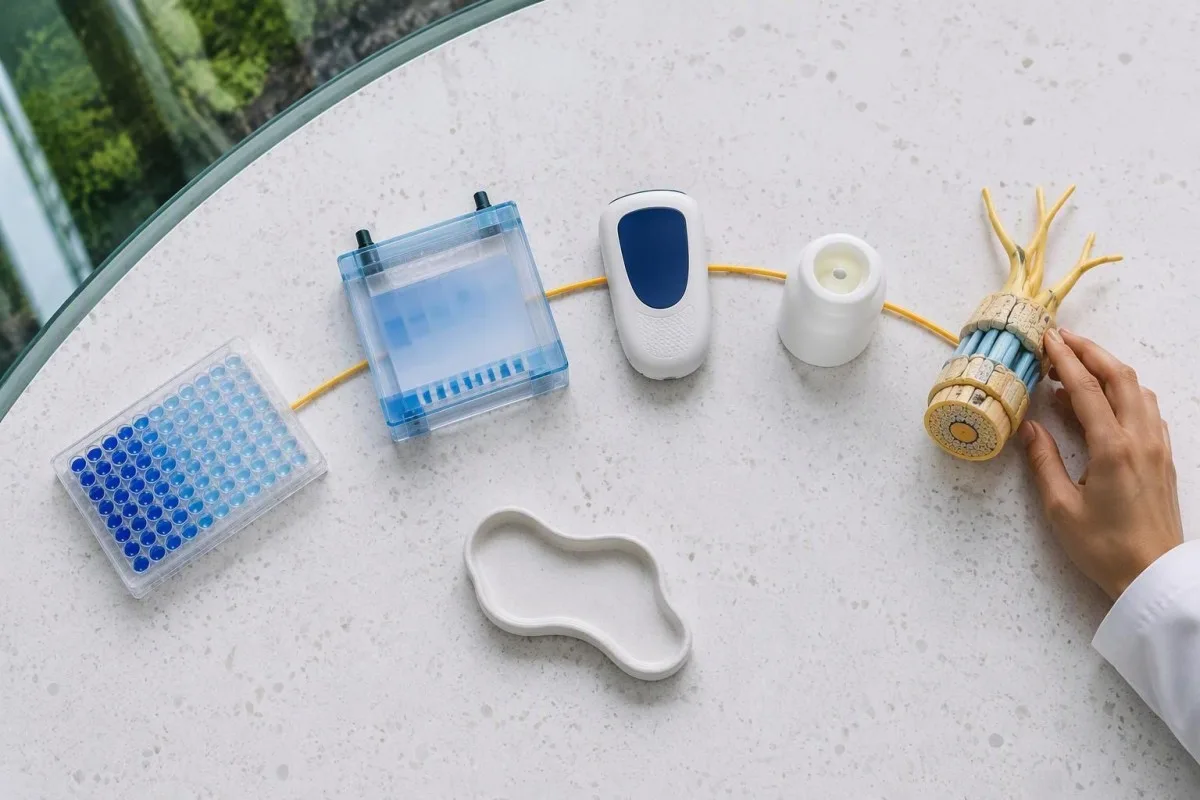
In HbA1c fan 6.5% of 48 mmol/mol is a diagnostic threshold for diabetes, usually requiring confirmation when unequivocal hyperglycemia is absent. That number identifies a metabolic disorder, not proof that diabetes caused the neuropathy; prominent demyelination, unusual asymmetry or rapid progression may still require a separate neurological explanation.
Vitamin B12 below approximately 200 pg/mL, equivalent to 200 ng/L, commonly suggests deficiency, although diagnostic cutoffs vary by assay and guideline. Borderline values may need methylmalonic acid interpretation; reduced kidney function can elevate methylmalonic acid independently, so an abnormal confirmatory marker should not be interpreted without creatinine or eGFR.
Protein measurements need similar context: a normal total protein or albumin/globulin ratio cannot exclude a small monoclonal band. Our gids foar it ynterpretearen fan serumeiwiten explains why a broad protein measurement and a targeted immunofixation study answer different questions, even when they come from the same sample.
Kantesti is an AI blood test interpretation platform that can help arrange these laboratory findings into a readable review, while the requesting clinician decides which tests are appropriate. Our gids foar bloedtest-biomarkers provides background terminology; a missing test should be identified as missing evidence, not silently treated as normal.
What more common conditions can cause numb feet?
Diabetes, vitamin B12 deficiency, alcohol-related nerve injury, medication effects and nerve compression are more common explanations for numb feet than anti-MAG neuropathy. The symptom distribution and nerve-study pattern guide the comparison, and identifying one possible cause does not exclude a second contributor.
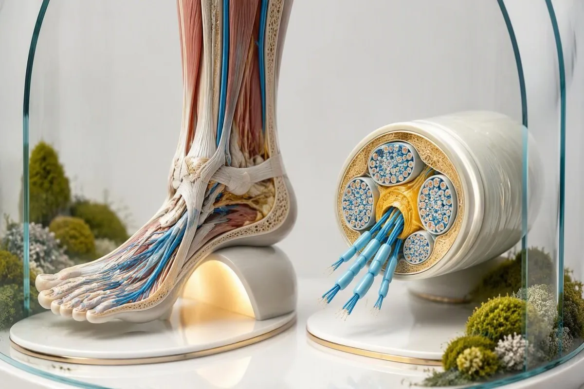
Diabetic distal polyneuropathy is often length-dependent and predominantly axonal, although mixed findings occur and mild slowing is not automatically immune demyelination. A hypothetical person with 12 years of diabetes, reduced sensory amplitudes and a weak anti-MAG signal needs careful correlation, not a diagnosis chosen by whichever laboratory result looks most unusual.
B12-related neurological problems can occur without anemia or a markedly raised MCV. Our borderline B12 assessment guide explains why a normal complete blood count cannot rule out a potentially treatable deficiency, especially when balance or position sense is affected.
Supplements can also complicate the picture: excessive vitamin B6 can cause sensory neuropathy, while excessive zinc can contribute to copper deficiency. A hypothetical daily combination of 3 supplements may duplicate ingredients, so bring the actual product labels; our explanation of zinc-related nerve symptoms covers one avoidable source of harm.
Compression or spinal nerve-root disease becomes more plausible when symptoms are one-sided, follow a specific nerve territory or accompany radiating back pain. Conversely, warskôgingssymptomen foar hege glukoaze can justify metabolic assessment, but the absence of thirst or frequent urination does not exclude diabetes or diabetic neuropathy.
What do contrasting anti-MAG results look like in practice?
Contrasting examples show why the same antibody flag can lead to different next steps. A high titer with a matching distal demyelinating neuropathy supports specialist evaluation for anti-MAG disease; a weak titer with a clearly different neurological pattern should trigger diagnostic reconsideration rather than automatic immunotherapy.
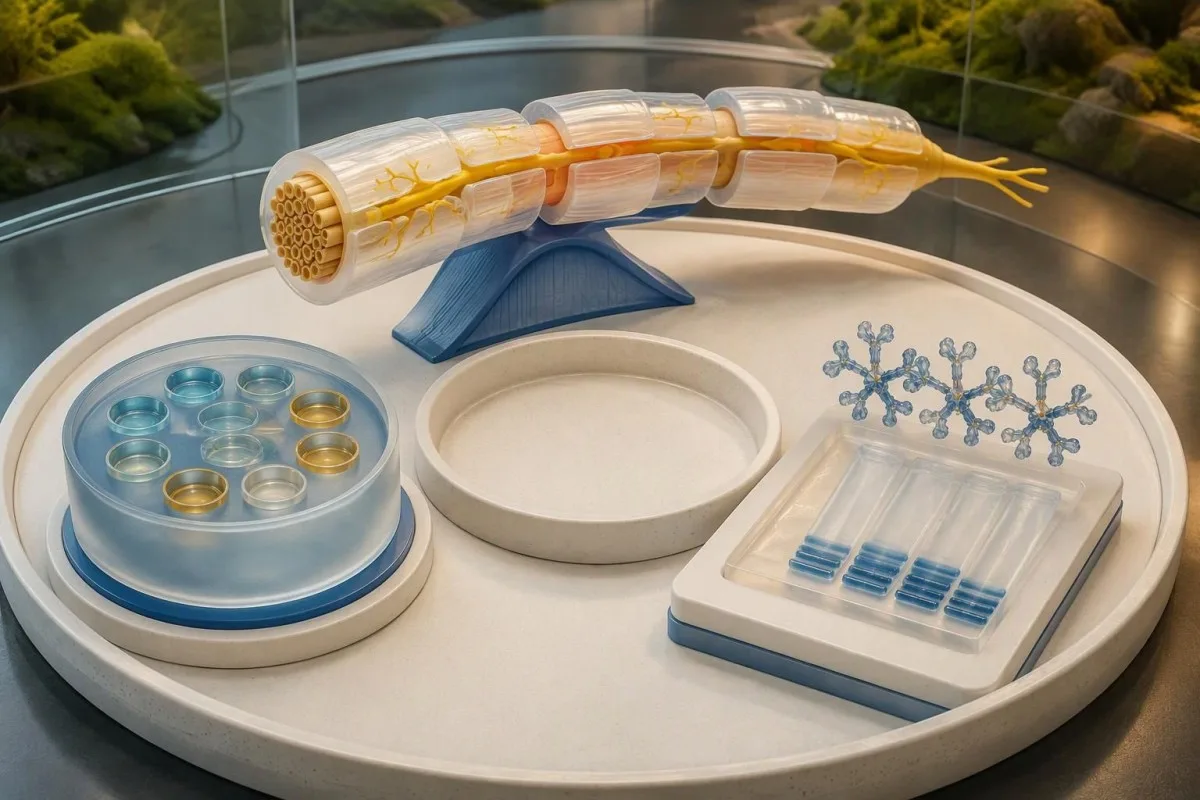
Example A is hypothetical: a 71-year-old has progressive symmetric foot numbness over 4 years, worsening balance in darkness, distal-predominant demyelination and anti-MAG antibodies of 28,000 BTU. Immunofixation identifies monoclonal IgM; together, these findings justify coordinated neuromuscular and hematology assessment, although treatment still depends on disability and progression.
Example B is also hypothetical: a 59-year-old has HbA1c of 9.1%, burning feet, an axonal nerve-study pattern and anti-MAG antibodies of 1,300 BTU. Diabetes is a stronger initial explanation than the low-positive antibody, but the clinician should still investigate atypical features or additional deficiencies rather than assuming every symptom has one cause.
Example C involves a hypothetical 64-year-old with 18,000 BTU but no numbness, weakness, balance change or abnormal neurological examination. The result merits review of why testing was ordered and whether an IgM clone is present; it does not justify treatment to prevent a neuropathy that has not been established.
Kantesti AI can help organize the differences between these laboratory profiles, but the decisive nerve-study and examination findings must come from the clinical record. A measurable M-spike result adds useful information about a clone; it still cannot establish that the clone is responsible for a particular person's sensory symptoms.
Does anti-MAG neuropathy always need immune treatment?
Anti-MAG neuropathy does not always require immediate immune treatment. Specialists generally weigh functional impairment, documented progression, fall risk and the associated IgM-producing clone; mild stable symptoms may be monitored, whereas progressive disability can justify discussion of clone-directed treatment despite uncertainty about the size and timing of benefit.

Rituximab is commonly considered by specialists, but the randomized evidence is honestly mixed. In a 54-participant trial, Léger et al. (2013) found no significant benefit on the primary sensory outcome at 12 months, although some secondary analyses suggested benefit; those secondary findings do not overturn the negative primary endpoint.
That trial used 375 mg/m² weekly for 4 weeks, a research regimen rather than a self-treatment recommendation. Clinical protocols vary, and an individual decision must address infection risk, vaccination planning, hepatitis B screening and baseline immunoglobulins; lowering antibody production is not the same as immediately repairing previously injured axons.
Neurological improvement, when it occurs, may take several months, and stabilization can be a meaningful outcome in progressive disease. Transient worsening after rituximab has also been reported; rapidly increasing symptoms after treatment should be reviewed promptly rather than dismissed as an expected laboratory fluctuation.
IVIG, corticosteroids and plasma exchange do not have the same established role in typical anti-MAG disease as in some other immune neuropathies. Before antibody-directed therapy, a history of frequent infections requiring assessment may influence safety planning; physiotherapy, balance support and symptom control remain useful regardless of whether immune treatment is chosen.
Do falling antibody levels mean the nerves are recovering?
Falling anti MAG antibody levels do not reliably prove that nerve function is recovering. Antibody production, myelin function and axonal survival can change on different timelines, so specialists track symptoms, examination and function alongside laboratory results rather than treating a percentage reduction as a neurological outcome.
A hypothetical fall from 30,000 to 15,000 BTU represents a 50% numerical reduction if the same assay measured both values within its reliable range. It does not mean sensation has improved by 50%, walking is 50% safer or half the nerve impairment has resolved.
Comparisons are strongest when the laboratory, assay, dilution approach and treatment timing are documented. Results above an analytical ceiling, or values from different assay systems, can create misleading trends; repeat nerve conduction testing is likewise most informative when comparable nerves and technical conditions are assessed.
Functional tracking might include a standardized disability scale, grip strength when relevant and a consistent walking task such as a 10-meter walk if the specialist selects it. Falls, footwear problems and difficulty using stairs often communicate meaningful change better than a daily tingling score, especially when sensory symptoms fluctuate.
Kantesti is an AI biomarker interpretation platform that can help present laboratory trends while keeping assay differences visible. Our technology-útlis describes the service's approach, and our listet de resultaatferoarings dy't de muoite wurdich binne om te dokumintearjen. explains why numerical change still needs clinical interpretation rather than an automatic treatment decision.
When do numb feet require urgent medical assessment?
Rapidly increasing weakness, breathing or swallowing difficulty, and new bladder or bowel dysfunction require urgent assessment regardless of an anti-MAG result. These are not features to attribute casually to a usually slow neuropathy; sudden one-sided neurological symptoms or a suddenly cold, painful foot also need immediate evaluation.

Weakness progressing over days rather than years raises concern for other neurological disorders, including Guillain–Barré syndrome. Our rapid weakness warning guide explains why someone who suddenly struggles to stand or climb stairs should not wait for a repeat anti-MAG titer to decide whether help is needed.
New saddle-area numbness with difficulty passing urine or loss of bladder control can indicate spinal nerve compression requiring emergency assessment. A pre-existing antibody-positive neuropathy does not protect someone from a second urgent condition, and a reassuring laboratory report cannot rule out a spinal emergency.
Reduced foot sensation increases the chance of unnoticed pressure injury or burns. Inspect both feet daily, check shoe interiors for foreign objects and test bath water safely before stepping in; avoiding direct heat pads on numb feet is practical prevention, not a treatment for the underlying antibody process.
Balance support should match actual fall risk rather than the height of the antibody result. For a hypothetical person who has fallen twice in 1 month, a mobility assessment, appropriate walking aid and home-hazard review may be more immediately useful than another titer measurement while specialist care is being arranged.
How can you prepare for review and assess our research?
A useful specialist review needs the original antibody report, nerve conduction interpretation, monoclonal protein results and a clear symptom timeline. Our research publications address blood-test interpretation methodology; they do not establish that an AI system can diagnose anti-MAG neuropathy or replace neurological and hematological assessment.

Bring 4 groups of information: the complete anti-MAG report with units, the electrophysiology report, protein studies including immunofixation, and a list of medications and supplements. A one-page timeline of symptom onset, balance change and falls helps distinguish slowly progressive neuropathy from a recent superimposed problem.
Ask whether the nerve pattern is genuinely demyelinating, whether the IgM finding is monoclonal and whether another treatable cause remains plausible. If treatment is proposed, agree on what success would mean—such as fewer falls or stable walking ability—and how it will be assessed over the next several months.
Kantesti's listed technical benchmark concerns 100.000 syntetyske testgefallen, while the accompanying validation framework describes a framework for evaluating interpretation and oversight. Synthetic-case performance is not equivalent to prospective clinical validation in people with antibody-positive neuropathy, and neither publication should be cited as evidence that an anti-MAG result alone establishes disease.
The 2 research publications are listed below separately from the external medical references supporting this article. Our technyske validaasje en tafersjoch page provides methodological context; the practical next step for persistent numb feet remains a clinician-led examination and targeted testing, not a diagnosis generated from one flagged biomarker.
Faak stelde fragen
Betsjutte hege anty-MAG-antistoffen dat ik anty-MAG-neuropaty haw?
High anti MAG antibodies support anti-MAG neuropathy only when symptoms and nerve conduction findings are compatible. A level above 7,000 BTU on the relevant ELISA provides stronger diagnostic support than a weak-positive result, but it is not a stand-alone diagnosis. Clinicians also assess monoclonal IgM and alternative causes such as diabetes or B12 deficiency. A very high result without neurological symptoms does not automatically justify immune treatment.
What anti MAG antibody level is considered high?
Anti MAG antibody thresholds depend on the assay and reporting units. On one commonly used commercial ELISA, approximately 1,000 BTU is the positivity threshold, while results above 7,000 BTU provide stronger support for anti-MAG neuropathy in the right clinical setting. Lower-positive results overlap with other conditions and require careful correlation. These thresholds cannot be transferred directly to dilution titers, index values or another laboratory's arbitrary units.
What are the first symptoms of anti-MAG neuropathy?
Anti-MAG neuropathy usually begins with slowly progressive, symmetric foot numbness, reduced vibration sense or impaired balance. Symptoms commonly develop over months to years, with weakness sometimes appearing later. A hypothetical history of 3 years of worsening steadiness in darkness fits better than numbness appearing suddenly over 3 hours. Rapid weakness, breathing difficulty or new bladder dysfunction requires urgent assessment for other causes.
Can I have anti-MAG neuropathy with a normal IgM level?
A normal total IgM level does not exclude a small monoclonal IgM band or anti-MAG neuropathy. A common adult IgM reference interval is approximately 0.4–2.3 g/L, but the interval varies by laboratory and measures quantity rather than clonality. Serum immunofixation can detect a small monoclonal band that is not obvious on total IgM testing or routine electrophoresis. Neurological findings still need to support the diagnosis.
Does a positive anti-MAG antibody result mean cancer?
A positive anti-MAG antibody result does not automatically mean cancer. Anti-MAG neuropathy can be associated with an IgM monoclonal gammopathy, while some people require evaluation for an underlying lymphoproliferative disorder such as Waldenström macroglobulinemia. Even a hypothetical monoclonal protein of 0.5 g/L may be neurologically relevant, but its concentration alone cannot classify the clone. Hematology assessment considers the full laboratory pattern, symptoms and additional testing when appropriate.
Can rituximab cure anti-MAG neuropathy?
Rituximab is not a guaranteed cure for anti-MAG neuropathy, and the randomized evidence is mixed. A 54-participant trial reported no significant improvement in its primary sensory outcome at 12 months, although some secondary analyses suggested benefit. Specialists may still consider treatment for progressive functional impairment after discussing safety and uncertainty. Antibody reduction does not guarantee recovery of nerve fibers that have already been lost.
Should anti-MAG antibodies be repeated to monitor numb feet?
Repeat anti-MAG testing can be useful when a specialist has a defined diagnostic or treatment-monitoring question, but frequent testing is not automatically helpful. A fall from 30,000 to 15,000 BTU is a 50% numerical reduction only when the same assay provides comparable measurements. That reduction does not establish a 50% improvement in sensation or walking. Functional change, examination findings and selected repeat nerve studies remain necessary parts of follow-up.
Lit jo bloedtest resultaten útlizze
Laad jo laboratoariumrapport op en de AI fan Kantesti leit elke biomarker binnen sekonden út yn gewoane taal. Brûkt troch mear as 2 miljoen minsken wrâldwiid.
Referinsjes
- Klein, T., Mitchell, S., & Weber, H. (2026). In pre-registered, rubric-basearre automatyske technyske benchmark fan de Kantesti Blood-Test Interpretation Engine op 100.000 syntetyske testgefallen. Kantesti AI Medysk Undersyk. DOI
- Klein, T., Mitchell, S., & Weber, H. (2026). Klinysk Falidaasjekader v2.0 (Medyske Falidaasjeside). Kantesti AI Medysk Undersyk. DOI
- Van den Bergh PYK et al. (2021). European Academy of Neurology/Peripheral Nerve Society guideline on diagnosis and treatment of chronic inflammatory demyelinating polyradiculoneuropathy: Report of a joint Task Force—Second revision. European Journal of Neurology. DOI
- Liberatore G et al. (2020). Sensitivity and specificity of a commercial ELISA test for anti-MAG antibodies in patients with neuropathy. Journal of Neuroimmunology. DOI
- Léger JM et al. (2013). Placebo-controlled trial of rituximab in IgM anti-myelin-associated glycoprotein neuropathy. Neurology.
- 2M+Analysearre tests
- 127+Lannen
- 100+Talen
Oer it medyske team


E-E-A-T fertrouwensignalen
Ûnderfining
Dokter-oandreaune klinyske resinsje fan lab-ynterpretaasje-wurkprosessen.
Ekspertize
Fokus fan laboratoariummedisyne op hoe’t biomarkers har gedrage yn in klinyske kontekst.
Autoriteit
Medysk beoardiele troch Dr. Thomas Klein, mei bydragen fan Dr. Sarah Mitchell en prof. dr. Hans Weber.
Betrouberens
Bewiis-basearre ynterpretaasje mei dúdlike ferfolchpaadkes om alarm te ferminderjen.



Medyske disclaimer
Dit artikel is allinnich foar ûnderwiisdoelen en foarmet gjin medysk advys. Rieplachtsje altyd in kwalifisearre soarchferliener foar besluten oer diagnoaze en behanneling.
Publisearre · Nim kontakt op mei it redaksjeteam
Publisearre troch Kantesti Ltd, registrearre yn Ingelân en Wales, bedriuwsnûmer. 17090423, Londen, Feriene Keninkryk.
