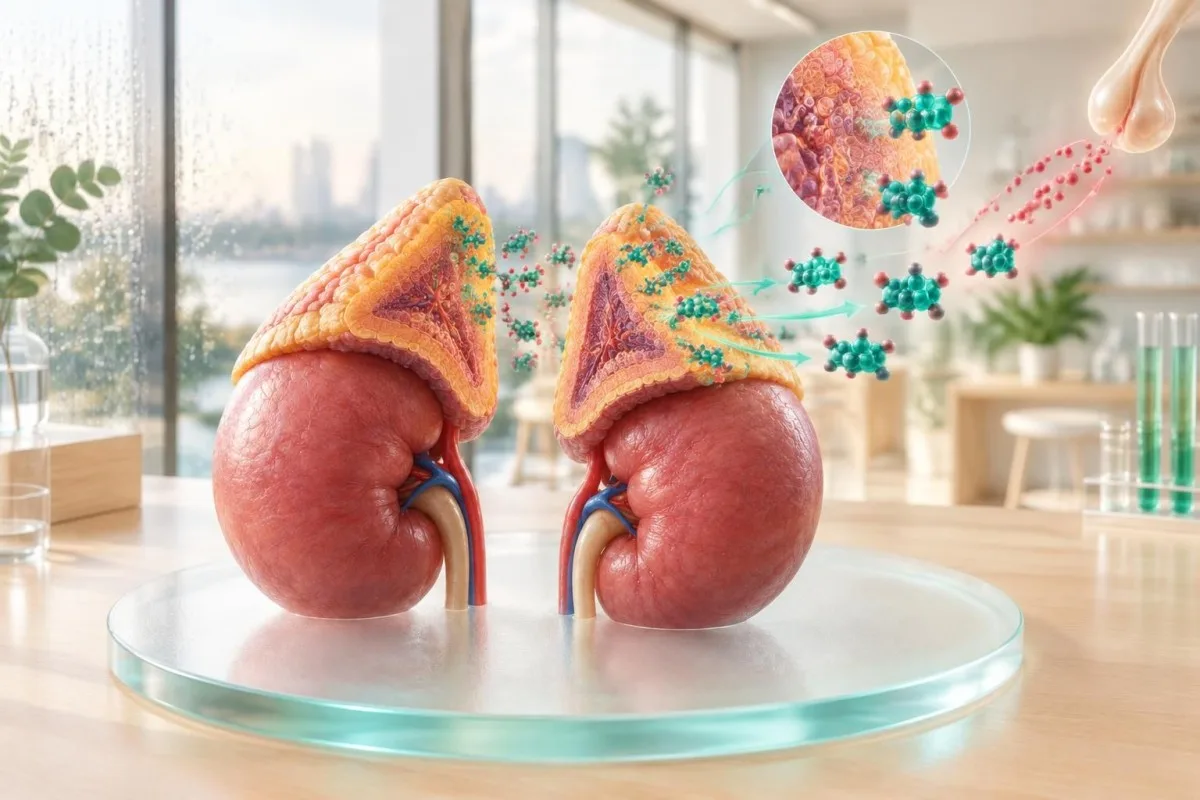
Low DHEA usually reflects normal ageing, reduced ACTH stimulation, adrenal disease, pituitary dysfunction, chronic illness, or medication effects. A low DHEA-S result alone cannot identify the cause; pairing it with morning cortisol, ACTH, electrolytes, renin and other pituitary hormones provides a much clearer clinical pattern.

Βασικά συμπεράσματα
- Most common causes include age-related decline, oral contraceptives or oral estrogen, glucocorticoids, chronic illness, obesity and reduced ACTH signalling.
- Low DHEA-S levels do not diagnose adrenal insufficiency because DHEA-S falls with age and varies by assay, sex and medication exposure.
- Πρωτοπαθής επινεφρική ανεπάρκεια usually combines low morning cortisol, high ACTH, low aldosterone and high renin; potassium may be high and sodium low.
- Central adrenal insufficiency usually combines low morning cortisol with low or inappropriately normal ACTH, while aldosterone and potassium are often preserved.
- Best first pairing is an 8-9 AM cortisol with ACTH; an indeterminate cortisol often leads to a 250 microgram cosyntropin stimulation test.
- Medication clues include systemic glucocorticoids, injected or potent inhaled steroids, opioids, some hormone contraceptives and steroid-synthesis inhibitors.
- Συμπτώματα such as fatigue, low libido, reduced exercise tolerance and low mood are nonspecific and should not be attributed to DHEA without a wider assessment.
- Επαναληπτικός έλεγχος is most useful when the sample timing, assay, illness status or medication history could have distorted the first result.
What a low DHEA or DHEA-S result actually means
Low DHEA causes include normal age-related steroid decline, reduced ACTH drive from the pituitary, adrenal injury, medication suppression and chronic disease. DHEA-S is made mainly by the adrenal cortex, but a low result is a clue rather than a diagnosis because the value depends on age, sex, assay method and recent treatment.
DHEA και DHEA-S are related adrenal androgens, but they are not interchangeable laboratory results. DHEA is more variable during the day, whereas DHEA-S is more stable because sulfate binding creates a circulating reservoir; many laboratories therefore prefer DHEA-S for screening.
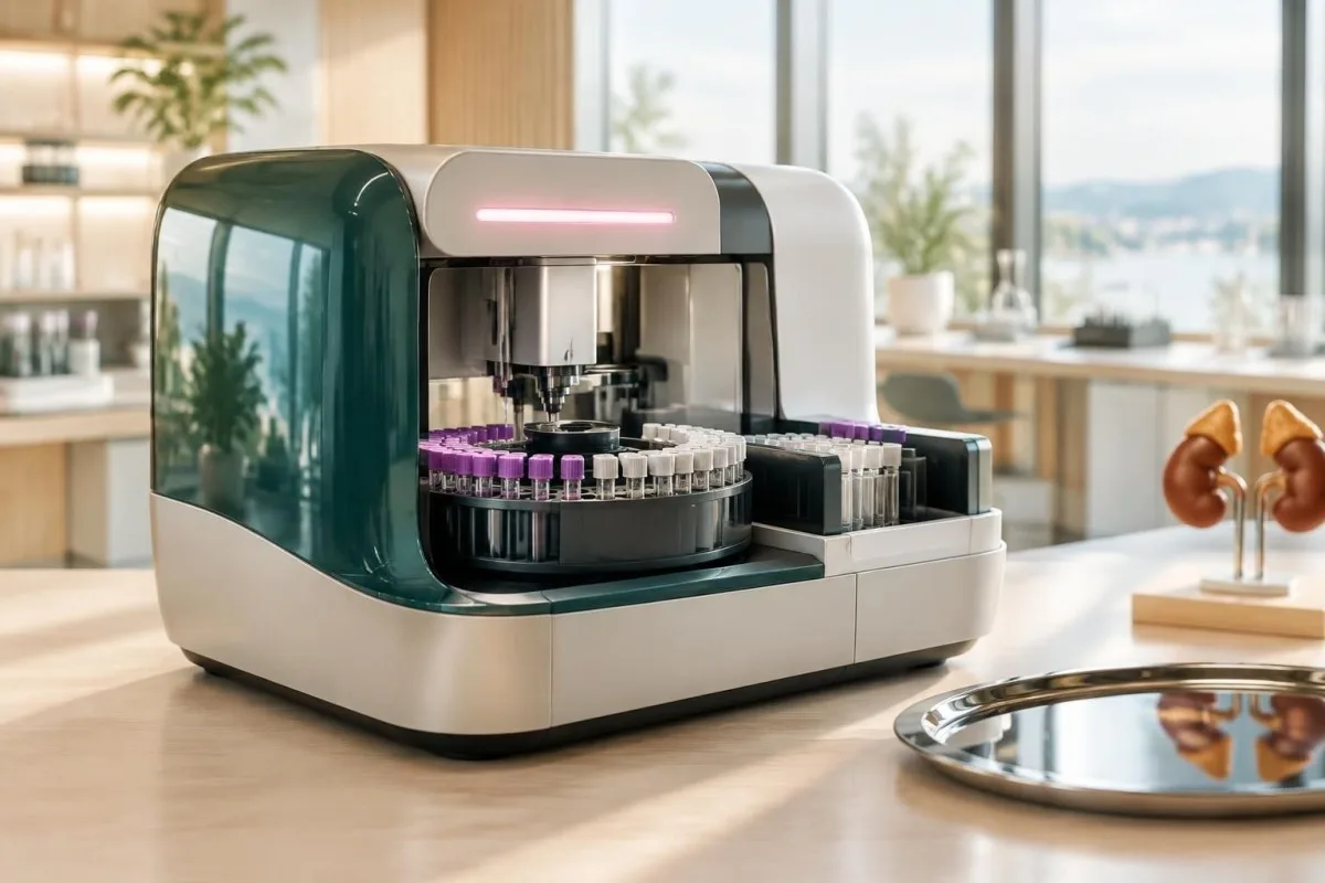
Το Καντέστι είναι ένα Αναλυτής εξέτασης αίματος AI that reads DHEA-S alongside cortisol, ACTH, sodium and potassium rather than treating one flagged result as a diagnosis. A DHEA-S value of 35 µg/dL may be low for a 25-year-old but expected for an older adult, depending on the laboratory interval.
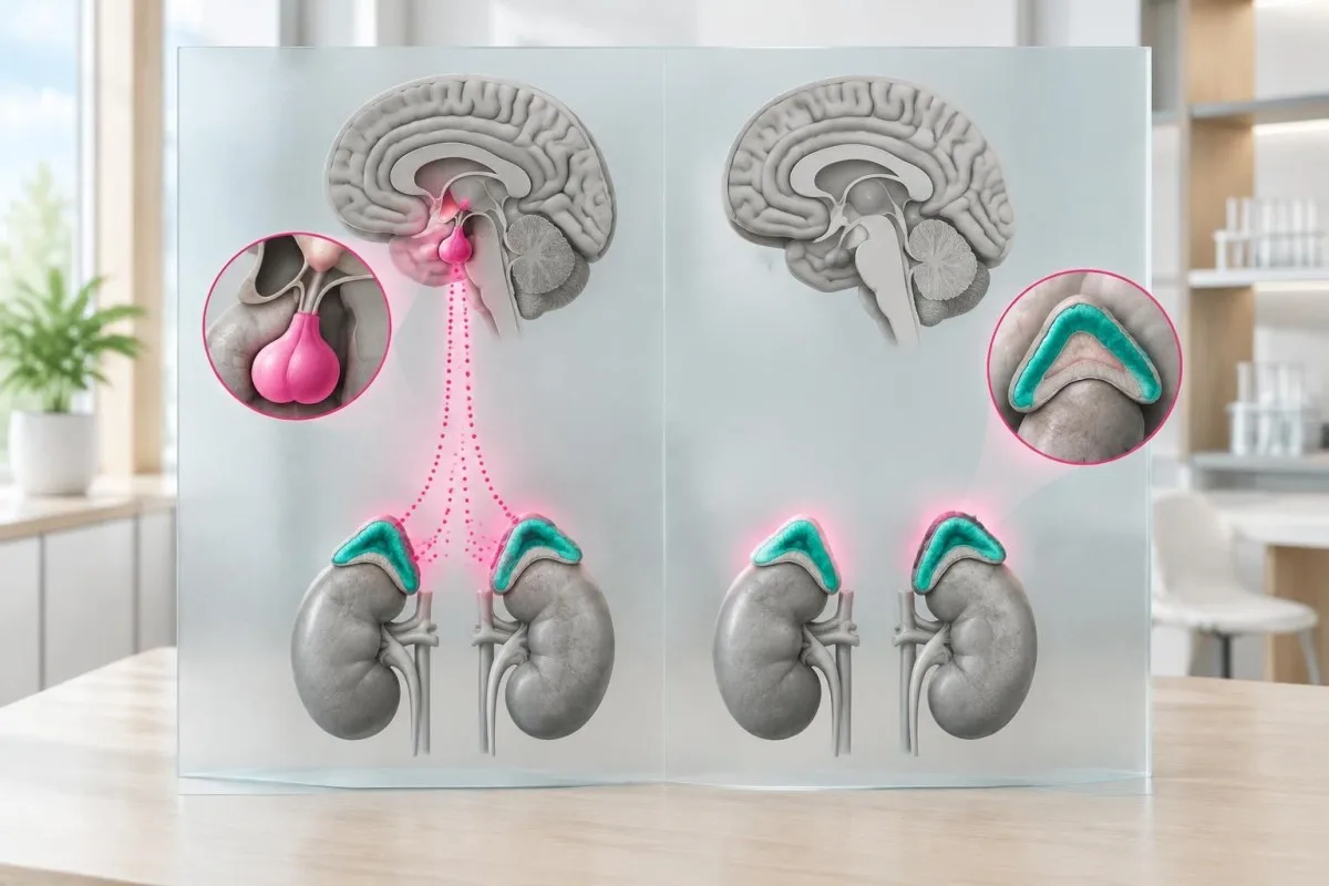
The adrenal cortex produces DHEA-S after stimulation by ACTH, the pituitary hormone that also supports cortisol production. This explains why low DHEA-S can appear in both primary adrenal disease and pituitary disease, even though ACTH moves in opposite directions.
A result below the printed range deserves context, not automatic supplementation. Review the collection time, age-specific interval, medication list and paired cortisol result before deciding whether the finding needs endocrine investigation.
How age changes DHEA-S levels and reference ranges
DHEA-S normally peaks in early adulthood and declines progressively after approximately age 30, so an adult result must be compared with an age- and sex-specific interval. Many laboratories report young-adult values around 100-600 µg/dL, but older-adult intervals can be markedly lower and assay ranges differ.
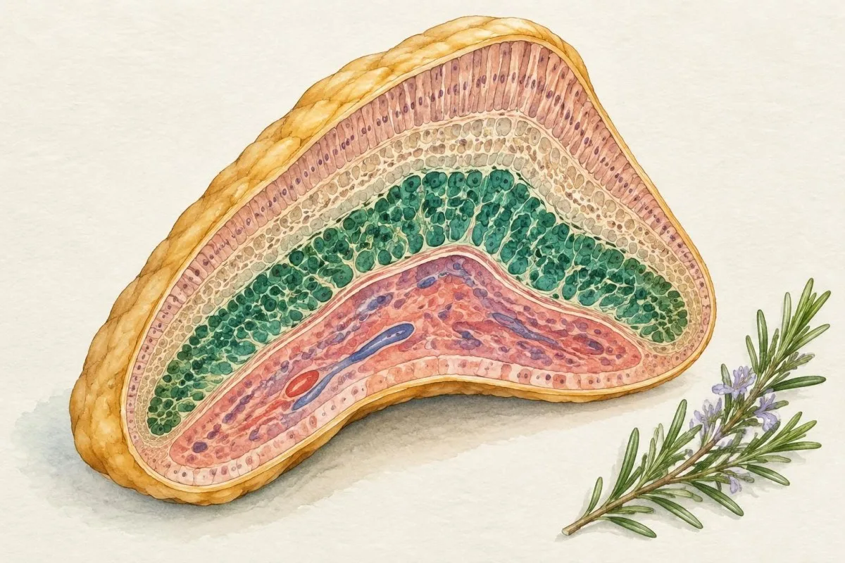
DHEA-S often reaches its highest concentration between ages 20 and 30, then declines by roughly 1-3% per year, although the slope varies substantially between people. By ages 70-80, concentrations may be only 10-30% of young-adult values without indicating adrenal failure.
A useful hypothetical example is a 72-year-old with DHEA-S of 42 µg/dL and normal morning cortisol, sodium and potassium. That value may be appropriate for the laboratory's age band, whereas the same number in a 24-year-old with weight loss and low blood pressure would deserve a different assessment.
Reference ranges are not universal. One European laboratory may report an adult female interval of 35-430 µg/dL, while another uses 65-380 µg/dL because the assay calibration and reference population differ; the reporting laboratory's interval should take priority.
Kantesti AI can place a DHEA-S value beside the reported reference interval and age information, but it cannot replace the clinician's review of symptoms, blood pressure or medication exposure. For related hormone timing principles, see our guide to morning hormone tests for men.
| Επίπεδο | Εύρος | Τι σημαίνει |
|---|---|---|
| Illustrative age-adjusted range | Laboratory-specific; often approximately 100-600 µg/dL in young adults | A result inside the interval is interpreted with age, sex and symptoms. |
| Απροσδόκητα υψηλή | Above the laboratory upper limit, often >500-700 µg/dL | May suggest androgen excess, adrenal overproduction or supplementation; the cutoff is assay-specific. |
| Lower than expected | Below the age-specific lower limit, such as <50 µg/dL | May reflect age, medication, illness or reduced ACTH signalling; pair with cortisol and ACTH. |
| Very low with concerning pattern | Often <20-30 µg/dL plus low cortisol or severe symptoms | Needs prompt clinical assessment, especially with vomiting, fainting, dehydration or confusion. |
Adrenal causes: when the gland itself is underactive
Primary adrenal insufficiency can cause low DHEA-S because adrenal tissue cannot produce normal steroid hormones, and it usually produces high ACTH as the pituitary attempts to compensate. The strongest laboratory pattern is low morning cortisol, high ACTH, low aldosterone, high renin and sometimes high potassium.
The most informative first test is usually an 8-9 AM serum cortisol paired with ACTH, not DHEA-S alone. Morning cortisol below approximately 3 µg/dL strongly supports adrenal insufficiency, while values above about 15-18 µg/dL make it less likely; intermediate results require local protocol and often stimulation testing.
The 2016 Endocrine Society guideline recommends evaluating suspected primary adrenal insufficiency with cortisol, ACTH and a corticotropin stimulation test when appropriate (Bornstein et al., 2016). A 250 microgram cosyntropin test measures whether cortisol rises adequately after direct adrenal stimulation.
A hypothetical 39-year-old with DHEA-S of 18 µg/dL, 8 AM cortisol of 2.4 µg/dL, ACTH of 310 pg/mL, sodium of 128 mmol/L and potassium of 5.8 mmol/L has a pattern far more concerning than a low DHEA-S result alone. The combination suggests impaired adrenal cortex function and needs urgent medical attention.
Autoimmune adrenalitis is a common cause in many higher-income settings, while tuberculosis, adrenal haemorrhage, metastatic disease, genetic disorders and certain drugs are other possibilities. A low DHEA-S does not identify the cause; 21-hydroxylase antibodies, imaging or infection testing may be selected after the hormone pattern is established.
Pituitary and hypothalamic clues behind low DHEA
Central adrenal insufficiency causes low DHEA-S through inadequate ACTH stimulation from the pituitary or hypothalamus, so ACTH is low or inappropriately normal rather than clearly elevated. Cortisol is low, but aldosterone and potassium often remain normal because mineralocorticoid regulation depends mainly on the renin-angiotensin system.
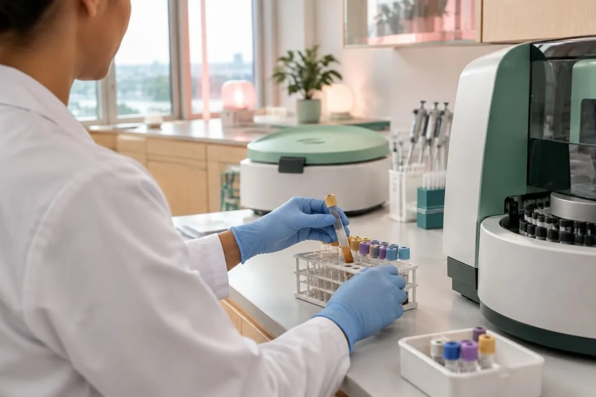
An ACTH of 12 pg/mL may look normal on a report, yet it is inappropriate if the paired 8 AM cortisol is 2.8 µg/dL. In primary adrenal failure, the same cortisol result would usually trigger a much higher ACTH response; interpretation depends on the pair, not either value in isolation.
Central causes include pituitary surgery, cranial irradiation, traumatic brain injury, pituitary inflammation, a sellar growth and prolonged glucocorticoid exposure. Other pituitary clues can include low free T4 with a normal or low TSH, low LH or FSH, low testosterone or estradiol, and raised prolactin.
A hypothetical 52-year-old taking prednisolone for 8 months has DHEA-S of 22 µg/dL, cortisol of 3.1 µg/dL, ACTH of 8 pg/mL, sodium of 139 mmol/L and potassium of 4.2 mmol/L. Preserved potassium does not rule out central adrenal suppression; the medication history is the major clue.
Low free T4 with a non-elevated TSH can support a broader pituitary pattern, although thyroid results require separate confirmation. Our related explanation of low free T4 with a normal TSH shows why a normal pituitary signal can still be biologically inadequate.
Medication clues that can lower DHEA-S
Systemic glucocorticoids are the clearest medication-related cause of low DHEA-S because they suppress hypothalamic CRH and pituitary ACTH. Oral estrogen-containing contraceptives can also lower measured androgen availability, while opioids, steroid-synthesis inhibitors and some long-term treatments may alter adrenal or pituitary signalling.

Prednisone, prednisolone, dexamethasone and hydrocortisone can suppress ACTH even at doses that patients may not consider high. Risk depends on dose, duration, route and individual susceptibility; the 2024 joint Endocrine Society and European Society of Endocrinology guideline treats prolonged exposure equivalent to roughly 4-6 mg prednisolone daily for 3-4 weeks or more as potentially suppressive (Beuschlein et al., 2024).
Inhaled, injected, topical and intra-articular steroids can contribute when exposure is substantial or repeated, although the risk is less predictable than with oral treatment. Never stop a prescribed glucocorticoid abruptly after prolonged use because withdrawal can precipitate adrenal insufficiency.
Combined oral contraceptives and oral estrogen may reduce DHEA-S in some people and alter sex-hormone binding proteins, making androgen symptoms and laboratory values harder to interpret. A useful companion resource is our explanation of DHEA-S results on birth control.
Opioids can suppress the hypothalamic-pituitary-adrenal and gonadal axes, while ketoconazole and related steroid-synthesis inhibitors can reduce cortisol and androgen production. Include dose, route, start date and last dose time when reviewing a result; a medication list without timing may miss the main explanation.
Low DHEA symptoms and why they are nonspecific
Low DHEA symptoms may include fatigue, reduced libido, low mood, poorer exercise tolerance, decreased muscle strength and reduced sense of wellbeing, but none is specific for DHEA deficiency. Sleep loss, anaemia, thyroid disease, depression, menopause, chronic infection and medication effects can produce the same complaints.
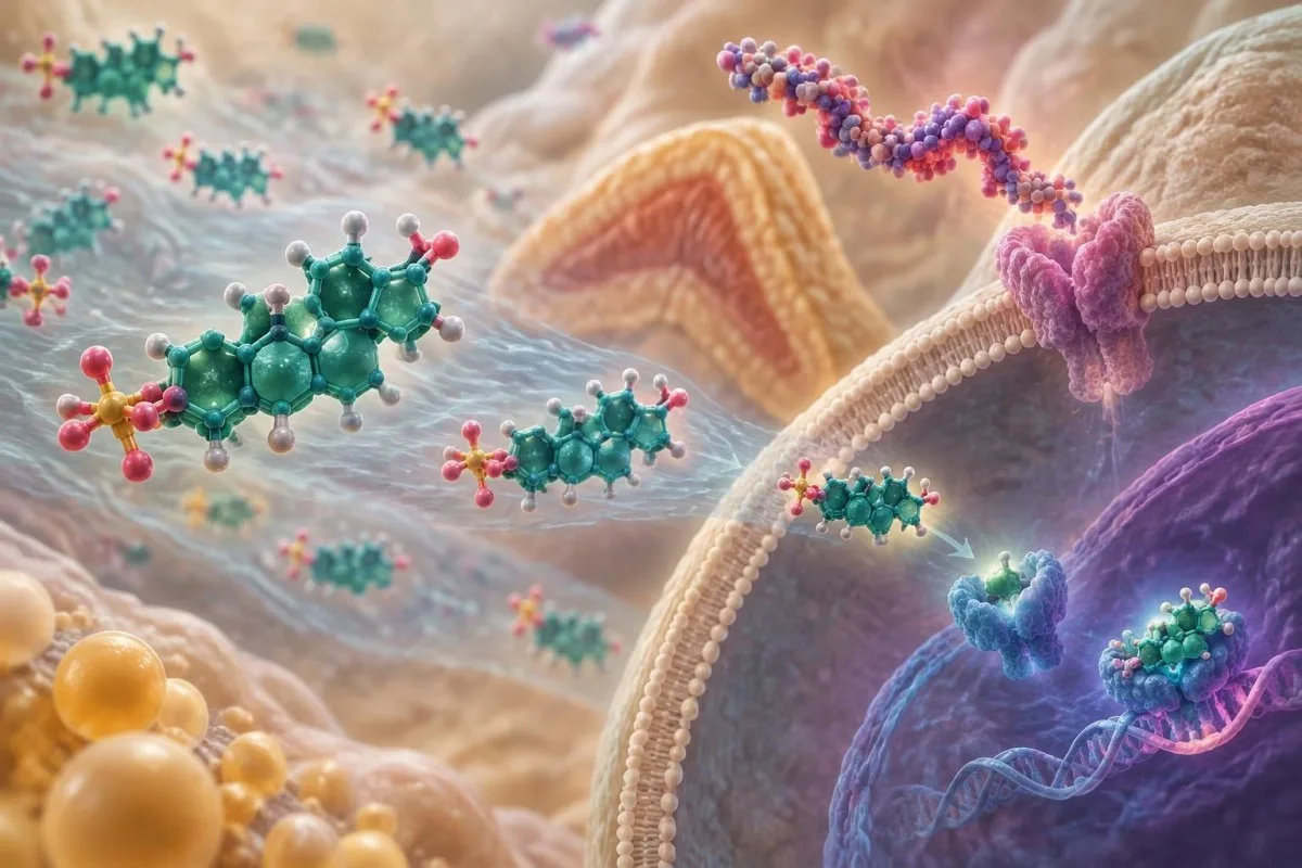
Fatigue is especially unreliable as a marker: in population studies, it is reported by a substantial minority of adults, while low DHEA-S is much less common in younger people. A person with DHEA-S of 48 µg/dL and ferritin of 9 ng/mL may feel exhausted because of iron depletion rather than adrenal androgen deficiency.
Reduced libido can reflect relationship stress, sleep deprivation, antidepressants, menopause, low testosterone, pain or vascular disease as well as adrenal hormones. A low result should prompt a structured review rather than a supplement recommendation based on symptoms alone.
A hypothetical 46-year-old with low mood, DHEA-S of 32 µg/dL, normal morning cortisol, TSH of 6.2 mIU/L and free T4 of 0.8 ng/dL has a thyroid clue that may be more actionable than the DHEA-S value. This is why paired and competing explanations matter.
DHEA supplementation is not a universal treatment for nonspecific fatigue. The Endocrine Society has advised against routine DHEA use for women with normal adrenal function, and even selected adrenal-insufficiency cases require clinician-supervised discussion; see our guide to εξετάσεις αίματος για χαμηλή λίμπιντο.
Which paired tests separate adrenal from pituitary causes?
The core paired test is an early-morning cortisol measured at the same time as ACTH; electrolytes, renin, aldosterone and a cosyntropin test then help separate primary from central disease. DHEA-S adds supportive information because it reflects ACTH-dependent adrenal androgen production, but it cannot localise the problem by itself.
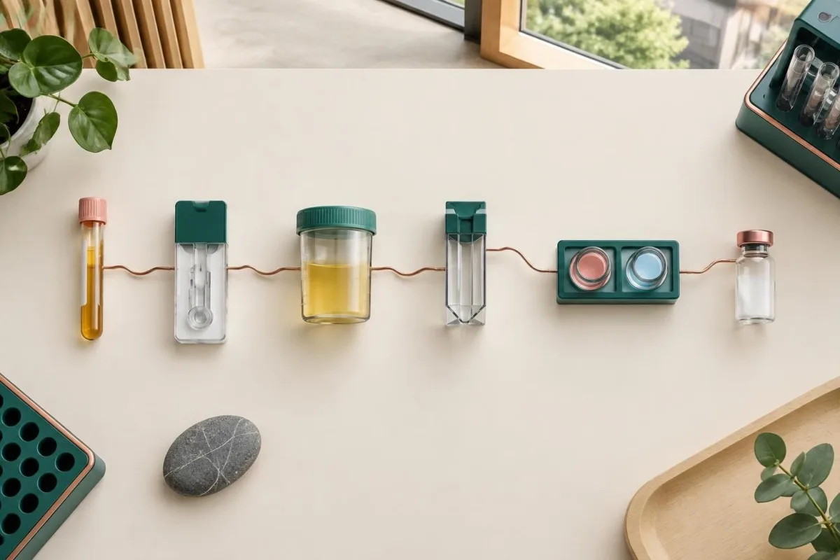
An 8-9 AM cortisol below 3 µg/dL is strongly suspicious, while a value above 15-18 µg/dL often makes clinically significant adrenal insufficiency unlikely; assays and guidelines differ, so local thresholds matter. Values between these boundaries commonly lead to a 250 microgram cosyntropin stimulation test.
ACTH above roughly twice the upper limit of normal with low cortisol supports primary adrenal insufficiency, whereas low or inappropriately normal ACTH supports central disease. Renin and aldosterone add a second axis: high renin with low aldosterone suggests mineralocorticoid loss, while central disease usually preserves this relationship.
The following pattern is more useful than any single low DHEA-S number: low cortisol plus high ACTH plus low sodium suggests primary disease; low cortisol plus low ACTH plus normal potassium suggests central suppression; normal cortisol with low DHEA-S usually points away from overt adrenal failure.
For broader test selection, our guide to free cortisol testing explains why saliva and urine tests answer different questions from an 8 AM serum cortisol.
| Επίπεδο | Εύρος | Τι σημαίνει |
|---|---|---|
| Likely adequate adrenal reserve | 8 AM cortisol often >15-18 µg/dL | Overt adrenal insufficiency becomes less likely, though assay-specific guidance applies. |
| Primary adrenal pattern | Low cortisol with ACTH >2 times the upper limit | Suggests adrenal gland failure; renin, aldosterone, sodium and potassium refine the pattern. |
| Indeterminate reserve | 8 AM cortisol approximately 3-15 µg/dL | Often requires cosyntropin stimulation testing rather than interpretation from DHEA-S alone. |
| Possible acute adrenal crisis | Low cortisol with vomiting, fainting, confusion or severe weakness | Urgent medical assessment is required; do not wait for a DHEA-S result. |
How cosyntropin testing adds information
A cosyntropin stimulation test checks whether the adrenal cortex can increase cortisol after synthetic ACTH, usually at baseline and 30 or 60 minutes. A peak cortisol below the laboratory's validated cutoff can support adrenal insufficiency, but modern assays often use cutoffs near 14-18 µg/dL rather than one universal number.
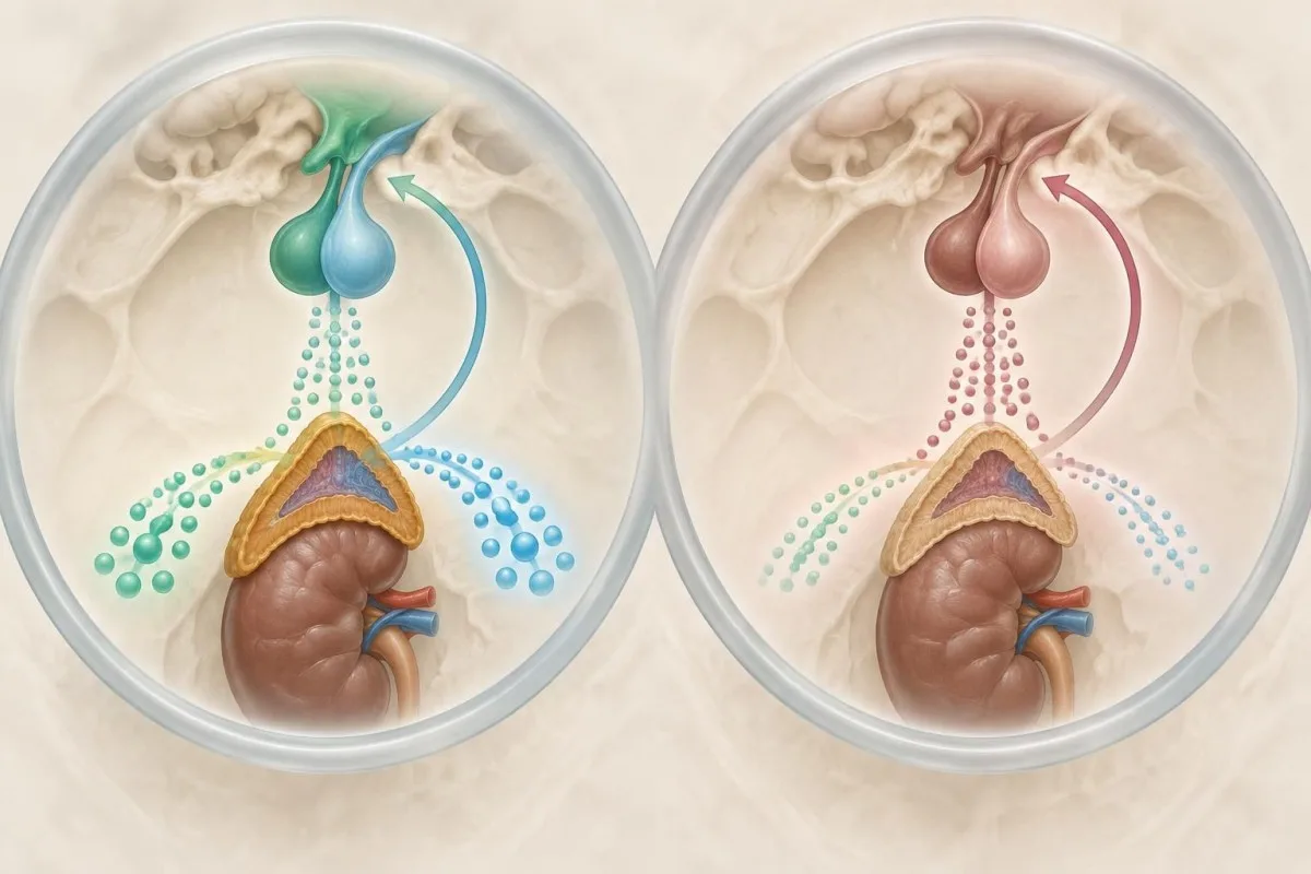
The traditional 250 microgram test is widely used, but interpretation depends on the cortisol assay and protocol. Older immunoassay cutoffs near 18 µg/dL may overdiagnose insufficiency with newer, more specific assays, for which validated thresholds can be closer to 14-15 µg/dL.
A low DHEA-S with a normal stimulated cortisol does not prove that DHEA replacement is needed. It may reflect age, contraception, chronic illness or a selective change in adrenal androgen output rather than inadequate cortisol reserve.
A hypothetical patient has baseline cortisol of 7 µg/dL, 30-minute cortisol of 13 µg/dL and 60-minute cortisol of 16 µg/dL. Whether this is adequate depends on the assay's validated cutoff and clinical context; reporting only the word abnormal can create unnecessary alarm.
Cosyntropin testing may be falsely reassuring early in recent-onset central adrenal insufficiency because the adrenal glands have not yet fully atrophied. If pituitary disease is strongly suspected, the endocrinology team may combine repeat testing with other pituitary-axis results and imaging; our οδηγός ελέγχου για σύνδρομο Cushing explains related cortisol-test limitations.
Why sodium, potassium, renin and aldosterone matter
Electrolytes and mineralocorticoid tests help distinguish primary adrenal failure from central ACTH deficiency because aldosterone is mainly controlled by renin and potassium rather than ACTH. Low sodium and high potassium raise concern for primary disease, while normal potassium does not exclude central adrenal insufficiency.

A sodium concentration below 135 mmol/L can occur in adrenal insufficiency, but it is not specific; vomiting, diuretics, heart failure and SIADH can produce the same finding. Potassium above 5.0 mmol/L is more suggestive of mineralocorticoid deficiency when kidney function and medications do not provide another explanation.
Plasma renin activity or direct renin may rise before potassium becomes abnormal, making renin an early clue in primary adrenal disease. Aldosterone below the laboratory interval with elevated renin strengthens that interpretation, whereas central disease usually leaves aldosterone relatively preserved.
A hypothetical 60-year-old taking an ACE inhibitor has sodium 133 mmol/L, potassium 5.4 mmol/L and high renin. The medication itself can raise potassium and renin, so the pattern cannot be read as primary adrenal failure without reviewing kidney function, blood pressure and drug effects.
Kantesti's biomarker view can place sodium, potassium, creatinine and DHEA-S in one trend, but treatment decisions require clinical oversight. For a related explanation of sodium interpretation, read our urine sodium results guide.
Other pituitary hormones that strengthen the diagnosis
Low DHEA-S is more suggestive of central disease when it appears with abnormalities in two or more pituitary-dependent axes. Helpful companion tests include free T4 with TSH, prolactin, LH, FSH, testosterone or estradiol, IGF-1 and sometimes serum osmolality, selected according to symptoms.
A low free T4 with TSH below 10 mIU/L does not exclude central hypothyroidism because TSH can be biologically weak or inappropriately normal. Similarly, low testosterone with low-normal LH and FSH is different from low testosterone with clearly high gonadotropins.
Prolactin can rise after pituitary stalk disruption, with some medicines, during pregnancy or with stress; a mildly high value around 25-40 ng/mL is not automatically evidence of a pituitary mass. Macroprolactin and repeat sampling may be relevant when the clinical picture is discordant.
A hypothetical 44-year-old has DHEA-S of 27 µg/dL, cortisol of 4.2 µg/dL, free T4 of 0.7 ng/dL, TSH of 1.1 mIU/L and prolactin of 38 ng/mL. That cluster is more concerning for multi-axis pituitary dysfunction than any isolated hormone result.
Gonadotropin interpretation changes with age, menstrual stage and hormone contraception. Our guide to μοτίβα LH και FSH explains why a single apparently normal result may be physiologically inappropriate.
Why sex, menopause and hormonal contraception change interpretation
DHEA-S interpretation differs by sex and reproductive stage because adrenal androgens interact with ovarian or testicular hormones, binding proteins and exogenous estrogen. Menopause may lower DHEA-S gradually, while combined contraception can make androgen-related symptoms and measurements less straightforward.

DHEA-S is not a direct measure of ovarian or testicular function, and the same concentration can have different clinical meaning across life stages. In a postmenopausal adult, DHEA-S of 40 µg/dL may be age-appropriate, while new hirsutism with DHEA-S above 700-800 µg/dL raises a separate concern about adrenal androgen excess.
Combined oral contraceptives often increase sex hormone-binding globulin and suppress ovarian androgen production, while measured DHEA-S may also decrease. Testing during use can therefore obscure baseline androgen physiology; stopping medication solely to obtain a test should be discussed with the prescriber.
A hypothetical 51-year-old using ethinylestradiol has DHEA-S of 29 µg/dL, total testosterone of 18 ng/dL and SHBG of 160 nmol/L. The low DHEA-S may reflect medication and age rather than adrenal disease, especially if morning cortisol and electrolytes are normal.
The menopause transition can also cause sleep disruption, hot flushes, low mood and reduced libido, all of which overlap with proposed low DHEA symptoms. Our women's hormonal symptoms guide provides context for separating reproductive-stage symptoms from adrenal clues.
How DHEA-S relates to testosterone and binding proteins
DHEA-S is an adrenal precursor, not a substitute for testosterone testing, and low DHEA-S may coexist with normal total testosterone or altered free testosterone. SHBG, albumin, age, liver function, thyroid status and medication exposure can change the amount of active sex hormone without changing DHEA-S in parallel.
A low SHBG concentration can make total testosterone look low while free testosterone remains reasonable, whereas high SHBG can produce the opposite pattern. SHBG may rise with oral estrogen and hyperthyroidism and fall with obesity, insulin resistance and hypothyroidism.
A hypothetical 38-year-old man has DHEA-S of 105 µg/dL, total testosterone of 290 ng/dL, SHBG of 14 nmol/L and normal albumin. The low SHBG may explain the borderline total testosterone; calculating free testosterone with a validated method may be more informative than treating DHEA-S.
DHEA supplements can raise DHEA-S and alter downstream androgens, but a higher laboratory value does not demonstrate improved energy or disease prevention. The evidence for routine supplementation remains mixed, and long-term safety data are limited for many populations.
Kantesti AI interprets DHEA-S with SHBG, albumin and testosterone when those values are available, helping identify discordant patterns rather than labelling every low androgen result as adrenal failure. See our explanation of low SHBG causes for related binding-protein clues.
Chronic illness, nutrition and physiological stress
Chronic illness, undernutrition, obesity, diabetes and severe physiological stress can lower DHEA-S without causing permanent adrenal failure. These effects may reflect altered adrenal prioritisation, inflammation, insulin signalling, reduced binding proteins or temporary suppression of the hypothalamic-pituitary-adrenal axis.
DHEA-S can be lower in people with chronic inflammatory disease, advanced liver disease, kidney disease or poorly controlled diabetes, but the direction and size of change vary. A low albumin concentration can also change hormone transport and complicate interpretation of total hormone measurements.
Severe calorie restriction and rapid weight loss may suppress reproductive and adrenal androgen pathways, while obesity is often associated with lower DHEA-S in adults. A 10% body-weight loss over 3 months is clinically meaningful and should be included when interpreting a new hormone result.
A hypothetical 29-year-old endurance athlete has DHEA-S of 44 µg/dL, normal cortisol, low energy availability and a 7 kg weight loss over 10 weeks. The immediate priority is nutritional and training assessment, not assuming autoimmune adrenal disease from DHEA-S alone.
Stress does not reliably explain every low DHEA-S result, and the popular phrase adrenal fatigue is not a recognised endocrine diagnosis. Review the full blood-test trend, medication changes and nutritional markers; our baseline blood-test guide explains how to obtain a more interpretable comparison.
Sampling errors and assay limitations that create misleading results
A low DHEA result can be misleading when the test uses the wrong analyte, an inappropriate age range, a poorly timed sample or an assay affected by supplementation. DHEA-S is generally more stable than DHEA, but neither result should be interpreted without the method, units and laboratory interval.

DHEA has a stronger diurnal pattern than DHEA-S, so a late-afternoon DHEA result may not be comparable with a morning value. DHEA-S is less variable but can still shift with age, acute illness, pregnancy, hormone therapy and recent DHEA use.
Immunoassays and liquid chromatography-tandem mass spectrometry can produce different results, particularly at low concentrations. A reported value of 0.9 µmol/L cannot be compared directly with 90 µg/dL without unit conversion; 1 µmol/L of DHEA-S is approximately 36.8 µg/dL.
Biotin supplements can interfere with some immunoassays, although the effect is assay-specific and often more important for thyroid or steroid-related tests than for every DHEA-S method. Tell the laboratory about high-dose biotin, usually 5-10 mg daily, and follow its advice about temporary withholding.
Kantesti's trend analysis can compare repeated results only when units, analyte and laboratory method are aligned. For a practical guide to recognising timing-related changes, read our οδηγός χρονοδιαγράμματος αιματολογικών εξετάσεων.
When to repeat a low DHEA-S result
Repeat DHEA-S testing is reasonable when the result is mildly low, symptoms are nonspecific, the sample was poorly timed or the medication history is unclear; urgent assessment is needed when low cortisol symptoms are present. Repeating a single low DHEA-S without adding cortisol and ACTH often provides little extra diagnostic value.
For a stable outpatient with DHEA-S only slightly below range, clinicians may repeat the test after acute illness resolves, often within several weeks to a few months. The repeat should use the same laboratory where possible and include the collection time, current medication doses and any DHEA supplement exposure.
A result that falls by 20-30% between draws may reflect biological and analytical variation rather than a new disease, especially when the values remain near the lower boundary. Trends become more persuasive when the change is sustained and accompanied by cortisol, ACTH or electrolyte abnormalities.
A hypothetical patient has DHEA-S of 56 µg/dL during pneumonia and 61 µg/dL three months later after recovery, with normal 8 AM cortisol and ACTH. The stable low-normal pattern is less concerning than a fall from 120 to 18 µg/dL with new hypotension and weight loss.
Do not delay care for repeat testing if there is persistent vomiting, severe weakness, fainting, confusion, dehydration or very low blood pressure. Our πληροφορίες ιατρικής επικύρωσης explains why automated interpretation should support, not replace, clinical review of potentially urgent patterns.
When low DHEA may signal an urgent adrenal problem
Low DHEA becomes urgent when it accompanies symptoms or laboratory findings suggesting acute adrenal insufficiency, especially vomiting, severe abdominal pain, fainting, confusion, profound weakness, dehydration, low blood pressure or low blood glucose. The DHEA-S value itself does not determine urgency; cortisol, ACTH, circulation and electrolytes do.
Adrenal crisis can occur with low or normal potassium, particularly in central adrenal insufficiency or early illness. A blood pressure below 90/60 mmHg with sodium of 128 mmol/L, glucose of 55 mg/dL and low cortisol is far more concerning than a DHEA-S value of 25 µg/dL by itself.
People taking long-term glucocorticoids are at particular risk during infection, surgery, trauma or prolonged vomiting because their stress response may be suppressed. They should follow their prescribed sick-day plan and seek urgent medical advice when they cannot keep steroid medication down.
A hypothetical 67-year-old with DHEA-S of 19 µg/dL develops diarrhoea, dizziness on standing, sodium of 124 mmol/L and glucose of 58 mg/dL. This combination requires urgent assessment for adrenal crisis and other causes of shock; waiting for a repeat DHEA-S test would be unsafe.
Kantesti can highlight combinations such as low sodium, low glucose and abnormal cortisol for follow-up, but emergency symptoms require local emergency services or urgent clinical care. Do not self-treat a suspected crisis with over-the-counter DHEA.
A practical workflow for interpreting low DHEA causes
The safest workflow is to confirm the analyte and age range, review medications, pair DHEA-S with morning cortisol and ACTH, check electrolytes and then consider stimulation or pituitary testing. This sequence prevents a common error: treating a low adrenal androgen as proof of adrenal insufficiency.
Step one is to record whether the report measures DHEA or DHEA-S, then check units, collection time, age-specific interval and assay method. Step two is to list glucocorticoids, opioids, hormone contraception, estrogen, steroid-synthesis inhibitors and supplements with dose and timing.
Step three is an 8-9 AM cortisol with ACTH, sodium, potassium, glucose, creatinine and sometimes renin and aldosterone. Step four is a cosyntropin test when morning cortisol is indeterminate; step five is broader pituitary testing when ACTH is low or other axes are abnormal.
A hypothetical 50-year-old has DHEA-S of 33 µg/dL, cortisol of 11 µg/dL, ACTH of 22 pg/mL, sodium of 140 mmol/L and potassium of 4.1 mmol/L. This is not an adrenal crisis pattern, and the next decision may be repeat testing or cosyntropin testing rather than immediate DHEA therapy.
Το Καντέστι είναι ένα Εργαλείο ανάλυσης αιματολογικών εξετάσεων με AI-powered that can organise the workflow across uploaded reports, trends and reference intervals, while the final diagnosis remains with a qualified healthcare professional. Our οδηγός τεχνολογίας AI describes how contextual pattern analysis works.
Συχνές ερωτήσεις
Ποιες είναι οι συχνότερες αιτίες χαμηλών επιπέδων DHEA;
The most common low DHEA causes are normal ageing, oral contraceptives or estrogen, systemic glucocorticoids, chronic illness, obesity, undernutrition and reduced ACTH signalling. DHEA-S often declines by about 1-3% per year after age 30, so a value of 40 µg/dL may be normal in an older adult but unexpectedly low in a young adult. A low DHEA-S result should be interpreted with morning cortisol, ACTH, medications and the laboratory's age-specific range.
Μπορεί τα χαμηλά επίπεδα DHEA-S να υποδηλώνουν επινεφριδιακή ανεπάρκεια;
Τα χαμηλά επίπεδα DHEA-S μπορεί να συνηγορούν υπέρ επινεφριδιακής ανεπάρκειας, αλλά δεν αρκούν από μόνα τους για τη διάγνωσή της. Η πρωτοπαθής επινεφριδιακή ανεπάρκεια συνήθως εμφανίζει χαμηλή πρωινή κορτιζόλη, υψηλή ACTH, χαμηλή αλδοστερόνη, υψηλή ρενίνη και, μερικές φορές, κάλιο πάνω από 5.0 mmol/L, ενώ η κεντρική ανεπάρκεια συνήθως εμφανίζει χαμηλή κορτιζόλη με χαμηλή ή απρόσφορα φυσιολογική ACTH. Κορτιζόλη στις 8 π.μ. κάτω από περίπου 3 µg/dL εγείρει ισχυρή υποψία, ενώ τα ενδιάμεσα αποτελέσματα συχνά απαιτούν δοκιμασία κοσυντροπίνης 250 μικρογραμμαρίων.
Ποια είναι η διαφορά μεταξύ χαμηλής DHEA και χαμηλής DHEA-S;
Η DHEA είναι ένα επινεφριδιακό ανδρογόνο που κυκλοφορεί στο αίμα και παρουσιάζει μεγαλύτερες ημερήσιες διακυμάνσεις, ενώ η DHEA-S είναι η θειική, πιο σταθερή μορφή αποθήκευσής της και συνήθως προτιμάται για την ερμηνεία των συνήθων εξετάσεων αίματος. Μια μέτρηση DHEA που γίνεται αργότερα μέσα στην ημέρα μπορεί να είναι χαμηλότερη από μια πρωινή μέτρηση λόγω της ημερήσιας διακύμανσης, ενώ η DHEA-S παρουσιάζει μικρότερη μεταβλητότητα, αλλά εξακολουθεί να εξαρτάται από την ηλικία, το φύλο, τη μέθοδο μέτρησης και τη φαρμακευτική αγωγή. Οι μονάδες δεν είναι εναλλάξιμες· περίπου 1 µmol/L DHEA-S αντιστοιχεί σε 36,8 µg/dL.
Which medications lower DHEA-S?
Systemic glucocorticoids such as prednisone, prednisolone, dexamethasone and hydrocortisone can lower DHEA-S by suppressing ACTH, especially after several weeks of treatment. Opioids, steroid-synthesis inhibitors and some estrogen-containing contraceptives can also alter adrenal androgen results or their interpretation. Do not stop a prescribed steroid abruptly; a clinician may need to assess an 8 AM cortisol, ACTH and adrenal reserve.
What symptoms can low DHEA cause?
Possible low DHEA symptoms include fatigue, reduced libido, low mood, poorer exercise tolerance and reduced muscle strength, but these symptoms are nonspecific. Iron deficiency, thyroid disease, menopause, depression, sleep disorders, chronic infection and medication effects can produce the same complaints. DHEA-S of 30-50 µg/dL does not establish the cause of symptoms without age-adjusted interpretation and paired adrenal testing.
Should I take DHEA supplements for a low result?
A low DHEA-S result does not automatically justify DHEA supplementation, and routine use is not recommended for people with normal adrenal function. Supplements can raise DHEA-S, alter testosterone or estrogen pathways and cause acne, unwanted hair growth, mood changes or adverse lipid effects; product dose and purity also vary. Discuss the cause, morning cortisol, ACTH and alternative explanations with a clinician before taking DHEA.
Δείτε πώς να διαβάσετε εξετάσεις αίματος
Ανεβάστε την εργαστηριακή σας αναφορά και η τεχνητή νοημοσύνη της Kantesti εξηγεί κάθε βιοδείκτη με απλή γλώσσα μέσα σε λίγα δευτερόλεπτα. Την έχουν χρησιμοποιήσει περισσότεροι από 2 εκατομμύρια άνθρωποι παγκοσμίως.
Αναφορές
- Klein, T., Mitchell, S., & Weber, H. (2026). Διάρροια μετά από νηστεία, μαύρα στίγματα στα κόπρανα και οδηγός γαστρεντερικού 2026. Ιατρική έρευνα της τεχνητής νοημοσύνης Kantesti. DOI
- Klein, T., Mitchell, S., & Weber, H. (2026). Οδηγός για την υγεία των γυναικών: ωορρηξία, εμμηνόπαυση και ορμονικά συμπτώματα. Ιατρική έρευνα της τεχνητής νοημοσύνης Kantesti. DOI
- Bornstein SR et al. (2016). Diagnosis and Treatment of Primary Adrenal Insufficiency: An Endocrine Society Clinical Practice Guideline. Journal of Clinical Endocrinology & Metabolism. DOI PubMed
- Beuschlein F et al. (2024). European Society of Endocrinology and Endocrine Society Joint Clinical Guideline: Diagnosis and Therapy of Glucocorticoid-Induced Adrenal Insufficiency. Journal of Clinical Endocrinology & Metabolism.
- Arlt W et al. (2008). Dehydroepiandrosterone Replacement in Women with Adrenal Insufficiency. New England Journal of Medicine. DOI PubMed
- 2+ εκατομμύριαΕξετάσεις που αναλύθηκαν
- 127+χωρών
- 100+Γλώσσες
Σχετικά με την ιατρική ομάδα


Σήματα εμπιστοσύνης E-E-A-T
Εμπειρία
Κλινική ανασκόπηση από ιατρό για τις ροές εργασίας ερμηνείας εργαστηριακών αποτελεσμάτων.
Πραγματογνωμοσύνη
Εστίαση στην εργαστηριακή ιατρική στο πώς συμπεριφέρονται οι βιοδείκτες στο κλινικό πλαίσιο.
Αυθεντικότητα
Ιατρική αξιολόγηση από τον Δρ. Thomas Klein, με συνεισφορές από τη Δρ. Sarah Mitchell και τον Καθ. Δρ. Hans Weber.
Αξιοπιστία
Ερμηνεία βασισμένη σε τεκμηριωμένα δεδομένα με σαφείς οδούς παρακολούθησης για τη μείωση του συναγερμού.

Ιατρική αποποίηση ευθύνης
Αυτό το άρθρο προορίζεται μόνο για εκπαιδευτικούς σκοπούς και δεν αποτελεί ιατρική συμβουλή. Συμβουλεύεστε πάντα έναν κατάλληλο επαγγελματία υγείας για αποφάσεις σχετικά με τη διάγνωση και τη θεραπεία.
Δημοσιεύθηκε · Επικοινωνήστε με τη συντακτική ομάδα
Εκδίδεται από την Kantesti Ltd, εγγεγραμμένη στην Αγγλία και την Ουαλία, αριθμός εταιρείας. 17090423, Λονδίνο, Ηνωμένο Βασίλειο.