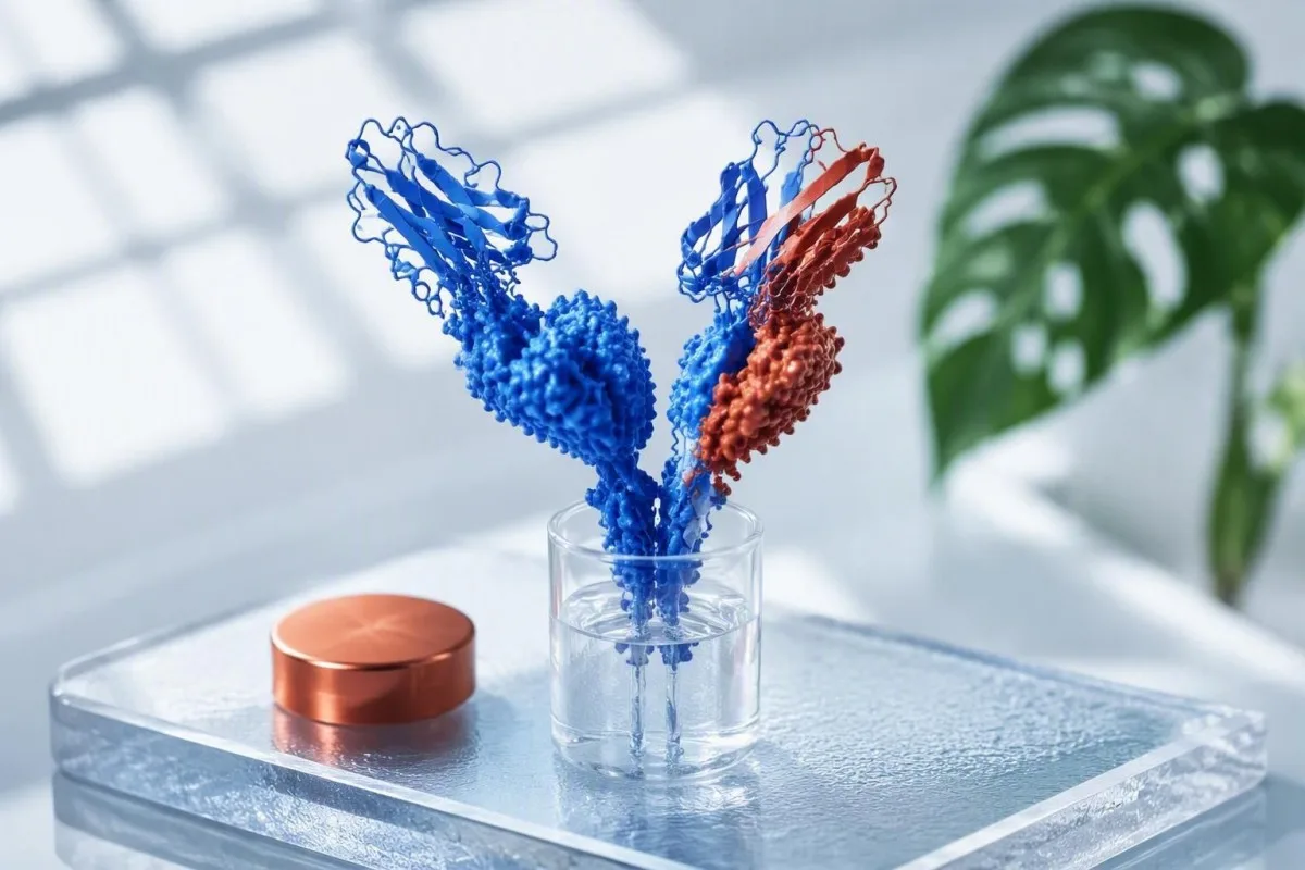
Ein Kappa/Lambda-Verhältnis außerhalb des Laborbereichs bedeutet nicht automatisch ein Myelom. Die absoluten Leichtkettenwerte, eGFR, Serumproteinuntersuchungen, Symptome und die zeitliche Veränderung bestimmen das weitere Vorgehen.


- Standardverhältnis liegt üblicherweise bei 0,26-1,65 bei normaler Nierenfunktion, aber die Labore müssen ihr eigenes Assay-spezifisches Intervall verwenden.
- Nierenerkrankung erhöht beide freien Leichtketten im Serum; ein Verhältnis von 2,4 kann bei chronischer Nierenerkrankung erwartet werden und ist für sich genommen nicht diagnostisch.
- Hohes Verhältnis freier Leichtketten bedeutet eine Kappa-dominante Produktion, während ein niedriges Verhältnis eine Lambda-dominante Produktion bedeutet.
- Myelom-definierende Schwelle ist ein Verhältnis von betroffen/nicht betroffen von mindestens 100 plus betroffene freie Leichtketten von mindestens 100 mg/L.
- MGUS-Nachsorge umfasst in der Regel wiederholte CBC, Kreatinin, Kalzium, SPEP, Immunfixation und freie Leichtketten nach 6 Monaten, bevor längere Intervalle entschieden werden.
- Dringende Überprüfung ist angebracht bei neuen Knochenschmerzen, Anämie, ansteigendem Kreatinin, Kalzium über 2,75 mmol/L, wiederkehrenden Infektionen oder sich schnell erhöhenden betroffenen Leichtketten.
- Ein auffälliges Ergebnis ist weniger aussagekräftig als ein bestätigter Aufwärtstrend, der von demselben Labor und demselben Assay gemessen wurde.
Was freie Leichtketten im Serum und das Verhältnis tatsächlich messen
Freie Leichketten im Serum sind überschüssige Antikörper-Proteine, die von Plasmazellen produziert werden, und das Verhältnis von freien Leichketten vergleicht Kappa mit Lambda. Ein hohes oder niedriges Ergebnis weist auf ein Ungleichgewicht hin, nicht auf eine Diagnose; der erste praktische Schritt ist, die beiden absoluten Werte zusammen mit der Nierenfunktion zu betrachten.
Plasmazellen produzieren normalerweise etwas mehr Kappa- als Lambda-Fragmente. Viele Labore, die den Freelite-Assay verwenden, geben Kappa 3,3-19,4 mg/L, Lambda 5,7-26,3 mg/L und ein Kappa/Lambda-Verhältnis von 0,26-1,65 an, obwohl diese Intervalle Assay- und laborspezifisch sind. Ein Verhältnis ist einfach Kappa geteilt durch Lambda.
Ein Kappa-Verhältnis von 2,0 kann entstehen, weil Kappa 20 mg/L bei Lambda 10 mg/L ist, oder weil Kappa 200 mg/L bei Lambda 100 mg/L ist. Diese Muster haben sehr unterschiedliche Auswirkungen. Wenn ich eine Panel überprüfe, frage ich zuerst, ob eine Klon übermäßig eine Leichkettenart zu produzieren scheint, ob beide Werte zusammen ansteigen und ob Serum-Protein-Elektrophorese zeigt ein M-Protein.
Kantesti ist ein KI-Bluttestanalysator das das Verhältnis der freien Leichketten mit Kreatinin, eGFR, Kalzium, Hämoglobin, Gesamtprotein und vorherigen Werten liest, anstatt das Verhältnis isoliert zu kennzeichnen. Nach meiner Erfahrung vermeidet diese musterbasierte Sichtweise eine beträchtliche Menge unnötiger Alarmmeldungen nach einem einzigen, leicht abnormalen Ergebnis.
Was ein hohes Verhältnis freier Leichtketten bedeuten kann
Ein hohes Verhältnis von freien Leichketten bedeutet normalerweise, dass Kappa-Leichketten unverhältnismäßig erhöht sind, aber eine verminderte Nieren-Clearance ist die häufigste nicht-krebsbedingte Erklärung. Der Grad der Erhöhung und der absolute Wert des involvierten Kappa sind wichtiger als die alleinige Kennzeichnung.
Kappa-Fragmente sind kleiner und sammeln sich oft früher an als Lambda-Fragmente, wenn die Filtration abnimmt. Ein Verhältnis von 1,8 mit Kappa 31 mg/L, Lambda 17 mg/L und eGFR 38 mL/min/1,73 m² wird normalerweise ganz anders interpretiert als ein Verhältnis von 18 mit Kappa 360 mg/L und Lambda 20 mg/L.
Polyklonale Immunaktivität kann beide Ketten bei Autoimmunerkrankungen, Lebererkrankungen, chronischen Infektionen oder kürzlichen signifikanten Entzündungen erhöhen, im Allgemeinen mit einem Verhältnis nahe dem relevanten renalen Intervall. Der Grund, warum sich Kliniker über ein sehr hohes Verhältnis in Kombination mit einem diskreten M-Spike Sorgen machen, ist, dass sie zusammen auf eine einzelne Plasmazellpopulation und nicht auf eine breite Immunantwort hindeuten. Überprüfung Muster mit hohem Gesamtprotein ob Globulin oder Gesamtprotein ebenfalls erhöht war.
A persistent kappa/lambda ratio above 1.65 with normal renal function deserves planned follow-up, not panic. Dr. Thomas Klein would usually repeat the assay with SPEP and serum immunofixation if the result is newly abnormal, particularly after age 50 or when anemia, proteinuria, neuropathy, or unexplained bone symptoms are present.
How high is concerning?
There is no universal “danger” ratio of 2, 5, or 10. A ratio above 8 in a person with preserved eGFR is more specific for a clonal process than 1.7, but diagnostic interpretation still requires immunofixation, quantitative immunoglobulins, and clinical context.
Was ein niedriges Kappa-Lambda-Verhältnis bedeuten kann
A low kappa lambda ratio below 0.26 means lambda light chains predominate, and it requires the same careful evaluation as a high ratio. Lambda clones can produce little or no visible M-spike, so the ratio may provide an early clue rather than a final answer.
A low ratio is most clinically meaningful when lambda is genuinely elevated, such as lambda 95 mg/L with kappa 12 mg/L and a ratio of 0.13. A low ratio caused by low-normal kappa with normal lambda is less persuasive and should be checked for analytical and biological variation.
Lambda-associated disorders merit attention to urine protein, albumin, kidney function, numbness or tingling, bruising, and symptoms suggesting cardiac or nerve involvement. These findings do not prove amyloidosis, but they change the urgency and breadth of testing; persistent foamy urine is one reason to examine Protein im Urin.
Some people assume a low ratio means “low immunity.” It does not. The test does not measure total immune strength; it measures the balance of circulating free fragments, while immunoglobulin levels, vaccine response, and infection history answer different questions.
Warum sich die eGFR auf die Interpretation von freien Leichtketten auswirkt
Chronic kidney disease raises serum free light chains because the kidneys normally clear them, so the standard ratio of 0.26-1.65 can overcall abnormality. Kidney-adjusted intervals are particularly useful when eGFR is below 60 mL/min/1.73 m².
The widely used “renal range” for the Freelite kappa/lambda ratio is 0.37-3.10. In the iStopMM CKD analysis, eGFR-specific 99% intervals were 0.46-2.62 for eGFR 45-59, 0.48-3.38 for eGFR 30-44, and 0.54-3.30 for eGFR below 30 mL/min/1.73 m² (Long et al., 2022).
This adjustment matters. Among people with eGFR below 60, using the standard range produces many abnormal ratios that disappear when renal reference intervals are applied; the ratio usually remains broadly balanced because both kappa and lambda rise. Read the basics of chronische Nierenerkrankungsstadien verfolgen before interpreting a renal-range result.
Kantesti ist ein KI-Labor-Befundinterpretationsservice that checks whether creatinine and eGFR were drawn on the same date as the light-chain test. Acute dehydration, a recent hospital stay, or an evolving kidney injury can make one result a poor baseline, so a repeat after clinical stabilization is often more informative.
Assay-Unterschiede, Zeitpunkt der Probe und irreführende Veränderungen
Free light chain values from different assays are not interchangeable, and serial monitoring should use the same laboratory whenever possible. A 20% numerical change may reflect method variation, kidney function, or a short-lived immune event rather than disease progression.
Freelite and N Latex assays use different antibodies and reference ranges, especially at high concentrations and in kidney disease. A report should never be judged against a range copied from another laboratory. The report’s own reference interval has priority.
I have seen an alarming apparent rise after a patient changed hospitals during a move, only to find a different assay was used. Record the test date, laboratory, kappa, lambda, ratio, creatinine, eGFR, recent infection, and steroids; our lab result tracking checklist makes this mundane detail easier to save.
Free light chains do not require fasting. However, avoid interpreting an elective surveillance result taken during an acute febrile illness or immediately after major surgery without repeating it later, because polyclonal production and transient renal impairment can distort the baseline.
Wie freie Leichtketten in die Überwachung von MGUS passen
An abnormal free light chain ratio is one of three major MGUS risk features, alongside non-IgG type and an M-protein of at least 1.5 g/dL. It predicts risk at a population level, but it cannot tell an individual exactly what will happen.
MGUS is common: roughly 3% of adults older than 50 years have it, and most never develop myeloma or another related disorder. The Mayo Clinic model described by Rajkumar and colleagues estimates 20-year progression risk from about 5% with no risk factors to about 58% with all three, although competing health risks reduce the practical risk for many older adults.
The International Myeloma Working Group advises repeat clinical review and laboratory testing at 6 months after diagnosis. Low-risk stable MGUS may then be checked every 2-3 years, while higher-risk disease is commonly reviewed annually; clinicians individualize that schedule for age, kidney function, and trajectory (Rajkumar et al., 2014).
Kantesti ist ein KI-Biomarker-Interpretationsplattform that can place each MGUS-era free light chain result on a time series, but surveillance decisions remain a clinician’s job. A rising involved light chain across three comparable measurements is more meaningful than a small one-off movement; see our Leitfaden zur Bluttest-Veränderung.
Leichtketten-MGUS im Vergleich zu einem vorübergehend abnormalen Verhältnis
Light-chain MGUS requires an abnormal ratio, an increased involved light chain, no heavy-chain M-protein on immunofixation, and no myeloma-defining organ damage. An isolated ratio abnormality does not meet that definition.
Light-chain MGUS is less common than conventional MGUS and can be missed if testing stops after a normal SPEP. Serum immunofixation and free light-chain testing complement each other because a small light-chain clone may not create an obvious electrophoresis spike.
The usual work-up includes CBC, calcium, creatinine/eGFR, SPEP, serum immunofixation, quantitative IgG/IgA/IgM, and often urine studies when there is kidney disease or proteinuria. Immunoglobulin results are useful because suppression of uninvolved immunoglobulins can increase concern.
A ratio of 0.22 with lambda 28 mg/L after a respiratory infection may normalize, whereas 0.08 with lambda 160 mg/L on repeat testing is a different situation. The repeat is not bureaucracy; it separates transient biology from reproducible clonal signal.
Wann ein abnormales Verhältnis zu einem myelomspezifischen Befund wird
A free light chain ratio becomes a myeloma-defining biomarker at an involved/uninvolved ratio of 100 or greater, with the involved chain at least 100 mg/L. This threshold applies only after confirming a clonal plasma-cell disorder and is not the same as a routine high ratio.
The 2014 International Myeloma Working Group criteria added this threshold because people meeting it had a very high short-term probability of progression to symptomatic organ damage. The criteria also require marrow evidence or a biopsy-proven plasmacytoma in the appropriate diagnostic framework; a lab result alone cannot diagnose myeloma (Rajkumar et al., 2014).
Clinicians look for CRAB features: calcium above 2.75 mmol/L or 0.25 mmol/L above the upper limit, renal impairment attributable to the clone, hemoglobin more than 20 g/L below normal or below 100 g/L, and bone lesions on imaging. A normal CBC does not exclude early disease, but it lowers immediate concern when the ratio is only mildly abnormal.
If free light chains are very high, assess renal function promptly because light chains themselves can injure kidney tubules. The complementary marker beta-2 microglobulin can contribute to staging once a diagnosis exists; our myeloma marker guide explains its limits.
Welche Ergebnisse eine hämatologische Nachuntersuchung erfordern
Hematology follow-up is appropriate for a persistent unexplained abnormal ratio with an elevated involved chain, an M-protein, immunoparesis, kidney injury, anemia, hypercalcemia, or suggestive symptoms. Same-day evaluation is needed for rapidly worsening kidney function, confusion from high calcium, or severe acute illness.
A practical referral trigger is a ratio outside the renal-adjusted interval on repeat testing, especially when kappa or lambda exceeds 100 mg/L. Many hematologists will also see a person with a lower abnormality if there is unexplained eGFR decline, albuminuria, recurrent infections, weight loss, persistent focal bone pain, or a first-degree relative with plasma-cell disease.
Urgent assessment is not dictated by the ratio alone. New reduced urine output, rapidly climbing creatinine, shortness of breath, marked weakness, confusion, or calcium above 3.0 mmol/L deserves immediate medical care, because complications can progress faster than routine referral pathways.
Dr. Thomas Klein’s practical advice is to bring every previous protein study to the appointment, not only the flagged result. A hematologist can then decide whether to repeat serum and urine tests, obtain low-dose whole-body imaging, or perform bone-marrow assessment; a Blutkrebs-Testpfad erklärt, warum diese Tests unterschiedliche Fragen beantworten.
Die begleitenden Tests, die Ärzte zusammen mit Leichtketten lesen
Free light chains are interpreted with SPEP, immunofixation, CBC, calcium, creatinine/eGFR, urine protein testing, and sometimes quantitative immunoglobulins. No single test can reliably distinguish MGUS, myeloma, kidney-related elevation, and inflammatory activation.
SPEP estimates and localizes a monoclonal protein, while immunofixation identifies its heavy- and light-chain type. A normal SPEP does not rule out light-chain disease, which is precisely why serum free light chains are useful in the first place.
CBC matters because hemoglobin below 100 g/L is one myeloma-defining anemia threshold when attributable to the plasma-cell disorder. Calcium and creatinine are not merely screening extras: they help identify organ involvement and decide whether a result needs days, weeks, or routine monitoring. For kidney context, compare BUN und Kreatinin rather than relying on one number.
Urine albumin-creatinine ratio detects albumin leakage, whereas urine protein electrophoresis can identify monoclonal light-chain excretion. That distinction is easy to miss in routine care, and it is one reason nephrology and hematology sometimes evaluate the same patient from different angles.
Drei Muster aus der Praxis, die zu unterschiedlichen nächsten Schritten führen
The same free light chain ratio can lead to reassurance, repeat testing, or urgent specialty work-up depending on the absolute values and eGFR. Pattern recognition is safer than using a universal cutoff.
Pattern one: kappa 42 mg/L, lambda 19 mg/L, ratio 2.21, eGFR 34 mL/min/1.73 m², stable creatinine, normal SPEP. This commonly fits renal retention and may be rechecked with the kidney clinician rather than treated as myeloma.
Pattern two: kappa 86 mg/L, lambda 10 mg/L, ratio 8.6, eGFR 92 mL/min/1.73 m², small IgG-kappa M-spike 0.7 g/dL, normal CBC and calcium. This needs hematology characterization and risk-based MGUS monitoring, but it is not automatically an emergency.
Pattern three: lambda 1,240 mg/L, kappa 9 mg/L, ratio 0.007, creatinine rising from 76 to 180 µmol/L in 4 weeks. That combination warrants urgent specialist contact because a light-chain process can threaten renal recovery; understand Warnzeichen für niedrige eGFR.
Wie man ein Verhältnis freier Leichtketten im Laufe der Zeit verfolgt
A meaningful trend uses the same assay, similar kidney function, and at least two or three measurements rather than reacting to one decimal point. Plotting involved chain concentration separately from the ratio prevents a deceptively stable ratio from hiding a large absolute increase.
If both kappa and lambda double during an eGFR decline, the ratio may hardly move while clearance has changed substantially. Conversely, the ratio may rise while total concentrations remain modest; both views are needed. This is why specialists often record the “involved” and “uninvolved” light chain explicitly.
For stable MGUS, a 6-month recheck establishes a personal baseline after the initial discovery. Thereafter, interval testing may be yearly or less frequent in low-risk disease, but a new 50% rise in the involved chain, symptoms, or renal deterioration warrants earlier review even if the ratio stays below 100.
Kantesti’s neural network can organize historical results across uploaded reports and flag mismatched units, but it cannot determine whether an individual has myeloma. Read our Anleitung für longitudinale Tests for the context worth recording between appointments.
Fragen, die Sie Ihren Arzt nach einem abnormalen Ergebnis stellen sollten
The most useful questions are whether the lab used a renal-adjusted ratio, which chain is involved, whether an M-protein is present, and when the result should be repeated. Asking for a concrete follow-up interval turns an ambiguous flag into a safe plan.
Ask: “What were my kappa and lambda values, not just the ratio?” and “Was my eGFR below 60 on that day?” Those two questions resolve a surprising number of misunderstandings. Ask whether SPEP and immunofixation were performed, because they are not always automatically ordered together.
Also ask what symptoms should prompt earlier contact. Most patients find it reassuring to hear a specific list: sustained new bone pain, unusual fatigue with anemia, recurrent infections, swelling or foamy urine, decreasing urine output, weight loss, or unexplained numbness merit a call rather than waiting for the next annual test.
Keep a copy of the actual PDF report. Kantesti’s medical validation approach emphasizes source-report review and clinical oversight, which are especially important when an automated flag could reflect an assay-specific or kidney-related reference interval.
Was Sie nach einem hohen oder niedrigen Verhältnis nicht tun sollten
Do not start supplements, “detox” regimens, or cancer-directed treatments to change a free light chain ratio. There is no diet, vitamin, or over-the-counter product proven to remove a monoclonal light-chain clone, and some products can worsen kidney function.
Avoid repeated testing every few days unless a clinician is monitoring acute kidney injury or known disease. Free light chains are biologically variable, and overly frequent testing produces noise that can look like progression. A planned 6-week to 6-month interval is often more useful, depending on the initial pattern.
Do not assume a normal ratio rules out every plasma-cell disorder, either. A clone can occasionally produce both chains or suppress the uninvolved chain in complicated ways, which is why an M-spike, symptoms, and organ tests still matter. For a broader explanation of unexpected flags, see außerhalb des Referenzbereichs liegenden Blutwerten.
As of September 13, 2026, the sensible next step after a mildly abnormal result is usually confirmation and context, not self-treatment. Kantesti is an KI-gestütztes Analyse-Tool für Bluttests used to help patients prepare focused questions for clinicians; our Medizinischer Beirat supports physician-led review of medically significant findings.
Häufig gestellte Fragen
Was ist das normale Verhältnis freier Leichtketten?
The commonly quoted normal free light chain ratio is 0.26-1.65 for the Freelite serum assay in adults with preserved kidney function. Laboratories use different methods and reference intervals, so the range printed on the report should take priority. In chronic kidney disease, many clinicians use a broader renal range of approximately 0.37-3.10, with eGFR-specific intervals sometimes being more precise. A result just outside 0.26-1.65 is not diagnostic without the kappa value, lambda value, eGFR, SPEP, and immunofixation.
Bedeutet ein hohes freies leichtes Kettenverhältnis Krebs?
A high free light chain ratio does not by itself mean cancer. Reduced eGFR commonly raises kappa and lambda light chains and can push the ratio above 1.65 without a plasma-cell cancer. A persistently high ratio with a clearly elevated involved chain, an M-protein, anemia, kidney injury, high calcium, or bone symptoms needs hematology assessment. An involved/uninvolved ratio of at least 100 plus involved free light chain of at least 100 mg/L is a myeloma-defining biomarker only in the proper diagnostic setting.
Kann Nierenerkrankung ein abnormales Kappa-Lambda-Verhältnis verursachen?
Yes, chronic kidney disease can cause abnormal serum free light chain values and a mildly high kappa lambda ratio because the kidneys clear these proteins. For eGFR below 60 mL/min/1.73 m², a ratio up to roughly 3.10 may be compatible with renal retention, depending on the assay. The iStopMM study reported eGFR-specific upper ratio limits between 2.62 and 3.38 across CKD categories. A ratio outside kidney-adjusted limits, especially with a rising involved chain or M-protein, still requires further evaluation.
Wie oft sollten MGUS freie Leichtketten überprüft werden?
Most newly diagnosed MGUS cases are reassessed with CBC, creatinine, calcium, protein studies, and serum free light chains at about 6 months. If low-risk MGUS remains stable, follow-up can often be every 2-3 years; higher-risk MGUS is commonly monitored annually. A new symptom, eGFR decline, anemia, calcium elevation, or substantial involved light-chain rise should bring the next test forward. The exact interval should be set by the clinician who knows the M-protein type and overall risk profile.
Welches Verhältnis gilt bei Myelom als gefährlich?
A ratio is especially concerning when the involved-to-uninvolved free light chain ratio is at least 100 and the involved light chain is at least 100 mg/L. This is not a general “danger cutoff” for every person with an abnormal result; it is a myeloma-defining event within International Myeloma Working Group diagnostic criteria when clonal plasma cells or a plasmacytoma are also established. Lower ratios can still justify hematology review when paired with organ abnormalities. Ratios of 2 or 3 are often explained by kidney function or non-clonal immune activity.
Kann eine Entzündung die Serum-Frei-Leichtketten erhöhen?
Inflammatory and immune conditions can raise both kappa and lambda serum free light chains because many plasma-cell populations are activated at once. In this polyclonal pattern, the ratio often remains within the standard or kidney-adjusted range, unlike a strongly one-sided monoclonal pattern. Acute infection, autoimmune disease, liver disease, and reduced kidney clearance can overlap, so repeat testing after recovery may be appropriate. Persistent one-sided elevation should not be dismissed as inflammation without protein studies.
Hol dir heute eine KI-gestützte Bluttest-Analyse
Schließe dich über 2 Millionen Nutzern weltweit an, die Kantesti für sofortige, genaue Laboranalysen vertrauen. Lade deine Blutwerte Ergebnisse hoch und erhalte in Sekunden eine umfassende Interpretation von 15,000+-Biomarkern.
📚 Referenzierte Forschungsveröffentlichungen
Klein, T., Mitchell, S., & Weber, H. (2026). Nipah-Virus-Bluttest: Leitfaden zur Früherkennung und Diagnose 2026. Kantesti KI-Medizinische Forschung.
Klein, T., Mitchell, S., & Weber, H. (2026). Leitfaden zu Blutgruppe B negativ, LDH-Bluttest & Retikulozytenzahl. Kantesti KI-Medizinische Forschung.
📖 Externe medizinische Referenzen
Long TE et al. (2022). Defining new reference intervals for serum free light chains in individuals with chronic kidney disease: results of the iStopMM study. Blood Cancer Journal.
⚕️ Medizinischer Haftungsausschluss
Dieser Artikel dient nur zu Bildungszwecken und stellt keine medizinische Beratung dar. Wende dich für Diagnose- und Behandlungsentscheidungen immer an eine qualifizierte medizinische Fachkraft.
E-E-A-T Vertrauenssignale
Erfahrung
Ärztlich geleitete klinische Überprüfung von Labor-Interpretations-Workflows.
Sachverstand
Fokus der Labormedizin darauf, wie Biomarker sich im klinischen Kontext verhalten.
Autorität
Verfasst von Dr. Thomas Klein, überprüft von Dr. Sarah Mitchell und Prof. Dr. Hans Weber.
Vertrauenswürdigkeit
Evidenzbasierte Interpretation mit klaren nächsten Schritten zur Reduzierung von Alarm.