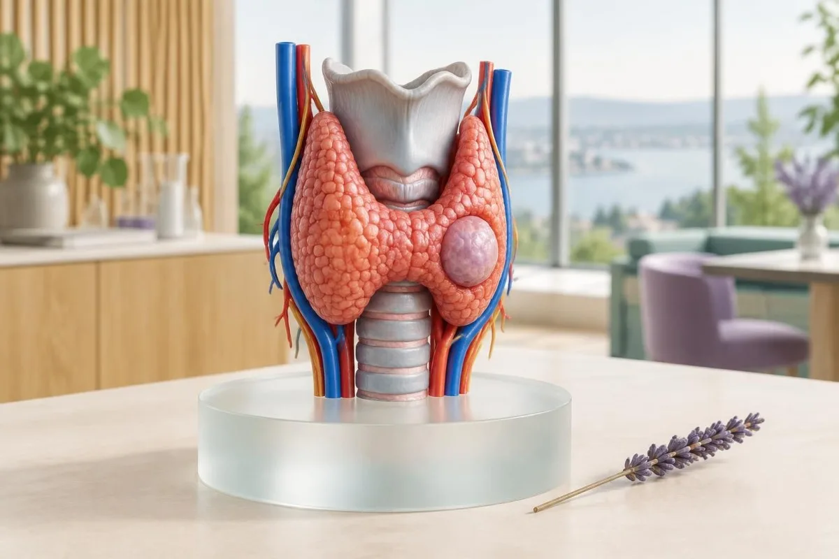
TSH helps decide whether a nodule may be hormone-producing, but bloodwork rarely shows whether it is benign or malignant. Ultrasound supplies the risk pattern; ultrasound-guided tissue examination supplies cellular evidence when indicated.
মূল বিষয়গুলো
- টিএসএইচ is the first blood test for most thyroid nodules; a suppressed result below 0.1 mIU/L usually prompts a radionuclide scan rather than immediate tissue sampling.
- স্বাভাবিক TSH does not rule out thyroid cancer because most nodules, including many malignant nodules, do not change hormone production.
- Free T4 and free T3 show thyroid function, not nodule structure; free T3 is usually most useful when TSH is low.
- TPO and thyroglobulin antibodies support autoimmune thyroid disease but cannot classify a solitary nodule as benign or malignant.
- ক্যালসিটোনিন may help detect medullary thyroid carcinoma in selected people, especially with a family history or MEN2 risk, but routine testing remains debated.
- আল্ট্রাসাউন্ড assesses composition, echogenicity, margins, shape, calcification and lymph nodes; these features determine whether follow-up or tissue examination is appropriate.
- Fine-needle aspiration is generally guided by ultrasound pattern and size, with common thresholds ranging from about 1 cm for high-suspicion nodules to 2.5 cm for low-suspicion nodules.
- আণবিক পরীক্ষা is mainly an add-on for indeterminate cytology; it does not replace ultrasound, clinical assessment or pathology review.
Which thyroid nodule results matter first?
TSH, ultrasound appearance and cytology matter most, but they answer different questions. TSH shows whether the thyroid is being over- or under-stimulated; ultrasound estimates structural risk; tissue examination evaluates cells. A normal TSH cannot classify most nodules, and a blood test alone cannot reliably distinguish a benign nodule from cancer.
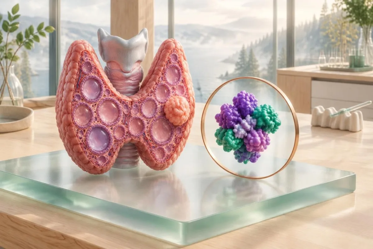
The average adult has a thyroid nodule risk of roughly 4% to 7% by palpation, while ultrasound detects nodules in far more people because it sees very small findings. Only about 5% to 15% of clinically recognized nodules prove malignant, depending on the population and referral setting.
The 2015 American Thyroid Association guideline recommends serum TSH as the initial laboratory test for a known nodule, then uses the result to decide whether radionuclide imaging is appropriate (Haugen et al., 2016). The reason is physiological: a nodule making thyroid hormone can appear different on a scan from a non-functioning nodule.
Kantesti is an AI blood test analyzer that can organize TSH, free T4, free T3, antibodies and calcitonin into a readable pattern, but our interpretation cannot replace a neck ultrasound or a clinician's examination. Start with the রক্ত পরীক্ষার বায়োমার্কার গাইড when you need help identifying which result belongs to which panel.
What does TSH reveal about a thyroid nodule?
TSH is the key first blood result because a low value may indicate an autonomous, hormone-producing nodule. Many laboratories use an adult reference interval near 0.4 to 4.0 mIU/L, although the interval printed on your report takes priority and may differ by assay, age and pregnancy.
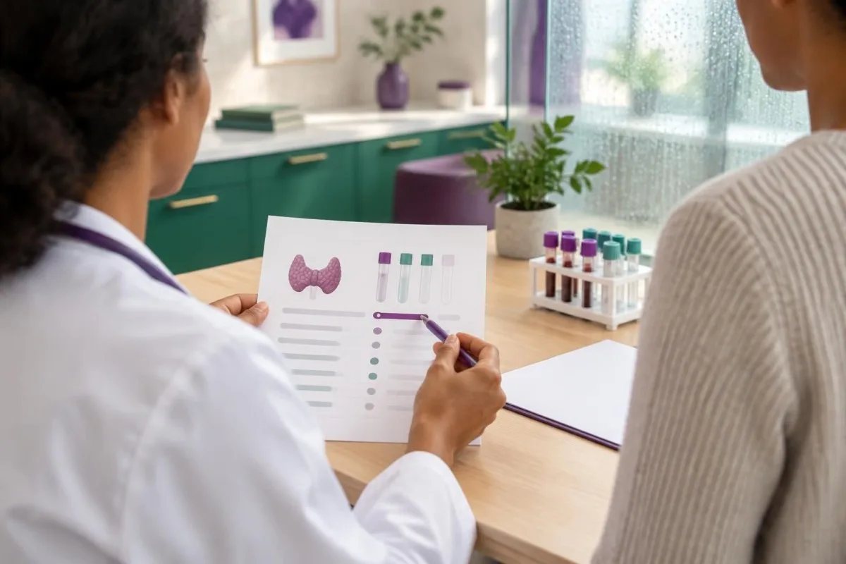
A TSH below 0.1 mIU/L is called suppressed and increases the likelihood that at least one nodule is autonomously functioning, particularly when free T4 or free T3 is high. A TSH between 0.1 and 0.39 mIU/L is mildly low and deserves context rather than an automatic diagnosis.
A high TSH does not make a nodule benign. In Hashimoto thyroiditis, TSH may rise while the gland becomes heterogeneous and nodular, and the inflammatory background can make ultrasound interpretation more difficult. A practical review of testing intervals is available in our guide to how often thyroid levels are checked.
Suppose a hypothetical 62-year-old has a TSH of 0.06 mIU/L, free T4 within range and a 1.8 cm nodule. The result suggests subclinical hyperthyroidism and may justify a radionuclide scan; it does not prove cancer, and it does not automatically justify tissue examination before functional imaging.
| মাত্রা | সীমা | এর অর্থ |
|---|---|---|
| এর মাধ্যমে পর্যালোচনার জন্য আপলোড করা যেতে পারে। | ০.৪-৪.০ এমআইইউ/লিটার | Thyroid stimulation is broadly within the laboratory interval; ultrasound still determines nodule risk. |
| সামান্য বৃদ্ধি পাওয়া | 4.1-10.0 mIU/L | May suggest subclinical hypothyroidism if free T4 remains normal; it does not identify nodule type. |
| উল্লেখযোগ্যভাবে বেড়েছে | 10-20 mIU/L | Often warrants clinical assessment for hypothyroidism, medication effects or autoimmune disease. |
| দমনকৃত (Suppressed) | <0.1 mIU/L | Suggests strong thyroid stimulation or excess hormone and commonly prompts radionuclide scanning. |
How do free T4 and free T3 change interpretation?
Free T4 and free T3 show whether the thyroid is producing excess or insufficient hormone, but neither test describes the nodule's ultrasound structure. Free T4 is usually the more useful companion to TSH; free T3 becomes especially informative when TSH is low and free T4 is still normal.
High free T4 with a suppressed TSH supports overt thyrotoxicosis, while normal free T4 with a suppressed TSH supports a subclinical pattern. T3 toxicosis occurs when free or total T3 is high despite a normal free T4, so a normal T4 does not always end the assessment.
Free hormone reference intervals vary substantially; one adult laboratory may report free T4 as approximately 10 to 22 pmol/L, while another uses 0.8 to 1.8 ng/dL. The conversion is not the main issue: assay method, pregnancy, binding proteins and biotin use can shift results, which is why T4 and T3 meanings should be read beside the local range.
Imagine a hypothetical result with TSH 0.08 mIU/L, free T4 15 pmol/L and free T3 8.1 pmol/L when the upper limit is 6.8. That pattern raises concern for T3-predominant hormone excess and may explain palpitations, but it still cannot tell whether the responsible nodule is structurally suspicious.
What can thyroid antibodies reveal?
TPO and thyroglobulin antibodies indicate autoimmune thyroid activity; they do not grade the cancer risk of an individual nodule. TPO antibody positivity supports Hashimoto disease or, in another clinical pattern, autoimmune hyperthyroidism, but antibody concentration does not translate into a nodule score.
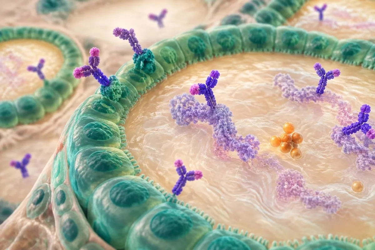
TPO antibody results are often reported as positive above a laboratory-specific cutoff such as 34 IU/mL, but cutoffs differ between assays. A positive result means immune recognition of thyroid proteins; it does not mean that a nodule is malignant, and a negative result does not exclude every form of thyroid disease.
Thyroglobulin antibodies can interfere with thyroglobulin measurement, sometimes producing a misleadingly low or occasionally unpredictable thyroglobulin result. This matters most after thyroid cancer treatment, when thyroglobulin is used for surveillance rather than for initial assessment of an untreated nodule.
Kantesti AI interprets thyroid antibody results beside TSH and free hormone values rather than treating a positive antibody as a cancer marker. If your report contains several antibody flags, compare the pattern with our explanation of পজিটিভ অ্যান্টিবডি ফলাফল and discuss ultrasound findings separately.
Does thyroglobulin diagnose a thyroid nodule?
Serum thyroglobulin is not recommended as a routine diagnostic blood test for an untreated thyroid nodule. Thyroglobulin is produced by normal thyroid tissue as well as differentiated thyroid cancer, so a high value usually reflects the amount of thyroid tissue, inflammation, iodine status or other context rather than a specific diagnosis.
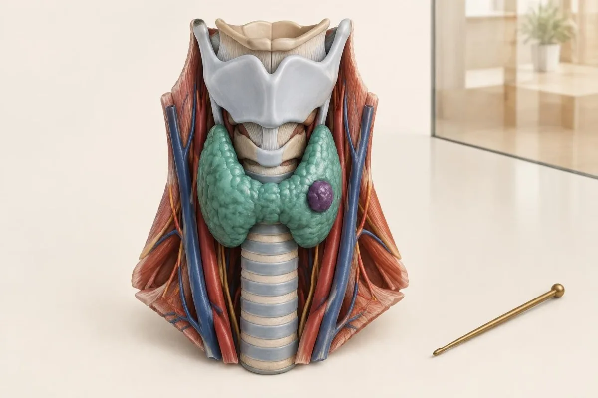
A thyroglobulin concentration of 40 ng/mL, for example, cannot be called cancerous without knowing the remaining thyroid volume, TSH, iodine exposure and antibody status. The number is more clinically useful after total thyroid removal and radioactive iodine treatment, when detectable thyroglobulin may signal residual or recurrent differentiated tissue.
The reverse mistake also occurs: a low thyroglobulin does not prove that a nodule is harmless. Small nodules may release little protein, and thyroglobulin antibodies can distort the assay. This is one reason clinicians prefer ultrasound risk features and, when indicated, cytology over a single serum marker.
The relationship between albumin, globulins and antibody interference is explained in this সিরাম প্রোটিন গবেষণা গাইড. The practical rule is simple: do not order thyroglobulin merely because a nodule was seen unless a specialist has identified a specific surveillance question.
When does a calcitonin thyroid test help?
A calcitonin thyroid test can support evaluation for medullary thyroid carcinoma, but routine testing of every nodule remains an area of guideline disagreement. A persistently elevated basal calcitonin above about 100 pg/mL is strongly concerning in the right clinical setting, whereas modest elevations require assay, kidney, medication and smoking context.
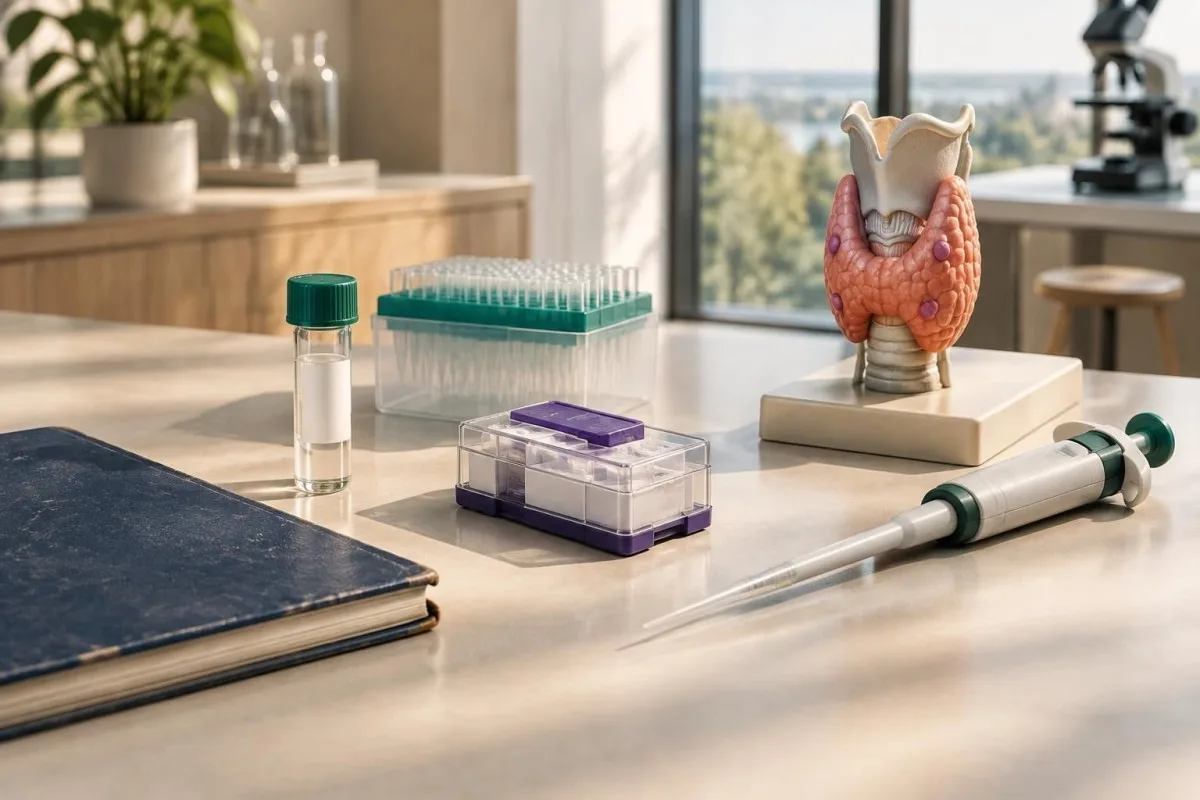
Calcitonin is produced by thyroid C cells, the cell type involved in medullary thyroid carcinoma. Values below 10 pg/mL are often reassuring, values from approximately 10 to 100 pg/mL are difficult to interpret, and values above 100 pg/mL are much more suspicious; these are practical guideposts, not universal diagnostic cutoffs.
The evidence is honestly mixed on population-wide screening because assay thresholds, false positives and downstream surgery differ across countries. Kidney impairment, autoimmune thyroid disease, proton-pump inhibitor use through gastrin effects, smoking and some neuroendocrine conditions can raise calcitonin without medullary cancer.
A family history of medullary thyroid carcinoma, multiple endocrine neoplasia type 2, a suspicious lymph-node pattern or a markedly elevated result makes specialist evaluation more urgent. Our মেডিক্যাল ভ্যালিডেশন তথ্য explains why an AI interpretation can organize this context but cannot confirm a rare thyroid cancer.
Why is ultrasound central to thyroid nodule tests?
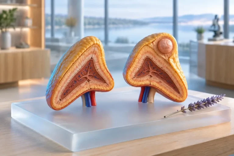
Ultrasound is the main test for classifying a thyroid nodule because it shows composition, echogenicity, margins, shape, calcification and cervical lymph nodes. Bloodwork describes thyroid function; ultrasound shows the physical pattern that determines whether monitoring or ultrasound-guided tissue examination is appropriate.
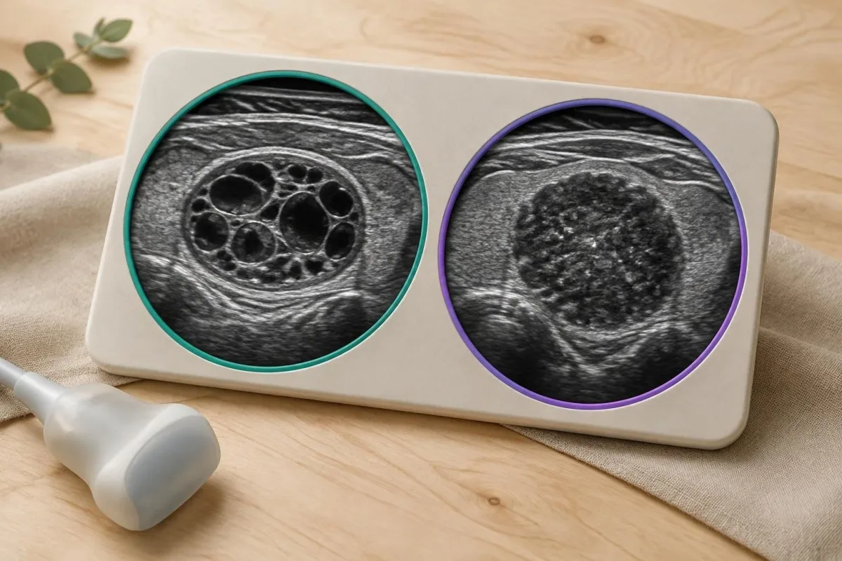
A solid hypoechoic nodule with irregular margins, punctate echogenic foci or a taller-than-wide shape carries more concern than a spongiform or purely cystic nodule. Suspicious lymph nodes may show rounded shape, loss of the fatty hilum, cystic change or abnormal vascular features, and they can change the sampling plan.
Size alone is not a cancer diagnosis. A 2.5 cm spongiform nodule may be monitored while a 0.9 cm nodule with extrathyroidal extension or suspicious nodes may require tissue examination; the reason for the difference is risk pattern, not simply diameter.
Kantesti is an AI blood test interpretation platform, not an ultrasound reader, so a blood report cannot substitute for the written imaging score and stored ultrasound images. Ask for the nodule's three dimensions, composition, echogenicity, margins, calcification description and lymph-node assessment rather than accepting only the word "nodule."
How do TI-RADS categories affect the next step?
TI-RADS systems convert ultrasound features into a risk category and size-based recommendation; they do not use TSH or antibody results to calculate the score. In the ACR system, a TR5 nodule commonly reaches tissue-examination criteria at 1.0 cm, while TR3 nodules generally reach that threshold at 2.5 cm.
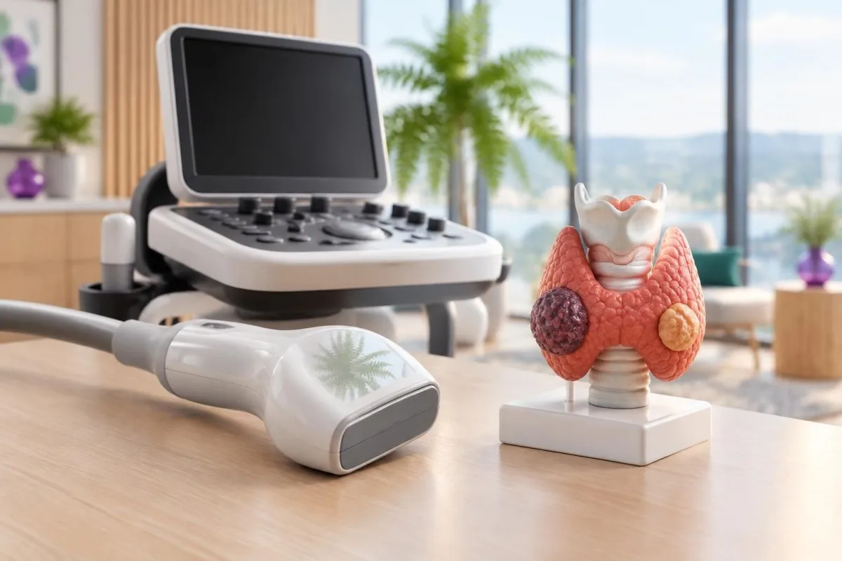
Typical ACR TI-RADS thresholds are TR5: tissue examination at 1.0 cm and follow-up at 0.5 cm; TR4: tissue examination at 1.5 cm and follow-up at 1.0 cm; and TR3: tissue examination at 2.5 cm and follow-up at 1.5 cm. TR1 and TR2 findings usually need no sampling.
The 2017 ACR TI-RADS publication by Tessler and colleagues describes this point-based approach, which aims to reduce unnecessary procedures while catching higher-risk patterns. Other systems, including ATA categories and European scoring methods, use different wording and thresholds, so two legitimate reports may recommend different follow-up for the same nodule (Tessler et al., 2017).
A hypothetical 1.2 cm TR5 nodule meets a common sampling threshold, while a hypothetical 1.2 cm TR3 nodule does not. You can read more about low-TSH patterns in our guide to সাবক্লিনিকাল হাইপারথাইরয়েডিজম, because functional status may determine whether scanning comes before sampling.
When is tissue examination or FNA needed?
Ultrasound-guided fine-needle aspiration is considered when the nodule's sonographic risk and size cross a guideline threshold, or when suspicious lymph nodes or symptoms change the risk calculation. It is not automatically required for every nodule, and a low TSH may lead to a radionuclide scan first.
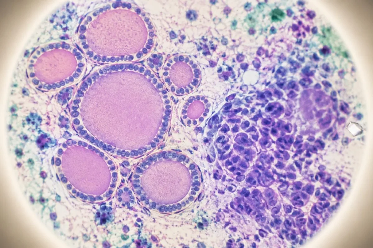
Common ATA size thresholds are about 1 cm for high- or intermediate-suspicion nodules, 1.5 cm for low-suspicion nodules and 2 cm for very-low-suspicion nodules, although patient preference and local practice matter. A purely cystic nodule often does not need diagnostic aspiration unless it causes pressure or requires drainage.
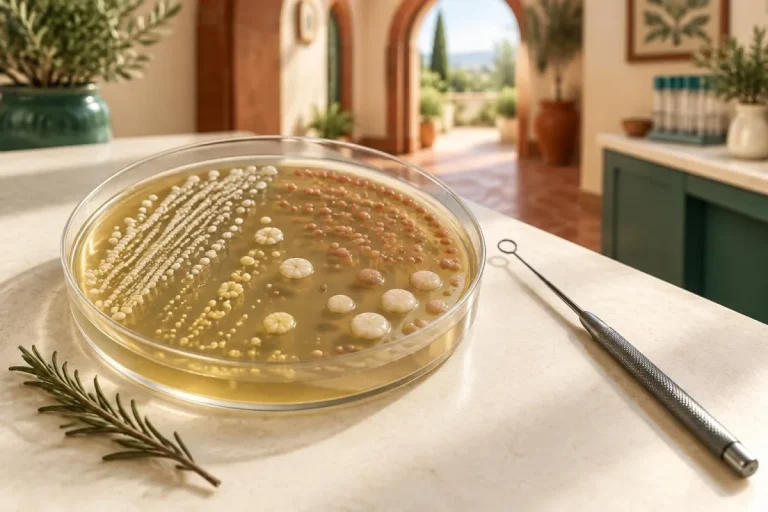
Cytology is reported using Bethesda categories. Bethesda I means nondiagnostic and may lead to repeat sampling; Bethesda III and IV are indeterminate; Bethesda V is suspicious; and Bethesda VI is malignant. Reported malignancy risks vary by institution and whether non-invasive follicular thyroid neoplasm is counted as cancer, so a category should never be interpreted without the laboratory's stated risk range.
A palpable rapidly enlarging mass, hoarseness, difficulty swallowing, breathing symptoms or a firm neck lymph node warrants prompt medical assessment even if TSH is normal. Our page on the চিকিৎসা উপদেষ্টা বোর্ড describes the role of clinical oversight, but only your treating team can decide whether sampling is safe and appropriate.
What do molecular thyroid nodule tests add?
Molecular thyroid nodule tests are mainly useful when cytology is indeterminate; they are not a replacement for ultrasound or an initial blood panel. Gene-expression or mutation panels can shift the estimated probability of cancer, helping some patients choose surveillance rather than diagnostic surgery, but performance depends on the test, prevalence and laboratory validation.
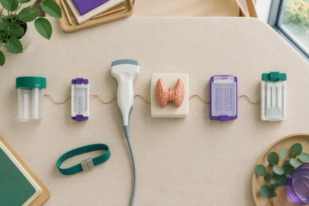
A negative molecular result may reduce the estimated cancer probability to a level compatible with surveillance, while a positive result may increase concern; neither result is an absolute yes-or-no diagnosis. A test with a 95% negative predictive value in one population may perform differently where the pre-test cancer prevalence is 10% rather than 30%.
The result must be matched to Bethesda category, ultrasound pattern, age, radiation history and family history. Molecular testing is often less useful for a clearly benign or clearly malignant cytology result because those results already provide stronger direction.
Kantesti AI can help place a molecular report beside the ordinary laboratory values and imaging summary, but our এআই প্রযুক্তি গাইড explains why algorithmic pattern recognition does not independently validate a proprietary genomic assay. Ask which genes were assessed, what population was studied and whether the lab reports sensitivity, specificity and predictive values.
When is a thyroid scan better than immediate FNA?
A radionuclide thyroid scan is especially useful when TSH is below the reference interval because it shows whether a nodule is hyperfunctioning, isofunctioning or nonfunctioning. A clearly hyperfunctioning nodule is rarely malignant and often does not need diagnostic tissue examination, although the final plan depends on the scan and ultrasound together.
A low TSH of 0.05 mIU/L with a focal area of increased tracer uptake suggests autonomy, while a normal or high TSH usually makes radionuclide scanning less urgent. Scan findings do not erase suspicious ultrasound features; a functioning nodule can still require review if its structure or lymph nodes are atypical.
Symptoms of hormone excess may include resting palpitations, heat intolerance, tremor or unexplained weight loss, but symptoms are not required. Persistent TSH suppression below 0.1 mIU/L matters more in adults over 65 and in people with osteoporosis or cardiac disease because untreated hormone excess can increase atrial-fibrillation and bone-loss risk.
Total T3 can reveal hormone excess when free T4 is normal, as explained in our total T3 testing guide. Do not stop thyroid medication or iodine-containing treatment before a scan without specific instructions, because preparation can alter tracer uptake and make the result harder to interpret.
How do pregnancy, age and history change testing?
Pregnancy, childhood, prior neck radiation and a family history of thyroid cancer lower the threshold for specialist assessment, but they do not make every nodule malignant. The same TSH number can carry different implications during pregnancy, in older adults or after exposure to medicines that alter thyroid function.
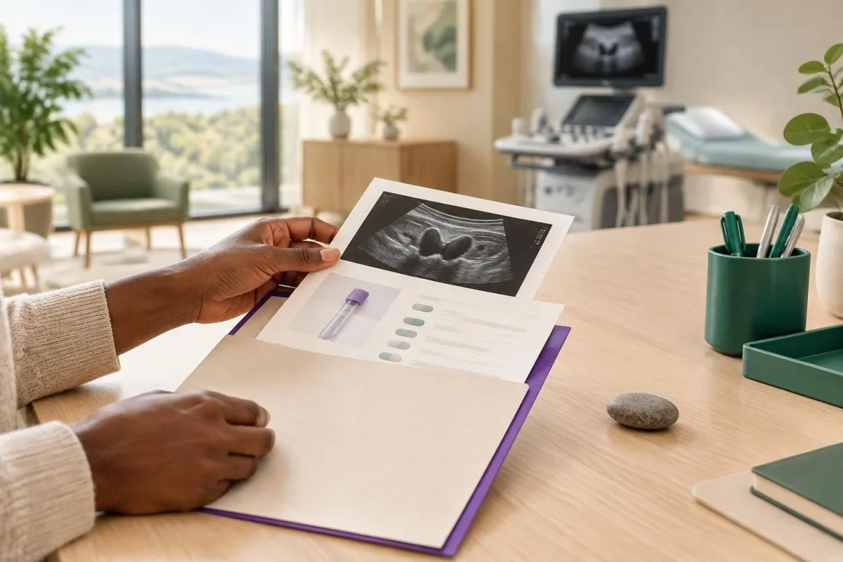
Pregnancy-specific TSH ranges are preferred, and radioactive iodine scanning is avoided during pregnancy. Ultrasound remains the main structural test, while free T4 interpretation requires a pregnancy-appropriate assay or reference interval because binding proteins change during gestation.
Children and adolescents with thyroid nodules generally receive more cautious evaluation because malignancy risk is higher than in adults, even though nodules are less common. A history of external neck radiation, familial medullary thyroid carcinoma or multiple endocrine neoplasia type 2 should be stated before testing because it can change the choice of calcitonin, genetic counselling and ultrasound follow-up.
A family history is not the same as proof of inherited disease. For practical thyroid nutrition context, see our diet and thyroid health guide, but avoid high-dose iodine or kelp supplements without advice because excess iodine can destabilize thyroid function in susceptible people.
Which symptoms make follow-up more urgent?
Rapid enlargement, persistent hoarseness, swallowing difficulty, breathing difficulty or a new firm lymph node requires prompt clinical assessment, regardless of a normal thyroid nodule blood test. These features can indicate compression, vocal-cord involvement or lymph-node disease, although benign conditions can also produce neck pressure.
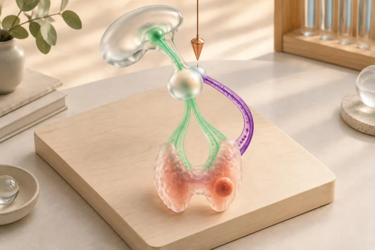
A normal TSH near 2.0 mIU/L does not reduce a suspicious ultrasound pattern to low risk. Conversely, a suppressed TSH of 0.08 mIU/L may point toward an autonomous nodule rather than cancer, so urgency depends on the combination of symptoms, examination, scan and ultrasound.
Follow-up intervals are risk-based rather than universal. A small, low-suspicion nodule may be re-imaged after 12 to 24 months, while a suspicious nodule or abnormal lymph node may be assessed sooner; meaningful growth is often defined as at least 20% increase in two dimensions with a minimum 2 mm increase, or more than 50% increase in volume.
আমাদের ব্যবহার করুন থাইরয়েড পরীক্ষার সময়সূচী as a preparation aid, not a substitute for a personalised plan. Seek urgent care for breathing difficulty, rapidly worsening neck swelling or inability to swallow fluids rather than waiting for a repeat blood result.
How should all thyroid nodule results be combined?
The safest interpretation combines TSH, free hormones, selected antibodies, ultrasound pattern, size, symptoms and cytology instead of ranking one abnormal number above everything else. Bloodwork answers whether the gland is functioning abnormally; imaging answers what the nodule looks like; tissue examination answers what cells are present.
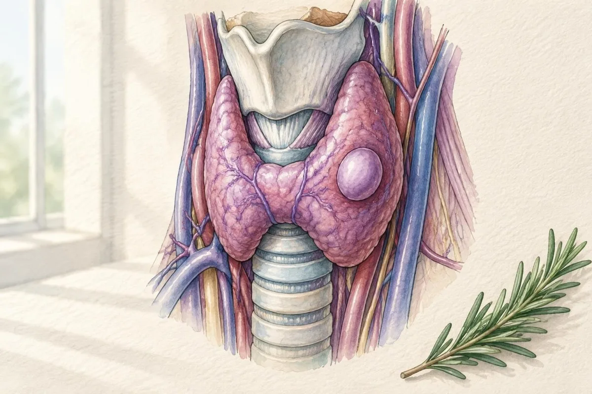
A practical sequence is: confirm the nodule and ultrasound details; measure TSH; add free T4 and sometimes free T3; consider antibodies when autoimmune disease is plausible; consider calcitonin when medullary cancer risk justifies it; then use scanning, tissue examination or surveillance according to the pattern.
Kantesti is an AI-powered blood test analysis tool that can compare current TSH and thyroid-marker results with prior reports, identify changes such as TSH falling from 1.8 to 0.12 mIU/L, and highlight questions for a clinician. It cannot inspect ultrasound images, determine cytology quality or make a cancer diagnosis from bloodwork.
For trustworthy interpretation, keep the laboratory reference ranges, medication list, supplement list, pregnancy status, prior imaging and exact nodule dimensions together. Our ক্লিনিকাল ওভারসাইট মান explain how medical information should be framed, while the research guide on serum proteins illustrates why assay context matters.
What can a thyroid nodule blood test not tell you?
A thyroid nodule blood test cannot reliably tell whether most nodules are benign, malignant, cystic, solid or compressive. Even an entirely normal panel can coexist with a suspicious ultrasound finding, while several abnormal blood results may reflect autoimmune disease or medication effects rather than a dangerous nodule.
TSH is a function marker, not a cancer screen. Thyroglobulin is a thyroid-tissue marker, not a general nodule test; TPO antibodies indicate immune activity, not malignancy; and calcitonin is a specialised marker whose interpretation depends on assay and pre-test risk.
Biotin supplements can interfere with some immunoassays, sometimes making TSH appear falsely low and free T4 or T3 falsely high. Ask the ordering clinician or laboratory whether biotin should be paused before testing; do not stop prescribed medicines independently, and disclose high-dose hair, skin and nail products.
Kantesti AI can explain why a result is flagged and compare it with previous values, but our platform cannot overrule an ultrasound recommendation or a cytology report. If the report is confusing, ask for the clinical question being answered: function, autoimmune activity, medullary cancer risk, structural risk or postoperative surveillance.
What should you bring to a thyroid nodule appointment?
Bring the complete laboratory report, ultrasound report, medication list, supplement list and relevant family or radiation history. A single number without its unit, reference interval and collection date can lead to a wrong conclusion, especially for calcitonin, free hormones and antibody assays.
Write down the exact TSH value, free T4 and free T3 units, antibody names, calcitonin assay and reference range. Include whether you take levothyroxine, antithyroid medication, lithium, amiodarone, iodine supplements or biotin because these can alter thyroid results or their interpretation.
Ask five focused questions: What is the ultrasound risk category? What are the nodule dimensions in millimetres? Are lymph nodes suspicious? Does TSH require a radionuclide scan? If cytology is indeterminate, would molecular testing change management? These questions often provide more useful information than asking whether a single blood result is "normal."
You can upload a laboratory PDF or photograph to Kantesti for an organised explanation of the blood results, then take the questions to your clinician. Keep privacy in mind and review our data security information before sharing health documents online.
What is the bottom line for thyroid nodule testing?
TSH is the most useful first blood result, but ultrasound usually determines structural risk and tissue examination confirms cellular diagnosis when indicated. A normal TSH is reassuring about thyroid function, not a guarantee that the nodule is harmless; a low TSH may redirect testing toward a radionuclide scan.
The most actionable pattern is not one isolated flag but a coherent combination: suppressed TSH plus a focal hot nodule; normal TSH plus a high-suspicion ultrasound pattern; positive TPO antibodies plus diffuse gland changes; or elevated calcitonin plus a family history of medullary thyroid carcinoma.
The two DOI-linked Kantesti research guides listed below concern broader laboratory interpretation rather than thyroid nodule diagnosis, so they should not be used as substitutes for thyroid guidelines. For questions about clinical review and responsible interpretation, consult the Kantesti মেডিকেল অ্যাডভাইজরি বোর্ড.
If your result is abnormal, the next step is usually clarification rather than panic: verify the assay and reference range, review medicines and supplements, obtain the full ultrasound description, and ask whether repeat bloodwork, scanning, follow-up imaging or tissue examination answers the remaining question.
প্রায়শই জিজ্ঞাসিত প্রশ্নাবলী
Can a blood test tell if a thyroid nodule is cancerous?
A blood test cannot reliably determine whether most thyroid nodules are cancerous. TSH, free T4 and free T3 assess thyroid function, while TPO and thyroglobulin antibodies assess autoimmune or assay-related context; none provides a general cancer diagnosis. Ultrasound evaluates structural risk, and ultrasound-guided tissue examination may be needed when a nodule meets size and appearance criteria, often around 1 cm for high-suspicion nodules.
What TSH level is concerning with a thyroid nodule?
A TSH below 0.1 mIU/L is suppressed and commonly prompts consideration of a radionuclide thyroid scan because the nodule may be producing hormone independently. TSH between 0.1 and 0.39 mIU/L is mildly low and needs free T4, free T3, symptoms and repeat testing for context. A normal TSH near 0.4 to 4.0 mIU/L does not rule out thyroid cancer or remove the need for ultrasound.
Should calcitonin be checked for every thyroid nodule?
Routine calcitonin testing for every thyroid nodule remains debated because assay thresholds and false-positive results vary. Calcitonin persistently above about 100 pg/mL is strongly concerning for medullary thyroid carcinoma, while results around 10 to 100 pg/mL require clinical context and repeat or specialist assessment. Testing is particularly relevant with a family history of medullary thyroid carcinoma, MEN2 risk or a suspicious clinical and ultrasound pattern.
Does a normal thyroid blood test mean a nodule is benign?
A normal thyroid blood test means thyroid hormone production is broadly within the laboratory interval; it does not prove that a nodule is benign. Many benign and malignant nodules occur with TSH around 0.4 to 4.0 mIU/L and normal free T4. Ultrasound features, nodule size, lymph nodes and cytology determine structural and cellular risk.
When is a thyroid nodule biopsy needed?
Tissue examination is usually considered when ultrasound risk and nodule size cross a guideline threshold or when suspicious lymph nodes or symptoms increase concern. Common thresholds range from about 1 cm for high-suspicion nodules to 2.5 cm for low-suspicion nodules, depending on the scoring system. A low TSH may lead to radionuclide scanning first, because a clearly hormone-producing nodule often does not need immediate diagnostic aspiration.
Do thyroid antibodies make a nodule more likely to be cancer?
TPO and thyroglobulin antibodies primarily indicate autoimmune thyroid activity and do not directly classify an individual nodule as malignant. A TPO antibody result above a laboratory cutoff such as 34 IU/mL can support autoimmune thyroid disease, but cutoffs vary by assay. The nodule still requires ultrasound-based assessment, and thyroglobulin antibodies can interfere with thyroglobulin measurement.
Can Kantesti interpret thyroid nodule blood test results?
Kantesti can organise and explain thyroid blood results such as TSH, free T4, free T3, TPO antibodies, thyroglobulin antibodies and calcitonin in relation to the laboratory reference ranges. It can compare trends, identify combinations that deserve clinical review and help prepare questions, but it cannot read ultrasound images, validate cytology or diagnose thyroid cancer. A result such as TSH below 0.1 mIU/L still requires clinician-directed assessment of medication, symptoms and possible functional imaging.
আপনার রক্ত পরীক্ষার রিপোর্ট কীভাবে পড়বেন
আপনার ল্যাব রিপোর্ট আপলোড করুন, আর Kantesti-এর AI কয়েক সেকেন্ডের মধ্যে প্রতিটি বায়োমার্কার সহজ ভাষায় ব্যাখ্যা করবে। বিশ্বজুড়ে 2 মিলিয়নেরও বেশি মানুষ ব্যবহার করেন।.
তথ্যসূত্র
- ক্লেইন, টি., মিচেল, এস., & ওয়েবার, এইচ. (২০২৬)।. সিরাম প্রোটিন নির্দেশিকা: গ্লোবুলিন, অ্যালবুমিন ও A/G অনুপাতের রক্ত পরীক্ষা।. Kantesti AI চিকিৎসাবিষয়ক গবেষণা।. ডিওআই
- ক্লেইন, টি., মিচেল, এস., & ওয়েবার, এইচ. (২০২৬)।. C3 C4 Complement Blood Test & ANA Titer Guide. Kantesti AI চিকিৎসাবিষয়ক গবেষণা।. ডিওআই
- Haugen BR et al. (2016). 2015 American Thyroid Association Management Guidelines for Adult Patients with Thyroid Nodules and Differentiated Thyroid Cancer. Thyroid। ডিওআই পাবমেড
- Tessler FN et al. (2017). ACR Thyroid Imaging, Reporting and Data System (TI-RADS): White Paper of the ACR TI-RADS Committee. Journal of the American College of Radiology। ডিওআই পাবমেড
- ২০ লক্ষ+বিশ্লেষণ করা পরীক্ষাগুলো
- 127+দেশগুলি
- 100+ভাষাসমূহ
চিকিৎসক দল সম্পর্কে
E-E-A-T বিশ্বাসযোগ্যতার সংকেত
অভিজ্ঞতা
চিকিৎসক-নেতৃত্বাধীন ল্যাব ব্যাখ্যা কর্মপ্রবাহের ক্লিনিক্যাল পর্যালোচনা।.
দক্ষতা
ক্লিনিকাল প্রেক্ষাপটে বায়োমার্কারগুলো কীভাবে আচরণ করে, সেটির ওপর ল্যাবরেটরি মেডিসিনের ফোকাস।.
কর্তৃত্ব
ডা. থমাস ক্লেইনের চিকিৎসাবিষয়ক পর্যালোচনায়, ডা. সারাহ মিচেল এবং প্রফেসর ডা. হান্স ভেবারের অবদানসহ।.
বিশ্বাসযোগ্যতা
প্রমাণভিত্তিক ব্যাখ্যা, যাতে সতর্কতা কমাতে স্পষ্ট পরবর্তী পদক্ষেপের পথ থাকে।.
চিকিৎসাবিষয়ক দাবিত্যাগ
এই প্রবন্ধটি কেবল শিক্ষামূলক উদ্দেশ্যে এবং এটি চিকিৎসা পরামর্শ হিসেবে গণ্য হয় না। রোগ নির্ণয় ও চিকিৎসা সিদ্ধান্তের জন্য সর্বদা একজন যোগ্য স্বাস্থ্যসেবা প্রদানকারীর সাথে পরামর্শ করুন।.
প্রকাশিত · সম্পাদকীয় দলের সঙ্গে যোগাযোগ করুন
Kantesti Ltd কর্তৃক প্রকাশিত, ইংল্যান্ড ও ওয়েলসে নিবন্ধিত, কোম্পানি নং. 17090423, লন্ডন, যুক্তরাজ্য।.