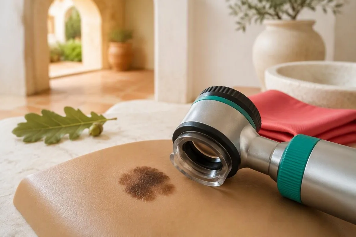
Melanomanın simptomlarına dəyişən xal, qeyri-adi yeni törəmə və ya dırnaqda, ovucda və ya ayaq dabanında uzun müddət qalan ləkə daxil ola bilər. Asimmetriyaya, qeyri-müntəzəm kənarlara, qeyri-bərabər rəngə və dəyişikliklərə diqqət yetirin, lakin bütün xəbərdarlıq əlamətlərinin yaranmasını və ya diametrin 6 mm-dən çox olmasını gözləməyin.


- Melanomanın ABCDE qaydası asimmetriya, qeyri-müntəzəm kənar, rəng müxtəlifliyi, diametr və dəyişikliklər deməkdir; narahatlıq doğuran bir əlamət belə müayinə üçün əsas ola bilər.
- Diametr 6 mm-dən çox olması xəbərdarlıq əlamətidir, lakin diaqnostik hədd deyil; daha kiçik melanomalar da olur.
- Yeni törəmələr əhəmiyyətlidir: 2017-ci il meta-analizinə görə, melanomaların 70.9%-i əvvəldən mövcud olan xalla əlaqəsi olmadan yaranıb.
- «Çirkin ördək balası» əlaməti o deməkdir ki, ləkə simmetrik görünsə belə, insanın öz adi xal naxışından fərqlənir.
- Düyünlü melanoma həftələr ərzində qabara, bərkiyə və böyüyə bilər; çəhrayı və ya dəri rəngində törəmələr də melanoma ola bilər.
- Dırnaq dəyişiklikləri məsələn, enən tünd zolaq, qonşu dəridə piqmentasiya və ya səbəbi bilinməyən dırnaq deformasiyası klinik qiymətləndirmə tələb edir.
- Ovuc içləri və ayaqaltılar hər dəri tonunda müayinə olunmalıdır; davamlı ləkələr və ya törəmələr göyərməyə, ziyilə və ya təzyiq zədəsinə bənzəyə bilər.
- Qan analizləri erkən melanomanı etibarlı şəkildə aşkar edə və ya istisna edə bilməz; diaqnoz üçün klinik müayinə və adətən dəri biopsiyası tələb olunur.
- Növbəti addım müntəzəm qan analizini gözləməkdənsə, dəyişən və ya qeyri-adi ləkə ilə bağlı dərhal həkimə müraciət etməkdir.
Melanomanın hansı simptomları dəri müayinəsinə əsas verməlidir?
Melanomanın simptomları ən çox görünən dəyişikliklə özünü göstərir: dəyişən xal, yeni qeyri-adi törəmə və ya davamlı anormal ləkə. Erkən melanoma ağrı və ya qaşınmaya səbəb olmaya bilər, şübhəli ləkənin ölçüsü isə 6 mm; -dən az ola bilər; diaqnoz üçün klinik qiymətləndirmə, çox vaxt ardınca biopsiya aparılır.
Ləkənin diqqətə layiq olması üçün ABCDE-nin bütün 5 əlamətinə malik olması vacib deyil. Aydın şəkildə böyüyən, kiçik və rəngi bərabər olan törəmə illərdir dəyişməyən iri xaldan daha əhəmiyyətli ola bilər; simptomlar bir fotoşəkillə deyil, insanın əvvəlki görünüşü ilə müqayisədə qiymətləndirilməlidir.
Melanoma əvvəllər tamamilə adi görünən dəridə yarana bilər. Dəri müayinəsi zamanı 2 ayrı sual verin: mövcud ləkə dəyişibmi və yeni, uyğun gəlməyən bir şey yaranıbmı? Yalnız tanış xallara baxmaq qarşısı alına bilən kor nöqtə yaradır.
Mənim, Thomas Klein, MD, şirkətinin baş tibb müşaviri kimi tövsiyəm Kantesti təşkilatının, növbəti addımı konkretləşdirməkdir: ləkə dəyişirsə, dəri müayinəsi təyin edin. Arxayınlaşdırıcı laboratoriya paneli başqa suala cavab verir və həmin törəmənin müayinəsini təxirə salmamalıdır.
Kantesti bir AI qan testi analizatoru, melanoma skrininq testi deyil; anormal göstərici olmayan 0 nəticə dəri xərçəngini istisna edə bilməz. Bizim rolumuz laboratoriya nəticələrini onların məhdudiyyətlərini nəzərə alaraq izah etməkdir, şübhəli dəri dəyişikliyini isə onu birbaşa müayinə edə bilən həkim qiymətləndirməlidir.
Melanomanın ABCDE qaydası necə işləyir?
The Melanomanın ABCDE qaydası asimmetriyanı, kənarların qeyri-müntəzəmliyini, rəng müxtəlifliyini, diametri və dəyişimi təsvir edir. Diametrin 6 mm -dən böyük olması faydalı xəbərdarlıq əlamətidir, lakin ölçü, forma, rəng və ya davranışdakı dəyişiklik — istənilən diametrdə — qiymətləndirməni əsaslandıra bilər.
A və B ölçüyə deyil, formaya aiddir: iki yarı fərqli görünə bilər və ya kənar daraqvari, çökəklikli, yaxud qeyri-dəqiq sərhədli ola bilər. Bu əlamətlərin heç biri melanomanı sübut etmir, çünki xoşxassəli atipik xallar da belə görünə bilər; həkim həmçinin anamnezi, yerləşmə yerini və insanın əvvəlki xal naxışını nəzərə alır.
C includes uneven shades of brown or black, but also areas of pink, white, red or blue. A spot containing 2 colors is not automatically cancerous, and a uniformly pink growth is not automatically safe; the distribution and evolution of color are more informative than simply counting shades.
D is often explained using a pencil eraser, roughly 6 mm across. That memory aid must not become a permission slip to ignore smaller growths: diameter describes surface width, whereas the pathological measurement that helps stage melanoma is its depth in millimeters.
E includes a new raised area, recurrent crusting or a change noticed across 2 dated photographs. Our tibbi məzmun standartları distinguish warning signs from diagnostic criteria; ABCDE helps people recognize a reason for assessment, not calculate whether a mole is definitely benign.
«Çirkin ördək balası» əlaməti nəyi göstərir?
The ugly-duckling sign identifies a spot that looks different from the other moles on the same person. It can flag 1 unusually dark, pale, large, small or raised spot even when that growth does not satisfy the usual ABCDE checklist.
Personal comparison matters because normal mole patterns differ substantially. Someone with 20 similar light-brown moles may have a conspicuous new black spot; another person with many dark moles may instead develop a pink growth that stands out. The outlier does not have to be the darkest mark.
Compare nearby spots as well as the overall pattern. A person may have 2 recurring mole patterns—for example, flatter moles on the limbs and raised moles on the trunk—so comparison works best when a clinician considers body site rather than assuming every mole should look identical.
Consider an illustrative scenario: a 42-year-old notices a symmetrical, 4 mm spot unlike every other mole on the shoulder. Its symmetry and small size do not cancel the unusual appearance; a clinician would ask when it appeared, whether it is changing and what dermoscopy shows.
An ugly duckling is a reason to investigate, not a diagnosis or an indication to remove every outlier. If melanoma runs in the family, discuss the actual relatives and diagnoses rather than requesting a generic cancer panel; our guide to family cancer testing limits explains why targeted assessment differs from broad laboratory screening.
Melanoma yeni törəmə kimi başlaya bilərmi?
Melanoma can arise as a new growth without a changing pre-existing mole. Pampena et al. reported that 70.9% of melanomas in their 2017 meta-analysis were de novo, while 29.1% were associated with a pre-existing nevus; these pooled estimates are not an individual's cancer probability.
The meta-analysis combined 38 studies and 20,126 melanomas, giving the new-growth message a stronger basis than an anecdote. Classification depended on the studies and tissue findings, however, so the percentages should not be treated as an exact split for every population or melanoma subtype.
A new spot is not automatically melanoma. Adults commonly develop benign seborrheic keratoses, angiomas and other growths; the useful distinction is whether a new mark persists, evolves or appears unlike the person's usual skin features, especially when it changes over several weeks.
Age changes the context, not the diagnosis. A new mole-like spot in a 65-year-old generally deserves more scrutiny than an otherwise typical new mole in a teenager, but there is no age at which new melanoma becomes impossible or every new growth must be malignant.
Checking only existing moles misses the central lesson of Pampena et al. (2017): surveillance should include previously unremarkable skin. Likewise, routine annual bloodwork is not a substitute for assessing a new growth, even when the last panel was obtained just 1 month ago.
Hansı melanomalar tünd xallara bənzəməyə bilər?
Düyünlü melanoma can appear as an elevated, firm growth that enlarges over weeks, while amelanotic melanoma may look pink, red or skin-colored. The 3-part EFG reminder—elevated, firm, growing— complements ABCDE, but neither checklist can reliably exclude melanoma.
A nodule can be symmetrical and have a smooth edge. That is why a growth enlarging over 4–6 weeks should not be dismissed simply because its photograph lacks an irregular outline; vertical growth may produce a different surface appearance from a spreading pigmented patch.
Pink growths have many benign explanations, including irritated follicles and vascular growths. The concerning combination is persistence plus enlargement, spontaneous bleeding or repeated crusting at the same site—not pink color alone. A clinician may need tissue examination because appearance overlaps among several skin conditions.
EFG does not mean every raised mole is dangerous. A soft raised mole unchanged for 10 years is a different clinical problem from a newly firm bump that grows noticeably between photographs; recent behavior supplies the missing context that a static checklist cannot capture.
The distinction between depth and diameter is especially useful here: 1 small surface measurement cannot tell you how deeply a growth extends. Our guide to AI hesabat mənbəyi yoxlamaları explains why a confident-looking summary must never replace the actual examination or pathology report.
Qaşınma, qanama və ya qabıqlanma melanoma deməkdirmi?
Itching, bleeding and crusting can occur with melanoma, but none is specific to it. Persistent symptoms in 1 changing spot, particularly spontaneous or recurrent bleeding, deserve assessment; generalized itching or a clearly traumatized stable mole requires a different clinical interpretation.
Bleeding after catching a mole with a razor has an obvious possible explanation. Bleeding repeatedly without that trigger, or returning after the surface appears healed, is more concerning; record the circumstances of each episode rather than describing every event simply as 'it bled once.'
An itchy mole can reflect friction, eczema or local irritation. Itch affecting 1 evolving growth is more relevant to melanoma assessment than itch affecting both legs with dry skin; our explanation of itching and laboratory limits separates skin examination from tests used for selected systemic causes.
Many itchy bumps appearing over 1–2 weeks suggest a different pattern from a solitary changing growth. Conditions such as scabies and nighttime itching can involve multiple sites and household contacts, but a widespread rash does not automatically explain away an unrelated unusual mole.
Repeatedly scratching or treating a spot makes its original appearance harder to interpret. Take 1 clear photograph before applying a non-prescribed treatment, and avoid home acids, freezing kits or cosmetic removal of a suspicious growth; destroying its surface can delay diagnosis and compromise useful tissue assessment.
Dırnaqlardakı hansı dəyişikliklər melanomaya işarə edə bilər?
Nail melanoma may cause a new or widening brown-black longitudinal band, irregular pigmentation, nail distortion or pigment extending onto nearby skin. A changing band affecting 1 nail deserves assessment, although benign pigmentation, trauma and other nail disorders are much more common.
A band's width is not a stand-alone diagnostic test. Width above approximately 3 mm is sometimes used as a warning clue, but a narrower evolving band can still need evaluation; irregular color, widening toward the nail base and change over time add more context than width alone.
Pigment extending onto the cuticle or surrounding nail-fold skin is called the Hutchinson sign. It increases concern but is not exclusive to melanoma; clinicians also distinguish genuine skin pigmentation from pigment visible through a translucent nail fold, sometimes called a pseudo-Hutchinson sign.
A subungual bruise often moves toward the fingertip as the nail grows. Fingernails grow roughly 3 mm per month and toenails more slowly, so dated photographs can help document movement; however, a trauma history or some outward movement cannot independently rule out a persistent underlying growth.
A new unexplained band on 1 nail in an adult should not be managed with repeated antifungal courses without reassessment. Keep previous photographs and reports together using consent-based clinical record sharing, but send nail photographs through the examining clinician's approved system rather than assuming a laboratory app can diagnose them.
Melanoma ovuclarda və ayaq dabanlarında necə görünə bilər?
Acral melanoma can develop on palms, soles and within the nail unit, sometimes resembling a bruise, wart or persistent pressure-related patch. A mark that enlarges, changes color or fails to resolve over several weeks needs assessment, even when it is smaller than 6 mm.
Pressure and friction make the sole a particularly confusing site. A person may explain a changing patch as damage from a shoe for 2 months, yet persistence despite reduced friction weakens that explanation; the clinician needs both the mechanical history and an examination of the actual mark.
Dermoscopy can reveal whether pigment follows skin ridges or furrows on palms and soles. A parallel-ridge pattern can raise suspicion, but these specialized patterns are not reliable do-it-yourself rules; small, early or unpigmented acral melanomas may not display a classic pattern.
A persistent foot growth should not automatically be treated as a wart. If 1 area keeps enlarging, changes after treatment or repeatedly breaks down, ask for reassessment before another destructive procedure; photographs taken before treatment can preserve information that the altered surface no longer shows.
Not every slow-healing foot area is melanoma. Diabetes, pressure injury and impaired circulation are alternative explanations, and foot circulation warning signs require their own assessment; more than 1 condition can coexist, so an abnormal vascular test does not explain every pigmented growth.
Kimlər melanoma simptomlarına diqqət yetirməlidir?
Melanoma can occur in every skin tone, although overall incidence and the distribution of melanoma subtypes vary. Personal melanoma history, a close family history, numerous atypical moles and ultraviolet exposure increase concern; no skin color makes a changing 1-nail band or sole growth automatically safe.
Darker skin reduces some ultraviolet-related melanoma risk but does not eliminate melanoma. Acral melanoma represents a larger proportion of melanomas diagnosed in some darker-skinned populations, which is not the same as saying its absolute incidence is necessarily higher; percentages need the correct denominator.
A first-degree relative means a parent, sibling or child. If 2 or more close relatives have melanoma, or melanoma occurs alongside certain other cancers in a family, ask whether specialist risk assessment is appropriate; genetic testing is selective, not a routine response to having many moles.
Immunosuppression and a previous melanoma can change the surveillance plan. The relevant details include the medication, duration, previous pathology and number of affected relatives; a generic family-risk score should not determine whether a newly evolving spot receives examination.
At Kantesti, our approach to risk əsaslı ailə sağlamlığı yoxlamaları separates useful laboratory monitoring from examinations that bloodwork cannot replace. Whether the family has 1 affected relative or several, a current suspicious growth needs its own clinical assessment rather than waiting for a risk calculation.
Dərinizi necə yoxlamalı və şəklini çəkməlisiniz?
A useful skin self-check compares your current skin with its previous appearance and includes nails, palms, soles and hard-to-see areas. A monthly check is a practical routine for many people, but clinicians may recommend a different schedule according to individual risk and surveillance needs.
Use good light, a full-length mirror and a hand mirror, with help for the scalp or back if available. Inspect between fingers and toes and remember the soles; checking only sun-exposed skin leaves at least 2 commonly overlooked areas—the nail unit and the underside of the foot.
Take 2 photographs of a concerning spot: an overview showing its location and a close image showing detail. Keep lighting, distance and camera angle as consistent as possible, and place a ruler beside rather than across the spot; image-processing filters can distort the color you are trying to document.
Do not postpone an appointment to collect a longer photo series. A new firmly growing bump or widening nail band warrants assessment now; comparison across 4 weeks may be helpful when requested by a clinician, but it is not a mandatory waiting period.
Photographs and laboratory PDFs are different kinds of medical records. Our medical upload privacy checklist offers transferable privacy principles, but skin images should go only to a service designed and authorized to assess them; 1 attractive interface is not evidence of diagnostic suitability.
Şübhəli xal nə qədər tez müayinə olunmalıdır?
Contact a clinician promptly for a clearly evolving mole, a growing firm nodule, unexplained recurrent bleeding or a suspicious nail change. NICE's weighted 7-point checklist recommends a suspected-cancer pathway referral for a suspicious pigmented lesion scoring 3 or more, and also addresses suspected nodular melanoma.
The NICE checklist assigns 2 points each for change in size, irregular shape and irregular color. Diameter of 7 mm or more, inflammation, oozing and altered sensation each score 1 point; this is a referral aid for clinicians, not a home test that guarantees safety below a score of 3.
The 7 mm NICE checklist feature and the 6 mm ABCDE reminder serve different purposes. Neither says that a 5 mm growth is safe, and NICE recommends referral when dermoscopy suggests melanoma or the appearance suggests nodular melanoma regardless of a reassuring self-calculated score (NICE, NG12).
A suspicious mole usually requires urgent outpatient assessment rather than an emergency department visit. If bleeding does not stop after 10–15 minutes of firm direct pressure, or there are separate signs of acute illness, seek urgent medical help for that immediate problem while still arranging assessment of the growth.
When booking, say what changed and over what period: 'This bump has grown over 3 weeks' is more useful than 'I have a mole.' Thomas Klein, MD, recommends clear symptom descriptions rather than a self-diagnosis; our Tibbi Məsləhət Şurası page identifies the medical governance behind our laboratory education, not a substitute urgent referral service.
Melanoma diaqnozu üçün adətən niyə biopsiya tələb olunur?
Melanoma diagnosis requires clinical assessment and usually a biopsy because tissue examination determines whether abnormal melanocytes are malignant and how deeply they extend. Dermoscopy improves assessment, but 1 photograph, symptom checklist or blood result cannot supply the microscopic information needed for diagnosis and staging.
The appointment usually combines the growth's history, direct examination and dermoscopy, with a broader skin check when appropriate. Bring 2 useful pieces of information: when the spot first appeared and what has changed; previous photographs may reveal an evolution that is not obvious on the examination day.
When feasible, a suspicious growth is generally removed in its entirety with a narrow diagnostic margin so the pathologist can assess its architecture and depth. Larger growths and difficult sites, including nails and soles, may require a different sampling approach; the clinician chooses a method intended to obtain adequate diagnostic tissue.
Breslow thickness measures melanoma depth in millimeters, not its surface diameter. A report describing 0.8 mm thickness therefore answers a different question from a photograph showing a 6 mm-wide spot; ulceration and other pathological findings can also influence subsequent management.
NICE NG14 recommends trained dermoscopic assessment and pathology-led management rather than relying on routine laboratory screening. Our clinical methodology and validation information concerns laboratory interpretation; even a technically accurate explanation cannot turn an unbiopsied skin growth into a confirmed diagnosis.
Rutin qan analizləri melanomanı aşkar edə və ya istisna edə bilərmi?
Routine bloodwork cannot reliably detect or exclude early melanoma. A complete blood count, liver panel or LDH result may remain normal while melanoma is present; an LDH value such as ULN-i, if within the reporting laboratory's range, does not clear a suspicious mole.
LDH is a nonspecific marker of cellular injury and has prognostic relevance in some patients with advanced melanoma. Its reference interval varies by laboratory, and an elevated result has many possible explanations; our guide to LDH meaning and limitations explains why 1 abnormal enzyme result does not identify a skin cancer.
A normal white-cell count or CRP does not assess the microscopic structure of a mole. The broader principle appears in our discussion of discordant WBC and CRP: different tests answer different questions, so 2 reassuring laboratory values cannot cancel a concerning examination finding.
Kantesti bir AI qan analizi nəticələrinin şərhi platforması that can help explain laboratory reports, but it cannot assign a melanoma probability from a routine panel. If a clinician orders kidney or liver tests during treatment planning, those values address organ function or treatment safety—not whether a 4 mm spot is benign.
Selected blood-based technologies, including circulating tumor DNA, are used or studied in particular oncology settings; they are not interchangeable with initial assessment of an undiagnosed mole. Our guide to tumor-marker testing limits illustrates the same boundary: even 1 apparently reassuring marker does not replace the indicated diagnostic procedure.
Bundan sonra nə baş verir və bu tövsiyəni hansı mənbələr dəstəkləyir?
After assessment, the next step may be reassurance with a specific review plan, clinician-directed photographic monitoring or biopsy followed by pathology-based treatment. As of 14 oktyabr 2026, this article's advice draws on NICE NG12, NICE NG14 and Pampena et al. (2017), rather than a claimed new blood test for melanoma.
If melanoma is confirmed, treatment depends on its stage and pathological features rather than how frightening the photograph looks. A sentinel lymph-node biopsy is a staging procedure, not the initial skin biopsy; NICE NG14 considers it for melanomas thicker than 1.0 mm and selected 0.8–1.0 mm melanomas with additional risk features.
Ask for 3 practical details before leaving an appointment: what the working diagnosis is, when the pathology or review result should arrive, and whom to contact if the growth changes meanwhile. Sun protection reduces ultraviolet exposure but does not treat an existing suspicious growth, and nutrition supplements should not delay its assessment.
Kantesti bir Süni intellektlə işləyən qan testi analizi aləti for explaining laboratory findings, not examining skin tissue or staging melanoma. Our responsible AI principles support keeping those boundaries explicit: 1 normal report is never a reason to disregard an evolving mole or a clinician's biopsy recommendation.
The 2 Zenodo resources listed below are supplementary laboratory education, not melanoma studies or evidence that bloodwork detects skin cancer. The RDW yozum nəşri explains red-cell measurements; the kidney-function ratio publication explains renal laboratory context. ResearchGate and Academia.edu links are title searches, not claims of verified copies or peer review.
Tez-tez verilən suallar
Melanomanın ən erkən əlamətləri hansılardır?
0 Erkən melanomanın əlamətlərinə dəyişən xal, yeni və qeyri-adi törəmə və ya insanın digər xallarından fərqli görünən ləkə daxildir. ABCDE asimmetriyanı, sərhədin qeyri-müntəzəmliyini, rəng müxtəlifliyini, diametri və dəyişimi təsvir edir, lakin melanoma 6 mm-dən kiçik də ola bilər. Erkən melanoma qaşınmaya, ağrıya və ya qanamaya səbəb olmaya bilər. Şübhəli dəyişiklik klinik müayinə tələb edir və melanomadan şübhələnildikdə adətən biopsiya aparılır.
Melanomanı adi xaldan necə fərqləndirmək olar?
0 Normal xal adətən sabit qalır və insanın adi xal nümunəsinə uyğun olur, melanoma isə dəyişə və ya qeyri-adi yeni törəmə kimi meydana çıxa bilər. Melanomanı xoşxassəli xaldan etibarlı şəkildə fərqləndirən tək bir əlamət yoxdur və simmetrik 4 mm-lik ləkə dəyişirsə, yenə də müayinə olunmalıdır. «Çirkin ördək balası» əlaməti ləkəni internetdəki fotoşəkillə deyil, insanın digər xalları ilə müqayisə edir. Dermoskopiya və göstəriş olduqda biopsiya diaqnozu evdə aparılan müqayisədən daha etibarlı şəkildə müəyyən edir.
Melanoma mövcud xal olmadan yarana bilərmi?
0 Melanoma dəridə əvvəldən mövcud olan nevusla əlaqəsi olmadan inkişaf edə bilər. Pampena və digərlərinin 2017-ci il meta-analizində daxil edilmiş melanomaların 70.9%-nin de novo yarandığı, 29.1%-nin isə nevusla əlaqəli olduğu hesablanmışdır. Bu ümumiləşdirilmiş faizlər hər hansı yeni ləkənin xərçəng olma ehtimalını göstərmir. Dəri müayinələri zamanı tanış nevuslardakı dəyişikliklərlə yanaşı, yeni və qeyri-adi törəmələr də yoxlanmalıdır.
Dırnaq altında melanoma necə görünür?
Dırnaq melanoması yeni yaranan və ya eninə genişlənən qəhvəyi-qara zolaq, qeyri-müntəzəm piqmentasiya, dırnağın deformasiyası və ya piqmentasiyanın qonşu dəriyə yayılması şəklində görünə bilər. Bir dırnaqdakı dəyişən zolaq qiymətləndirilməlidir, baxmayaraq ki, xoşxassəli zolaqlar və göyərmələr daha çox rast gəlinir. Təxminən 3 mm-dən enli zolaq narahatlıq doğura bilər, lakin daha ensiz zolaqlar avtomatik olaraq təhlükəsiz sayılmır. Diaqnoz üçün müayinə və mütəxəssis tərəfindən dırnaq aparatının biopsiyası tələb oluna bilər.
Melanoma adi qan analizlərində aşkar olunurmu?
Adi qan analizləri erkən melanomanı etibarlı şəkildə aşkarlamır və ya istisna etmir. Normal tam qan sayımı, qaraciyər paneli və ya LDH göstəricisi — hətta həmin laboratoriyanın norma diapazonuna daxil olan 180 U/L LDH göstəricisi belə — xalın xoşxassəli olduğunu müəyyən edə bilməz. LDH göstəricisinin irəliləmiş melanomada müəyyən proqnostik məqsədlərlə istifadəsi var, lakin şübhəli dəri törəməsinin skrininq testi deyil. Diaqnostik məlumatı klinik müayinə və adətən biopsiya təmin edir.
Dəyişən xalımı nə qədər tez həkimə göstərməliyəm?
Xal dəyişdikdə, yeni bərk qabartı bir neçə həftə ərzində böyüdükdə və ya ləkə aydın bir zədə olmadan təkrar qanadıqda dərhal həkimə müraciət edin. Diametrinin 6 mm-i keçməsini və ya ABCDE xüsusiyyətlərinin hamısının meydana çıxmasını gözləməyin. NICE şübhəli piqmentli törəmələr üçün çəkili 7 ballıq yoxlama siyahısında 3 və ya daha yüksək nəticə olduqda şübhəli xərçəng üzrə yönləndirmə yoluna göndərişi tövsiyə edir; düyünlü melanomaya şübhə olduqda da yönləndirmə tələb olunur. Müayinənin nə vaxt təyin ediləcəyi klinik qiymətləndirmədən və yerli yönləndirmə yolundan asılıdır.
Bu gün AI ilə gücləndirilmiş qan analizi nəticələrinin şərhini əldə edin
Ani və dəqiq laborator analiz şərhinə güvənən dünya üzrə 2 milyondan çox istifadəçiyə qoşulun. Qan analizinin nəticələrini yükləyin və bir neçə saniyəyə 15,000+ biomarkerlərinin hərtərəfli şərhini alın.
📚 İstinad edilən tədqiqat nəşrləri
Klein, T., Mitchell, S., & Weber, H. (2026). RDW qan analizi: RDW-CV, MCV və MCHC üçün tam bələdçi. Kantesti AI Tibbi Tədqiqatlar.
Klein, T., Mitchell, S., & Weber, H. (2026). BUN/Kreatinin Nisbətinin İzahı: Böyrək Funksiyası Testi Təlimatı. Kantesti AI Tibbi Tədqiqatlar.
📖 Xarici Tibbi İstinadlar
Milli Sağlamlıq və Qulluq üzrə İnstitut (NICE) (2015). Xərçəng şübhəsi: tanınma və yönləndirmə (NG12). NICE guideline.
Milli Sağlamlıq və Qulluq üzrə İnstitut (NICE) (2015). Melanoma: assessment and management (NG14). NICE guideline; updated 2022.
Pampena R et al. (2017). A meta-analysis of nevus-associated melanoma: Prevalence and practical implications. Amerika Dermatologiya Akademiyasının jurnalı.
⚕️ Tibbi Məsuliyyətdən İmtina
Bu məqalə yalnız təhsil məqsədlidir və tibbi məsləhət təşkil etmir. Diaqnoz və müalicə qərarları üçün həmişə ixtisaslı səhiyyə işçisi ilə məsləhətləşin.
E-E-A-T Etibar Siqnalları
Təcrübə
Həkim rəhbərliyi ilə laborator şərh iş axınlarının klinik icmalı.
Ekspertiza
Biomarkerlərin klinik kontekstdə necə davrandığına dair laboratoriya təbabəti diqqəti.
Səlahiyyətlilik
Dr. Thomas Klein tərəfindən yazılıb; Dr. Sarah Mitchell və Prof. Dr. Hans Weber tərəfindən nəzərdən keçirilib.
Etibarlılıq
Aydın izləmə yolları ilə həyəcanı azaltmağa yönəlmiş sübuta əsaslanan şərh.