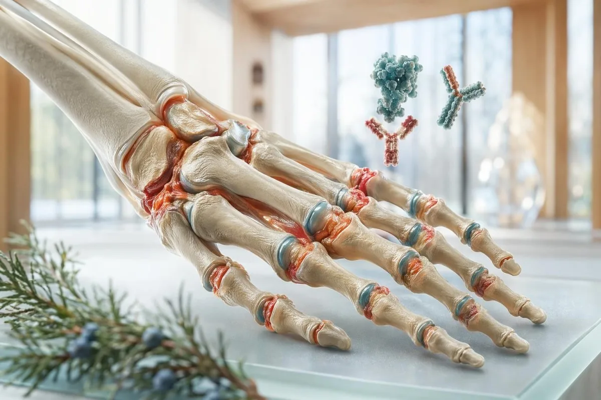
এটা নেগেটিভ ৰিউমেটয়েড ফেক্টৰ থকাটো আশ্বস্তকাৰী অনুভৱ হ’ব পাৰে, কিন্তু ই কেৱল ৰিউমেটয়েড আৰ্থ্ৰাইটিছৰ ধাঁধাটোৰ এটা অংশহে। ডায়াগন’ছিছ প্ৰায়ে anti-CCP, প্রদাহজনিত সূচক, ইমেজিং আৰু ফুলা গাঁঠিৰ ধৰণৰ ওপৰত নিৰ্ভৰ কৰে।.


- ৰিউমেটয়েড ফেক্টৰ নেগেটিভ সাধাৰণতে মানে RF লেবৰেটৰিৰ কাট-অফৰ তলত থাকে, বহু সময় <14 IU/mL, কিন্তু ই ৰিউমেটয়েড আৰ্থ্ৰাইটিছক নাকচ নকৰে।.
- seronegative ৰিউমেটয়েড আৰ্থ্ৰাইটিছ প্ৰায় 20–30% RA কেছৰ অংশ, বিশেষকৈ ৰোগৰ আৰম্ভণিৰ সময়ত।.
- Anti-CCP ৰিউমেটয়েড ফেক্টৰতকৈ অধিক নিৰ্দিষ্ট; RF নেগেটিভ থাকিলেও পজিটিভ ফলাফলে শক্তিশালীভাৱে RA সমৰ্থন কৰে।.
- anti-CCP নেগেটিভ RA তেতিয়াও ডায়াগন’ছিছ কৰিব পাৰি, যেতিয়া স্থায়ী সৰু গাঁঠিৰ synovitis, ইমেজিংৰ পৰিৱর্তন আৰু প্রদাহজনিত সূচকসমূহে সেই ধৰণৰ সৈতে মিল খায়।.
- ESR আৰু CRP প্রাৰম্ভিক RA-ত CRP স্বাভাৱিক হ’ব পাৰে; <5 mg/L আৰু বয়স-সমন্বয় কৰা ESR-ৰ ভিতৰত থাকিলে সক্ৰিয় গাঁঠিৰ ৰোগক বাদ নিদিয়ে।.
- ৬ সপ্তাহৰ ওপৰত স্থায়ী হোৱা লক্ষণসমূহ পুৱা কঁপনি (morning stiffness) >30–60 মিনিট আৰু MCP, PIP, কবজি বা MTP গাঁঠিত ফুলা থাকিলে ৰিউমেট’লজি পৰ্যালোচনাৰ বাবে উৎসাহিত কৰিব লাগে।.
- চিত্ৰায়ন (Imaging) আল্ট্ৰাছাউণ্ড বা MRI-এ X-ray-ত erosion দেখা দিয়াৰ আগতেই synovitis দেখুৱাব পাৰে, যিটো বিশেষকৈ seronegative ৰোগত উপযোগী।.
- পুনৰ পৰীক্ষা লক্ষণসমূহ বিকশিত হ’লে, প্ৰথম পৰীক্ষা অতি আৰম্ভণিত হ’লে, বা পৰীক্ষাৰ ফলাফল পৰীক্ষণৰ সৈতে মিল নাখালে উপযোগী; সাপ্তাহিকভাৱে পুনৰাবৃত্তি কৰাটো বিৰলভাৱে সহায় কৰে।.
- চিকিৎসা সময় গুৰুত্বপূৰ্ণ কাৰণ স্থায়ী ফুলা আৰম্ভ হোৱাৰ পিছৰ প্ৰথম ১২ সপ্তাহত আৰম্ভণিৰ inflammatory arthritis আটাইতকৈ বেছি চিকিৎসাযোগ্য।.
নেগেটিভ ৰিউমেটয়েড ফেক্টৰ থাকিলেও ৰিউমেটয়েড আৰ্থ্ৰাইটিছ থাকিব পাৰেনে?
হয়। এটা নেগেটিভ anti-CCP ৰিউমেট’ইড আৰ্থ্ৰাইটিছ (rheumatoid arthritis) বাদ নিদিয়ে; RA থকা প্ৰায় 20–30% লোক ডায়াগন’ছিছৰ সময়ত RF-negative হয়, আৰু কিছুমান আজীৱন negative হৈ থাকে। চিকিৎসকে anti-CCP, ESR/CRP, গাঁঠিৰ ফুলাৰ ধৰণ, আল্ট্ৰাছাউণ্ড বা MRI, আৰু ৬ সপ্তাহৰ ওপৰৰ লক্ষণৰ স্থায়িত্ব ব্যৱহাৰ কৰি তথাপিও RA ডায়াগন’ছ কৰিব পাৰে।.
মই আটাইতকৈ বেছি দেখা ভুলটো হ’ল “negative”ক “অসম্ভৱ” বুলি ধৰা। আমাৰ 2M+ আপলোড কৰা লেবৰেটৰী ৰিপ’ৰ্টৰ বিশ্লেষণত দেখা গৈছে, ৰোগীসকলে সাধাৰণতে 14 IU/mL-ৰ তলৰ RF-এ RA আলোচনাৰ অন্ত পেলায় বুলি ধৰি লয়; কিন্তু ক্লিনিকেলভাৱে ই কেৱল সম্ভাৱনা কমায় আৰু আমাক ধৰণটো আৰু বেছি ভালদৰে চাবলৈ বাধ্য কৰে।.
Kantesti হৈছে এটা AI ৰক্ত পৰীক্ষা বিশ্লেষক (blood test analyzer) যিয়ে anti-CCP কাষত RF, CRP, ESR, CBC আৰু medication-safety লেবৰেটৰীসমূহো পঢ়ে—এটা একক “হয়/নহয়” ফ্লেগ হিচাপে নহয়। আপুনি যদি RF assay-ৰ নিজস্ব গভীৰ কাৰিকৰী দিশ (mechanics) জানিব বিচাৰে, আমাৰ rheumatoid factor পৰীক্ষা গাইডে false positives, false negatives আৰু লেব কাটঅফসমূহ বিশদভাৱে ব্যাখ্যা কৰে।.
মই Thomas Klein, MD, আৰু ক্লিনিকত মই এটা একক নেগেটিভ RF-তকৈও বেছি চিন্তা কৰোঁ—ফুলা কবজি (swollen wrist) আৰু ৪৫ মিনিট পুৱা কঁপনি (morning stiffness) থাকিলে। কাৰণ সৰল: RA প্ৰথমে এটা clinical inflammatory arthritis, আৰু দ্বিতীয়তে এটা antibody pattern।.
আমাৰ সংস্থাটোৰ বিৱৰণ আছে আমাৰ কোম্পানী পেজত, কিন্তু চিকিৎসা নীতিটো যিকোনো চফ্টৱেৰৰ আগতেই পুৰণি: স্থায়ী synovitis-এ এটা ডায়াগন’ছিছৰ প্ৰয়োজন, প্ৰথম antibody ফলাফল শান্ত (quiet) থাকিলেও।.
ৰিউমেটয়েড ফেক্টৰ নেগেটিভ বুলি আচলতে কি বুজায়?
A ৰিউমাটয়েড ফ্যাক্টর নেগেটিভ ফলাফল মানে RF-ৰ ঘনত্ব সেই লেবৰেটৰীৰ positive cutoff-ৰ তলত, সাধাৰণতে <14 IU/mL বা <20 IU/mL (পদ্ধতি অনুসৰি) হয়। ইয়াৰ অৰ্থ এইটো নহয় যে immune system স্বাভাৱিক, আৰু ই আৰম্ভণিৰ বা seronegative rheumatoid arthritis-ও বাদ নিদিয়ে।.
Rheumatoid factor সাধাৰণতে IgG-ৰ Fc অংশৰ বিৰুদ্ধে নিৰ্দেশিত এটা IgM antibody। বহু লেবৰেটৰীয়ে RF-কে IU/mL-ত ৰিপ’ৰ্ট কৰে, কিন্তু cutoff পদ্ধতি-নিৰ্দিষ্ট; দুটা ৰিপ’ৰ্ট বেলেগ দেখা যাব পাৰে কাৰণ এটা বিশ্লেষকে latex agglutination ব্যৱহাৰ কৰে আৰু আন এটা ব্যৱহাৰ কৰে nephelometry বা immunoturbidimetry।.
এটা সাধাৰণ ব্যৱহাৰিক cutoff হ’ল RF <14 iuml as negative, 14–30 low-positive,and>৩০–৪০ IU/mL স্পষ্টভাৱে পজিটিভ বুলি ধৰা হয়, কিন্তু কিছুমান ইউৰোপীয় লেবোৰেটৰীয়ে ব্যৱহাৰ কৰে <20 iuml. if your result changed after switching labs, check units and methods before assuming biology changed; our unit changes এই লেখাটোৱে এই একে ফাঁদটো সামৰি লয়।.
RF ৰ RA-নিৰ্দিষ্টতা নাই। কম-পজিটিভ ৰিউমেটয়েড ফ্যাক্টৰ Sjögren’s disease, hepatitis C, ক্ৰনিক ফুসফুসৰ ৰোগ, subacute bacterial endocarditis আৰু RA নথকা বয়স্ক লোকৰ 5–10% ত দেখা যাব পাৰে।.
RF-নেগেটিভ RA “সংজ্ঞা অনুসৰি মৃদু” নহয়। মোৰ অভিজ্ঞতাত, RF-নেগেটিভ ৰোগ বহু সময়ত পিছত ধৰা পৰে, কিয়নো প্ৰথম রিপোর্টখন মিছাকৈ শান্তিদায়ক যেন লাগে, আৰু বিলম্বে কেৱল এন্টিবডিৰ অৱস্থাতকৈ বেছি গুৰুত্ব পাব পাৰে।.
RF নেগেটিভ হ’লে anti-CCP এ কেনেকৈ ছবিখন সলনি কৰে
Anti-CCP ৰিউমেটয়েড ফ্যাক্টৰৰ তুলনাত বহু অধিক তীক্ষ্ণভাৱে ৰিস্ক নিৰ্ণয় কৰিব পাৰে, কিয়নো ই RA-ৰ বাবে অত্যন্ত নিৰ্দিষ্ট। পজিটিভ anti-CCP ফলাফল, বহু সময়ত 20 U/mL-ৰ ওপৰত (পৰীক্ষা অনুসৰি), RF নেগেটিভ থাকিলেও ৰিউমেটয়েড আৰ্থ্ৰাইটিছক দৃঢ়ভাৱে সমৰ্থন কৰে।.
Anti-CCP-ক ACPA বুলিও কোৱা হয়, বা anti-citrullinated protein antibody। Nishimura et al. 2007 Annals of Internal Medicine-ৰ মেটা-এনালাইছিছত, anti-CCP-ৰ RA-ৰ বাবে প্ৰায় 67% sensitivity আৰু প্ৰায় 95% specificity আছিল, আনহাতে ৰিউমেটয়েড ফ্যাক্টৰ কম নিৰ্দিষ্ট আছিল (Nishimura et al., 2007)।.
বাস্তৱ জীৱনত এই নিৰ্দিষ্টতাই গুৰুত্বপূৰ্ণ। ৩৮ বছৰীয়া এজনৰ MCP গাঁঠি ফুলা, ৭০ মিনিটলৈকে চলা পুৱা কঁপনি, RF <10 IU/mL আৰু anti-CCP 86 U/mL থাকিলে কেৱল RF 22 IU/mL থকা অস্পষ্ট বিষত ভোগা এজনতকৈ RA-ৰ সৈতে বহু বেছি মিল থাকে।.
Anti-CCP টাইটাৰসমূহ লেবৰ মাজত একে ধৰণে সম্পূৰ্ণভাৱে বিনিময়যোগ্য নহয়। এটা পৰীক্ষাই >20 U/mL পজিটিভ বুলি ক’ব পাৰে, আন এটা পৰীক্ষাই >7 U/mL ব্যৱহাৰ কৰিব পাৰে, সেয়ে ব্যাখ্যা ৰিপ’ৰ্টত ছপা হোৱা reference range অনুসৰণ কৰিব লাগে; আমাৰ anti-CCP পৰীক্ষা পজিটিভ স্তৰসমূহ আৰু ভৱিষ্যৎ RA-ৰ ৰিস্ক ভাঙি দেখুৱায়।.
anti-CCP নেগেটিভ RA সম্ভৱ হৈ থাকে। লেবেল সাধাৰণতে মানে দুয়োটা—RF আৰু anti-CCP—নেগেটিভ, কিন্তু পৰীক্ষা আৰু ইমেজিং-এ স্থায়ী inflammatory synovitis দেখুৱায় যি RA-ৰ দৰে আচৰণ কৰে।.
seronegative RA ত ESR আৰু CRP এ কি দেখুৱাব পাৰে
ESR আৰু CRP ই systemic inflammation জুখে, নিজেই ৰিউমেটয়েড আৰ্থ্ৰাইটিছ নহয়। CRP বহু সময়ত 5 mg/L-ৰ তলত স্বাভাৱিক থাকে আৰু ESR বয়স আৰু লিংগ-নিৰ্ভৰশীল, সেয়ে স্বাভাৱিক inflammatory marker-এ seronegative rheumatoid arthritis নাকচ নকৰে।.
CRP দ্রুত বৃদ্ধি পায়, অধিকাংশ সময়েই কোনো প্রদাহজনক উদ্দীপনা ঘটার ৬–৮ ঘণ্টার মধ্যেই, এবং প্রদাহ থিতিয়ে এলে ESR-এর তুলনায় দ্রুত কমে। ESR ধীর, এবং ৰক্তাল্পতা, গর্ভাবস্থা, কিডনি ৰোগ, বয়স্ক বয়স আৰু উচ্চ ইমিউনোগ্লোবুলিনে ESR বৃদ্ধি পেতে পারে।.
ESR-এর এটা বাস্তৱসম্মত ঊর্ধ্বসীমা অনুমান হলো—পুৰুষৰ বাবে বয়স ভাগ ২, আৰু মহিলাৰ বাবে বয়স প্লাছ ১০ ভাগ ২; যদিও লেবৰেটৰিগুলো নিজৰ সময়সীমা ব্যৱহাৰ কৰে। ESR 34 mm/h থকা ৬২ বছৰ বয়সী এগৰাকী মহিলাৰ ক্ষেত্ৰত বয়স-সমন্বয়কৃত ঊর্ধ্বসীমাৰ ওচৰত হ’ব পাৰে, কিন্তু একে সংখ্যাটো ২৪ বছৰ বয়সী পুৰুষৰ ক্ষেত্ৰত অধিক মনোযোগৰ যোগ্য।.
হতাশাজনক অংশটো হলো—কিছুমান সক্রিয় RA থকা ৰোগীৰ CRP <5 mgl and esr under 20 mmh. if the joint exam shows true swelling, our প্রদাহৰ মাপকাঠী গাইড উপকাৰী, কিন্তু পৰীক্ষাই শেষত জিতে।.
১০০ mg/Lৰ ওপৰৰ CRP অনিয়ন্ত্রিত/অসুবিধাহীন আৰম্ভণিৰ RA-ত অস্বাভাবিক, আৰু চিকিৎসকসকলে সংক্রমণ, ক্ৰিষ্টাল আৰ্থ্রাইটিছ, ভাস্কুলাইটিছ বা অন্য কোনো প্রদাহজনক চালক খুঁজিবলৈ উচিত। এই সংখ্যাবোৰৰ ক্ষেত্ৰত চেকবক্সতকৈ প্ৰসংগ অধিক গুৰুত্বপূৰ্ণ।.
RF নেগেটিভ হ’লে কোনবোৰ লক্ষণৰ ধৰণে আটাইতকৈ বেছি গুৰুত্ব পায়?
RF-নেগেটিভ RA আটাইতকৈ সন্দেহজনক হয় যেতিয়া ফুলা ৬ সপ্তাহৰ অধিক সময় ধৰি সৰু গাঁটসমূহত সমমিতভাৱে (symmetrically) প্ৰভাৱিত কৰে। ৩০–৬০ মিনিটৰ অধিক সময় ধৰি থকা পুৱা কটকটনি (morning stiffness), ফুলা MCP/PIP গাঁট, কব্জিৰ (wrist) জড়িততা আৰু MTP-ত কোমলতা—সাধাৰণ গাঁটব্যথাৰ তুলনাত অধিক শক্তিশালী সূত্ৰ।.
মই এতিয়াও এটা ৪১ বছৰ বয়সী শিক্ষকক মনত ৰাখোঁ, যাৰ RF <10 IU/mL দুবাৰ হৈছিল আৰু কোৱা হৈছিল যে তেওঁ “সম্ভৱত মানসিক চাপত” আছে। তেওঁৰ সূত্ৰটো লেবৰেটৰিটো নাছিল; তেওঁ দ্বিতীয় কাপ কফি খোৱাৰ আগলৈকে মুঠি (fist) বানাব নোৱাৰিছিল, আৰু পৰীক্ষাত দুয়োখন কব্জি দৃশ্যমানভাৱে ফুলা দেখা গৈছিল।.
RA সাধাৰণতে MCP গাঁট, PIP গাঁট, কব্জি আৰু MTP গাঁটক লক্ষ্য কৰে, আনহাতে ক্লাছিক অষ্টিঅ’আর্থ্রাইটিছ (osteoarthritis) সাধাৰণতে DIP গাঁট, বুঢ়া আঙুলৰ (thumb) মূল অংশ আৰু হাঁঠুক লক্ষ্য কৰে। যদি আপুনি এপয়েন্টমেণ্টৰ আগতে লক্ষণসমূহ মিলাই চাইছে, আমাৰ গাঁঠিৰ বিষ লেব লেখাটোৱে দেখায় কোনবোৰ পৰীক্ষাই প্রদাহজনক বনাম যান্ত্ৰিক (mechanical) ব্যথা পৃথক কৰাত সহায় কৰে।.
১০ মিনিটৰ কম সময়ৰ পুৱা কটকটনিয়ে RA-ৰ সম্ভাৱনা কমায়, যদিও ই একেবাৰে নিৰ্দিষ্ট নহয়। ৬০ মিনিটৰ ওপৰৰ কটকটনি, চলাচলেৰে উন্নতি হোৱা ফুলা, আৰু দুয়োখন হাতে নিশাৰ বেদনা—এইবোৰে সম্ভাৱনাক পুনৰ প্রদাহজনক আৰ্থ্রাইটিছৰ দিশে ঠেলি দিয়ে।.
এটা সৰু বেডসাইড কৌশল: আঙুলত আঙঠিৰ টান লাগিছে নেকি, গাঁটৰ (knuckle) “উপত্যকা” কমি গৈছে নেকি, বা আগফালে (forefoot) জোতা খন সংকীৰ্ণ লাগিছে নেকি চাওক। ৰোগীসকলে বহু সময়ত “ফুলা” বুলি কোৱাৰ ২–৩ মাহ আগতেই এইবোৰ কথা কয়।”
তেজৰ পৰীক্ষা নেগেটিভ হ’লেও ইমেজিং কিদৰে RA নিশ্চিত কৰিব পাৰে
আল্ট্ৰাছাউণ্ড আৰু MRI-এ X-ray-এ স্থায়ী ক্ষতি দেখোৱাৰ আগতেই synovitis দেখুৱাব পাৰে। সেরোনেগেটিভ রিউমাটয়েড আর্থ্রাইটিস, ৰ ক্ষেত্ৰত, ইমেজিং বহু সময়তেই অস্পষ্ট লক্ষণক এটা যুক্তিসংগত (defensible) প্রদাহজনক আৰ্থ্রাইটিছ নিৰ্ণয়লৈ ৰূপান্তৰিত কৰা প্ৰমাণ হয়।.
সাধাৰণ X-ray-সমূহ বেছলাইন ক্ষতি (baseline damage)ৰ বাবে উপকাৰী, কিন্তু আৰম্ভণিৰ RA-ত কেইমাহমানলৈ ফিল্ম স্বাভাবিক থাকিব পাৰে। power Doppler-সহ আল্ট্ৰাছাউণ্ডে সক্রিয় synovial টিস্যুৰ সঁহাৰি, tenosynovitis আৰু সৰু erosions ধৰা পেলাব পাৰে যিবোৰ সাধাৰণ হাতৰ X-ray-ত অদৃশ্য থাকে।.
Annals of the Rheumatic Diseases-ত Colebatch et al. এ EULAR ইমেজিং পৰামৰ্শসমূহে সমর্থন কৰিছে—যেতিয়া ক্লিনিকেল পৰীক্ষা আৰু conventional radiography অনিশ্চিত হয়, তেতিয়া ultrasound আৰু MRI ব্যৱহাৰ কৰা উচিত (Colebatch et al., 2013)। বিশেষকৈ উপকাৰী লাগে যেতিয়া RF আৰু anti-CCP দুয়োটাই নেগেটিভ, কিন্তু গাঁটৰ বিন্যাসটো ক্লাছিক।.
MRI-এ হাড়ৰ মজ্জাৰ শোথ (bone marrow edema) দেখুৱাই আৰু এটা স্তৰ যোগ কৰে, যিয়ে পিছলৈ ক্ষয়জনিত (erosive) পৰিৱৰ্তন পূৰ্বানুমান কৰিব পাৰে। কিন্তু বাধা হৈছে অভিগম্যতা আৰু খৰচ; সকলো ফুলা আঙুলৰ বাবে MRI লাগে বুলি নহয়, কিন্তু স্থায়ী কবজি বা MCP synovitis আৰু এন্টিবডি নেগেটিভ থাকিলে বহু সময়ত ইমেজিং সম্পৰ্কে আলোচনা কৰাটো যোগ্য।.
এটা বহল “autoimmune screen” এ লক্ষ্যভিত্তিক ইমেজিংক প্ৰতিস্থাপন কৰিবলৈ নিদিব। আমাৰ autoimmune panel সীমিত কিয় পেনেলে এনে ৰোগ মিছ কৰিব পাৰে যাক এটা ভাল ultrasound-এ দেখা পায়—সেই কথা গাইড-এ ব্যাখ্যা কৰে।.
RF পজিটিভ নোহোৱাকৈ ডাক্টৰসকলে RA ক্ৰাইটেৰিয়া কেনেকৈ প্ৰয়োগ কৰে
২০১০ ACR/EULAR RA classification স্ক’ৰ ১০ ৰ ভিতৰত ৬ বা তাতকৈ অধিক পইণ্টত RA-এ শ্রেণীবদ্ধ কৰিব পাৰে, ৰিউমেটয়েড ফেক্টৰ পজিটিভ নাথাকিলেও। এন্টিবডি নেগেটিভ হ’লে joint count, লক্ষণৰ স্থায়িত্ব আৰু ESR/CRP-এ স্ক’ৰ পূৰণ কৰিব পাৰে।.
Aletaha et al. ২০১০ criteria-এ জড়িত গাঁঠ, serology, লক্ষণৰ স্থায়িত্ব আৰু acute-phase reactants (Aletaha et al., 2010) ৰ বাবে পইণ্ট দিয়ে। Serology কেৱল এটা ড’মেইন; এজন ৰোগীয়ে সকলো এন্টিবডি পইণ্ট হেৰুৱালেও বহুতো সৰু গাঁঠৰ জড়িততা আৰু ESR বা CRP অস্বাভাবিক হ’লে শ্রেণীবিভাগত উপনীত হ’ব পাৰে।.
উদাহৰণস্বৰূপে, কমেও এটা সৰু গাঁঠসহ ১০-তকৈ অধিক জড়িত গাঁঠে ৫ পইণ্ট আনে, ৬ সপ্তাহ বা তাতকৈ বেছি দিন ধৰি থকা লক্ষণে ১ পইণ্ট আনে, আৰু অস্বাভাবিক ESR বা CRP-এ ১ পইণ্ট আনে। এই মুঠ যোগফল RF বা anti-CCP নোহোৱাকৈও ইতিমধ্যে ৭ হয়।.
Classification criteria bedside diagnosis-ৰ সৈতে একে নহয়। ৰিউমেট’লজিষ্টে যদি পেটাৰ্নটো বিশ্বাসযোগ্য হয় তেন্তে আনুষ্ঠানিক স্ক’ৰ আগতেই ৰোগ নিৰ্ণয় কৰি চিকিৎসা আৰম্ভ কৰিব পাৰে, বা যদি ফুলা বস্তুগতভাৱে উপস্থিত নাথাকে তেন্তে অপেক্ষা কৰিব পাৰে।.
ৰোগীয়ে যেতিয়া পেটাৰ্ন চিনাক্ত নকৰাকৈ এটা একক flagged বা unflagged মান পঢ়ে তেতিয়াই সমস্যা হয়। আমাৰ তেজ পৰীক্ষাৰ সংখ্যা গাইড-এ দেখুৱায় কিয় এটা ক্লাষ্টাৰ (cluster) এটা একক মানতকৈ বেছি গুৰুত্বপূৰ্ণ।.
ৰিউমেটয়েড ফেক্টৰ বা anti-CCP কেতিয়া পুনৰ পৰীক্ষা কৰা উচিত?
লক্ষণসমূহ আৰম্ভণিৰ সময়ত, বিকাশমান (evolving) বা প্ৰথম ফলৰ সৈতে অসামঞ্জস্য (discordant) হ’লে পুনৰ RF বা anti-CCP পৰীক্ষা কৰাটো গুৰুত্বপূৰ্ণ। কেইদিনমানৰ ব্যৱধানত একে এন্টিবডি পৰীক্ষা পুনৰ কৰা সাধাৰণতে কম সহায়ক; সন্দেহ উচ্চ হৈ থাকিলে ৬–১২ সপ্তাহ বা ৩–৬ মাহৰ অন্তৰ ক্লিনিকেলভাৱে অধিক যুক্তিসংগত।.
Seroconversion হ’ব পাৰে, কিন্তু সাপ্তাহিকভাৱে ধাওয়া দি অনুসৰণ কৰিবলৈ যথেষ্ট সাধাৰণ নহয়। মই সাধাৰণতে RF আৰু anti-CCP পুনৰ কৰাৰ কথা ভাবোঁ যেতিয়া এজন ৰোগীক প্ৰথমবাৰ লক্ষণ আৰম্ভ হোৱাৰ সপ্তাহ ১–২ ত পৰীক্ষা কৰা হৈছিল, আৰু তাৰ পিছত সপ্তাহ ৮ বা ১২ ত স্পষ্টভাৱে ফুলা দেখা দিয়ে।.
নমুনা (sample) ভুলভাৱে হেণ্ডল কৰা হৈছিল, পৰীক্ষাৰ ফল পৰীক্ষা-নিৰীক্ষাৰ সৈতে তীব্ৰভাৱে বিৰোধী, বা লেব’ৰেটৰীয়ে অচিনাকি পদ্ধতি ব্যৱহাৰ কৰিছিল—এই ক্ষেত্ৰতো পুনৰ পৰীক্ষা কৰাটো যুক্তিসংগত। সাধাৰণ সময়-সম্পৰ্কীয় নীতিৰ বাবে আমাৰ পুনৰ অস্বাভাৱিক তেজ পৰীক্ষা গাইড-এ কেতিয়া পুনৰ পৰীক্ষাই সিদ্ধান্ত সলনি কৰে আৰু কেতিয়া কেৱল শব্দ (noise) সৃষ্টি কৰে—সেই কথা সামৰি দিয়ে।.
RF যদি <14 ৰ পৰা 18 IU/mL হয়—ই নিজে নিজাকৈ ডায়েগন’ষ্টিক নহয়। কবজি ফুলা থকা কোনো এজনৰ ক্ষেত্ৰত 120 U/mL ৰ এটা নতুন anti-CCP বেলেগ; সেই ফলৰ অৰ্থই সম্ভাৱনাক (probability) অৰ্থপূৰ্ণভাৱে সলনি কৰে।.
যদি আপুনি ইতিমধ্যে ষ্টেৰয়ড, NSAIDs বা disease-modifying drug লৈ আছে, তেন্তে ESR আৰু CRP কৃত্ৰিমভাৱে উন্নত যেন লাগিব পাৰে। এন্টিবডি পৰীক্ষা কম প্ৰভাৱিত হয়, কিন্তু ক্লিনিকেল ছবিখন (clinical picture) অস্পষ্ট হৈ পৰিব পাৰে।.
anti-CCP নেগেটিভ RA ৰ দৰে আৰু কি দেখা যায়?
anti-CCP নেগেটিভ RA সত্য, কিন্তু কেইবাটাও অৱস্থা ইয়াক অনুকৰণ (mimic) কৰিব পাৰে। Psoriatic arthritis, lupus, Sjögren’s disease, viral arthritis, gout, pseudogout, thyroid disease আৰু osteoarthritis—এই সকলোবোৰে নেগেটিভ ৰিউমেটয়েড ফেক্টৰসহ গাঁঠৰ বিষ (joint pain) সৃষ্টি কৰিব পাৰে।.
Psoriatic arthritis-এ DIP joints, tendon insertions, dactylitis বা গাঁঠৰ লক্ষণৰ পিছত দেখা দিয়া psoriasis ৰ ইতিহাস জড়িত কৰিব পাৰে। Lupus-এ বেদনাদায়ক ফুলা গাঁঠ সৃষ্টি কৰিব পাৰে, কিন্তু erosive RA-ৰ দৰে ক্ষতি সাধাৰণতে কম দেখা যায়, যদিহে overlap disease নাথাকে।.
Sjögren’s disease-এ RA নোহোৱাকৈও পজিটিভ RF সৃষ্টি কৰিব পাৰে, কিন্তু ই inflammatory arthritis-ৰ সৈতে সহ-অৱস্থান (coexist) কৰিবও পাৰে। শুকান চকু (dry eyes), শুকান মুখ (dry mouth), parotid ফুলা আৰু পজিটিভ SSA/SSB এন্টিবডি থাকিলে workup-টো পুনঃনিৰ্দেশ (redirect) কৰা উচিত।.
নেগেটিভ ANA-এ সকলো autoimmune disease নুই কৰিব নোৱাৰে, কিন্তু ই classic lupus-ৰ সম্ভাৱনা কম কৰে। লক্ষণসমূহ নেগেটিভ screening সত্ত্বেও স্থায়ী থাকিলে আমাৰ নেগেটিভ ANA গাইডে বৰ্ণনা কৰে যে চিকিৎসকে সাধাৰণতে পৰৱৰ্তীভাৱে কি কি পৰীক্ষা কৰে।.
বয়স্ক লোকৰ ক্ষেত্ৰত ক্ৰিষ্টাল আৰ্থ্ৰাইটিছ হৈছে এক মহান অনুকাৰী। CRP 80 mg/L থকা এটা গৰম, ফুলা কব্জি গাউট বা ছিউডোগাউট হ’ব পাৰে, আৰু আটাইতকৈ নিৰ্ণায়ক পৰীক্ষা আন এটা এন্টিবডি পেনেল নহৈ যৌথৰ ফ্লুইড মাইক্র’স্ক’পি হ’ব পাৰে।.
RF ৰ বাহিৰেও কোনবোৰ তেজৰ পৰীক্ষাই ডাক্টৰসকলক সিদ্ধান্ত ল’বলৈ সহায় কৰে?
চিকিৎসকে সাধাৰণতে RF-ৰ ব্যাখ্যা anti-CCP, ESR, CRP, CBC, লিভাৰ এনজাইম, কিডনি ফাংশন আৰু কেতিয়াবা ANA বা ইউৰিক এচিডৰ সৈতে কৰে। এই পৰীক্ষাসমূহে যৌথ পৰীক্ষাক প্ৰতিস্থাপন নকৰে, কিন্তু ইহঁতে প্রদাহ, ৰক্তহীনতা, ঔষধৰ সুৰক্ষা আৰু প্ৰতিদ্বন্দ্বী (competing) নিৰ্ণয়সমূহ উন্মোচন কৰে।.
এটা CBC-এ প্রদাহজনিত ৰক্তহীনতা দেখুৱাব পাৰে, সাধাৰণতে নৰ্ম’চাইটিক; মহিলাৰ ক্ষেত্ৰত হিম’গ্ল’বিন 12 g/dL-ৰ তলত বা পুৰুষৰ ক্ষেত্ৰত 13 g/dL-ৰ তলত থাকে। সক্ৰিয় প্রদাহৰ সময়ত প্লেটলেট 400 x 10^9/L-ৰ ওপৰলৈ উঠিব পাৰে, আৰু সেই প্লেটলেটৰ সূত্ৰটো কেতিয়াবা সকলো RF-ত মনোনিৱেশ কৰোঁতে এৰি দিয়া হয়।.
কম হিম’গ্ল’বিনৰ সৈতে উচ্চ ESR এটা বিশেষভাৱে উপযোগী সংযোজন, কাৰণ ৰক্তহীনতাই ESR বৃদ্ধি কৰিব পাৰে আৰু লগতে ক্ৰনিক প্রদাহজনিত বোজাও সংকেত দিব পাৰে। আমাৰ ESR আৰু ৰক্তহীনতা ব্যাখ্যা কৰে কিয় এই গোটটোৱে কেৱল এটা এটা ফলাফলতকৈ অধিক গুৰুত্ব দাবী কৰে।.
Methotrexate, leflunomide বা biologic থেৰাপিৰ আগতে baseline ALT, AST, albumin, creatinine আৰু eGFR গুৰুত্বপূৰ্ণ। creatinine-ভিত্তিক eGFR 60 mL/min/1.73 m²-ৰ তলত হ’লে ড’জিং সম্পৰ্কীয় আলোচনা সলনি হয় আৰু কেতিয়াবা ঔষধৰ পছন্দো সলনি হয়।.
Kantesti-ৰ neural network-এ এই মাৰ্কাৰসমূহক আমাৰ 15,000+ biomarker সংজ্ঞাসমূহৰ বিপৰীতে মানচিত্ৰ কৰে। বায়'মাৰ্কাৰ গাইড, কিন্তু আউটপুটে—প্ৰতিস্থাপন নহৈ—সেই চিকিৎসকক সহায় কৰিব লাগে যিয়ে আপোনাৰ যৌথসমূহ পৰীক্ষা কৰে।.
seronegative RA হলেও কিয় আৰম্ভণিৰ চিকিৎসা গুৰুত্বপূৰ্ণ
Seronegative RA-এ তথাপিও যৌথ ক্ষতি কৰিব পাৰে, সেয়েহে চিকিৎসা ৰিউমেটয়ড ফেক্টৰ পজিটিভ হ’বলৈ অপেক্ষা নকৰিব লাগে। বহু ৰিউমেট’লজি পথেই কেইসপ্তাহমানৰ ভিতৰত persistent inflammatory arthritis মূল্যায়ন কৰাৰ লক্ষ্য ৰাখে, কিয়নো প্ৰথম 12 সপ্তাহ এটা মূল্যৱান চিকিৎসা-সময় (treatment window)।.
Methotrexate সাধাৰণতে সপ্তাহত এবাৰকৈ 15 mg আৰম্ভ কৰা হয় আৰু উপযুক্ত হ’লে সপ্তাহে 20–25 mg লৈ সামঞ্জস্য কৰা হয়, সাধাৰণতে side effect কমাবলৈ folic acid-ৰ সৈতে। এই সংখ্যাসমূহ নিজে নিজে চিকিৎসা কৰাৰ পৰিকল্পনা নহয়; prescription লিখাৰ আগতে baseline CBC, ALT, AST আৰু creatinine কিয় গুৰুত্বপূৰ্ণ—সেই কাৰণেই এইবোৰ।.
চুটি সময়ৰ steroid course-এ ফুলা যৌথসমূহ দ্ৰুত শান্ত কৰিব পাৰে, কিন্তু ইহঁতে নিৰ্ণায়ক সূত্ৰসমূহো লুকুৱাব পাৰে আৰু CRP দমন কৰিব পাৰে। বিশেষজ্ঞৰ পৰ্যালোচনাৰ আগতে যদি steroid ব্যৱহাৰ কৰা হয়, মই পছন্দ কৰোঁ যে ড’জ আৰু সময় স্পষ্টকৈ ৰেকৰ্ড কৰা হওক।.
মনিটৰিং আকৰ্ষণীয় নহয়, কিন্তু ই ক্ষতি ৰোধ কৰে। আমাৰ ঔষধ নিৰীক্ষণ গাইডে ব্যাখ্যা কৰে কিয় কিছুমান disease-modifying drug আৰম্ভণিৰ আৰম্ভণিতে CBC আৰু লিভাৰ এনজাইম প্ৰায়ে প্ৰতি 2–4 সপ্তাহত পৰীক্ষা কৰা হয়, তাৰ পিছত স্থিৰ হ’লে ব্যৱধান বৃদ্ধি কৰা হয়।.
“seronegative” বুলি কেতিয়াও “erosion দেখা নোপোৱালৈ অপেক্ষা কৰা” বুজাব নালাগে। X-ray-ত erosion দেখা পোৱাৰ পিছত ৰোগে ইতিমধ্যে এটা ছাপ এৰি গৈছে।.
AI ব্যাখ্যাই কিদৰে RF-নেগেটিভ ধৰণসমূহ সংগঠিত কৰিব পাৰে
AI-ৰ ব্যাখ্যাই RF-negative লেবৰেটৰী পেটাৰ্নসমূহ সংগঠিত কৰাত সহায় কৰিব পাৰে, কিন্তু শাৰীৰিক যৌথ পৰীক্ষা নোহোৱাকৈ RA নিৰ্ণয় কৰিব নোৱাৰে। আটাইতকৈ সুৰক্ষিত ব্যৱহাৰ হ’ল RF, anti-CCP, ESR/CRP, CBC আৰু সুৰক্ষা-সম্পৰ্কীয় পৰীক্ষাসমূহক আপোনাৰ চিকিৎসকৰ বাবে এটা স্পষ্ট প্ৰশ্ন-তালিকাত সাৰাংশ কৰা।.
Kantesti হৈছে এটা AI blood test interpretation প্লেটফৰ্ম যিয়ে আপলোড কৰা লেবৰেটৰী PDF বা ফটো প্ৰায় 60 ছেকেণ্ডত প্ৰচেছ কৰে আৰু অসংগতি (contradictions) যেনে উচ্চ CRP আৰু ৰক্তহীনতাৰ সৈতে RF-negative ফলাফল—এইবোৰ হাইলাইট কৰে। এই workflow-ৰ পিছে থকা চিকিৎসা তত্ত্বাৱধান আমাৰ দলটোৱে প্ৰকৃত মেচিন বিকল হোৱাৰ তুলনাত প্ৰি-এনালাইটিকেল (pre-analytical) সমস্যাবোৰ বহু বেছি দেখা পায়।.
Kantesti AI-এ কোনো ৰোগীক ক’ব নোৱাৰে, “আপোনাৰ RA আছে।” ই অধিক সুৰক্ষিতভাৱে ক’ব পাৰে যে anti-CCP, ESR, CRP বা CBC পেটাৰ্নে উদ্বেগ তুলিলে <14 IU/mL-এ inflammatory arthritis বাদ নিদিয়ে। <14 IU/mL does not exclude inflammatory arthritis when anti-CCP, ESR, CRP or CBC patterns raise concern.
আমাৰ ইঞ্জিনিয়াৰিং দলে validation কাম প্ৰকাশ কৰিছে, য’ত এটা clinical benchmark আৰু এটা pre-registered যাচাই (validation) ডেটাসেট, কাৰণ লেবৰ ব্যাখ্যা কঠিন কেছৰ বিপৰীতে পৰীক্ষা কৰিব লাগিব, কেৱল সুন্দৰ উদাহৰণৰ বিপৰীতে নহয়।.
যদি আপোনাৰ ৰিপ’ৰ্ট এখন স্কেন কৰা ছবি হয়, তেন্তে ব্যৱহাৰিক সমস্যা প্ৰায়েই চিকিৎসাৰ তুলনাত পঢ়িব পৰা ক্ষমতা (readability) হয়। The PDF upload workflow ব্যৱহাৰ কৰে ব্যাখ্যা কৰে যে আমাৰ চিস্টেমে ফটো, ৰেঞ্জ, ফ্লেগ (flags) আৰু ইউনিট ৰূপান্তৰ (unit conversions) কেনেকৈ পৰিচালনা কৰে।.
নেগেটিভ RF থকাৰ পিছতো কেতিয়া ৰিউমেট’লজি যত্ন বিচাৰিব লাগে?
৬ সপ্তাহৰ অধিক সময় ধৰি গাঁঠিৰ ফুলা থাকিলে, পুৱা জড়তা (morning stiffness) ৩০–৬০ মিনিটৰ অধিক হলে, বা কেইবাটাও সৰু গাঁঠি জড়িত থাকিলে ৰিউমেট’লজি (rheumatology) যত্ন লওক। শাৰীৰিক ধৰণে যদি inflammatory arthritis সূচায়, তেন্তে negative rheumatoid factor এ ৰেফাৰেল (referral) পলম কৰাব নালাগে।.
একে সপ্তাহতে মূল্যায়ন যুক্তিসংগত, যদি জ্বৰসহ এটা গৰম (hot) একক গাঁঠি থাকে, তীব্ৰ বিষ (severe pain) থাকে, ভৰ দি থিয় হব নোৱাৰা হয়, বা CRP ১০০ mg/Lৰ ওপৰত থাকে—কাৰণ সংক্রমণ (infection) আৰু crystal arthritis-এ inflammatory arthritisৰ দৰে দেখা দিব পাৰে। চকুৰ বিষ (eye pain) ৰঙা (redness) সহ, নতুন বুকৰ লক্ষণ (new chest symptoms) বা স্নায়বিক লক্ষণ (neurologic signs) থাকিলেও তৎক্ষণাৎতা (urgency) সলনি হয়।.
দুয়োখন হাতৰ কবজি (bilateral wrist) বা MCP ফুলা, নতুন গুটি (new nodules), উচ্চ ESR সহ ব্যাখ্যাতীত ৰক্তহীনতা (unexplained anemia), বা anti-CCP পজিটিভ হ’লে নিয়মীয়া কিন্তু তৎক্ষণাৎ ৰেফাৰেল উপযুক্ত। যদি সুবিধা (access) ধীৰ হয়, তেন্তে ফুলা গাঁঠিৰ গণনা (swollen joint count) নথিভুক্ত কৰা এটা ভাল প্ৰাইমাৰী-কেৰ (primary-care) নোটে মাহৰ পিছত মাহৰ বৃত্তাকাৰ পৰীক্ষা (circular testing) ৰোধ কৰিব পাৰে।.
Virtual care এ লেবৰ পেটাৰ্ন (lab patterns) triage কৰিব পাৰে, কিন্তু synovitis স্পৰ্শ কৰি (palpate) পৰীক্ষা কৰিব নোৱাৰে। Our টেলিহেলথ পৰ্যালোচনা ব্যাখ্যা কৰে যে কেতিয়া দূৰৰ পৰা লেবৰ পৰ্যালোচনা (remote lab review) সহায়ক হয় আৰু কেতিয়া ব্যক্তিগত (in-person) পৰীক্ষা পৰৱৰ্তী সুৰক্ষিত পদক্ষেপ।.
ফ্লেয়াৰ (flares) সময়ত দৃশ্যমান ফুলাৰ ফটো তুলক, বিশেষকৈ যদি অ্যাপইণ্টমেণ্টৰ দিনত সেইটো নাইকিয়া হৈ যায়। ই সহজ যেন লাগে, কিন্তু সেই ফটোৱে ভিজিটটো সলনি কৰিব পাৰে।.
নেগেটিভ RF ৰ পিছত আপোনাৰ ডাক্টৰক কি সুধিব লাগে?
negative RFৰ পিছত, আপোনাৰ লক্ষণবোৰ inflammatory arthritisৰ লগত খাপ খায় নেকি সুধক, anti-CCP পৰীক্ষা কৰা হৈছিল নেকি সুধক, ESR/CRP আৰু CBC-এ inflammatory সমৰ্থন কৰে নেকি সুধক, আৰু ultrasound উপযুক্ত নেকি সুধক। ২৬ মে’ ২০২৬ তাৰিখলৈ, কেৱল এটা ৰক্ত পৰীক্ষাই নিজে নিজাকৈ RA নিৰাপদভাৱে বাদ দিব নোৱাৰে।.
এটা উপযোগী অ্যাপইণ্টমেণ্ট প্ৰশ্ন হ’ল: “আপুনি objective swelling দেখিছিল নে কেৱল বিষ (tenderness)?” কেৱল tenderness বহুতো কাৰণৰ পৰা আহিব পাৰে; >৬ সপ্তাহ ধৰি থকা objective synovitis হৈছে সিদ্ধান্ত লোৱাৰ মূল মোড় (fork in the road)।.
কেৱল “negative” বুলি নক’ৰি RFৰ সঠিক মান (exact value) আৰু ইউনিট (unit) বিচাৰক। RF <10 IU/mL, ১৪ৰ কাটঅফৰ ওচৰত RF 13.8 IU/mL, আৰু ভিন্ন assay-ত ৰিপ’ৰ্ট কৰা RF-এ একে ধৰণৰ অৰ্থ বহন নকৰে।.
Kantesti হৈছে এটা AI লেব টেষ্ট ব্যাখ্যা সেৱা (AI lab test interpretation service) যিয়ে আপোনাক এটা সংক্ষিপ্ত লেব সারাংশ (lab summary) প্ৰস্তুত কৰাত সহায় কৰিব পাৰে, কিন্তু চূড়ান্ত ডায়াগন’ছিছ সেই চিকিৎসকৰ (clinician) সৈতে থাকে যিয়ে গাঁঠিবোৰ পৰীক্ষা কৰে। Thomas Klein, MD আৰু আমাৰ চিকিৎসক পৰ্যালোচকসকলে এই সীমাৰেখা (boundary) through the মেডিকেল এডভাইজাৰী ব’ৰ্ড.
মূল কথা: rheumatoid factor negative এটা তথ্য বিন্দু (data point), ৰায় (verdict) নহয়। যদি হাত, কবজি বা ভৰিৰ গাঁঠিত ফুলা চলি থাকে, তেন্তে কোনোবাই সম্পূৰ্ণ পেটাৰ্নটো ব্যাখ্যা নকৰালৈকে সুধি থাকক।.
সঘনাই সোধা প্ৰশ্ন
ৰিউমেটয়েড ফ্যাক্টৰ নেগেটিভ হ’লেও কি ৰিউমেটয়েড আৰ্থ্ৰাইটিছ থাকিব পাৰে?
হয়, তবুও ৰিউমেটইড আৰ্থ্ৰাইটিছ (Rheumatoid arthritis) থাকিব পাৰে যদি ৰিউমেটইড ফেক্টৰ (rheumatoid factor) নেগেটিভ হয়। RA থকা লোকৰ প্ৰায় 20–30% জনে ডায়াগন’ছিছৰ সময়ত ছেৰ’নেগেটিভ (seronegative) হয়, অৰ্থাৎ RF নেগেটিভ আৰু কেতিয়াবা anti-CCP-ও নেগেটিভ হয়। চিকিৎসকে এই ক্ষেত্ৰসমূহ স্থায়ী গাঁঠৰ ফুলা (persistent joint swelling), ৬ সপ্তাহতকৈ অধিক দিন ধৰি থকা লক্ষণ (symptoms lasting more than 6 weeks), ESR/CRP, anti-CCP, আৰু আল্ট্ৰাছাউণ্ড বা MRI দৰে ইমেজিং (imaging) ব্যৱহাৰ কৰি চিনাক্ত কৰে।.
সেৰ’নেগেটিভ ৰিউমেটইড আৰ্থ্ৰাইটিছ মানে কি?
ছেৰ’নেগেটিভ ৰিউমেটয়েড আৰ্থ্ৰাইটিছ মানে ক্লিনিকেল ৰোগটোৱে RAৰ দৰে আচৰণ কৰে, কিন্তু সাধাৰণ এন্টিবডি, বিশেষকৈ ৰিউমেটয়েড ফেক্টৰ আৰু বহু সময়ত anti-CCP, নেগেটিভ থাকে। বহু লেবৰেটৰীয়ে RF-নেগেটিভ বুলি প্ৰায় ১৪ IU/mL তলত সংজ্ঞায়িত কৰে, যদিও কাট-অফসমূহ ভিন্ন হ’ব পাৰে। ছেৰ’নেগেটিভ RA-এ মূল্যায়ন আৰু চিকিৎসা নকৰিলে তথাপিও ছাইনোভাইটিছ, পুৱা কঁকাল-জঠৰতা, ক্ষয় (erosions) আৰু অক্ষমতা সৃষ্টি কৰিব পাৰে।.
অ্যান্টি-CCP নেগেটিভ RA বাস্তৱ হ’ব পাৰেনে?
হয়, anti-CCP নেগেটিভ RA বাস্তৱ হ’ব পাৰে যেতিয়া গাঁঠিৰ ধৰণ আৰু ইমেজিংয়ে প্ৰদাহজনিত আৰ্থ্ৰাইটিছক সমৰ্থন কৰে। Anti-CCP অতি উচ্চ নিৰ্দিষ্টতা সম্পন্ন, এটা ডাঙৰ মেটা-এনালাইছিছত প্ৰায় 95%, কিন্তু ইয়াৰ সংবেদনশীলতা প্ৰায় দুই-তৃতীয়াংশৰ ওচৰতহে, সেয়ে ই RA ৰ এক তাৎপৰ্যপূর্ণ সংখ্যালঘু কেছ মিছ কৰে। Anti-CCP নেগেটিভ RA সাধাৰণতে psoriatic arthritis, lupus, viral arthritis, gout আৰু osteoarthritis দৰে অনুকৰণকাৰী (mimics) ৰ ক্ষেত্ৰত অধিক সাৱধানতাৰে বাদ দিয়া প্ৰয়োজন হয়।.
ৰিউমেটয়েড ফেক্টৰ নেগেটিভ ফলাফলৰ পিছত পুনৰ পৰীক্ষা কৰা উচিত নে?
ৰিউমেটয়েড ফেক্টৰ পুনৰ পৰীক্ষা কৰিব পাৰি যেতিয়া লক্ষণসমূহ নতুনকৈ আৰম্ভ হয়, বিকশিত হয় বা প্ৰথম পৰীক্ষাৰ ফলৰ সৈতে অসামঞ্জস্য দেখা যায়, কিন্তু প্ৰতি কেইদিনমানৰ মূৰে মূৰে পুনৰ পৰীক্ষা কৰাটো সাধাৰণতে বেছি উপকাৰী নহয়। ব্যৱহাৰিক অন্তৰাল প্ৰায়ে ৬–১২ সপ্তাহ বা ৩–৬ মাহ হ’ব পাৰে, যদি গাঁঠিৰ ফুলি উঠা স্পষ্ট হৈ পৰে। প্ৰথম পৰীক্ষা অতি সোনকালে কৰা হৈছিল আৰু সন্দেহ উচ্চ হৈ থাকিলে anti-CCP পুনৰ পৰীক্ষা কৰাও সহায়ক হ’ব পাৰে।.
ৰিউমাটয়েড আৰ্থ্ৰাইটিছত ESR আৰু CRP স্বাভাৱিক হ’ব পাৰেনে?
হয়, ৰিউমাটয়েড আৰ্থ্ৰাইটিছত ESR আৰু CRP স্বাভাৱিক হ’ব পাৰে, বিশেষকৈ আৰম্ভণিৰ ৰোগত বা কেইটামান গাঁঠিত সীমাবদ্ধ ৰোগত। CRP সাধাৰণতে 5 mg/L ৰ তলত স্বাভাৱিক বুলি ধৰা হয়, আৰু ESR বয়স, লিংগ আৰু ৰক্তহীনতা (anemia)ৰ ওপৰত যথেষ্টভাৱে নিৰ্ভৰ কৰে। স্বাভাৱিক প্রদাহজনিত সূচক (inflammatory markers)ে সিস্টেমিক প্রদাহৰ সম্ভাৱনা কমায়, কিন্তু বস্তুগতভাৱে ফুলা গাঁঠ (objectively swollen joints) নথকাটো নিশ্চিতভাৱে নাকচ নকৰে।.
কোনবোৰ লক্ষণে RF নেগেটিভ হ’লেও RA হোৱাৰ সম্ভাৱনা বেছি কৰে?
লক্ষণসমূহে শৰীৰৰ দুয়োফাল জড়িত থকা, MCP, PIP, কবজি বা MTP গাঁঠিত প্ৰভাৱ পৰা, আৰু ৬ সপ্তাহৰ অধিক সময় ধৰি থকা অৱস্থাত RA হোৱাৰ সম্ভাৱনা বেছি। ৩০–৬০ মিনিটৰ অধিক সময় ধৰি থকা পুৱা কঁপনি (morning stiffness) ১০ মিনিটতকৈ কম সময়ৰ কঁপনিৰ তুলনাত অধিক চিন্তাজনক। কেৱল সাধাৰণ বিষৰ তুলনাত দৃশ্যমান ফুলা (visible swelling), গাঁঠিৰ আকৃতি স্পষ্টতা হেৰোৱা (loss of knuckle definition) আৰু চলাচলৰ সৈতে উন্নতি হোৱা (improvement with movement) অধিক শক্তিশালী সূত্ৰ।.
ৰিউমেটয়েড ফ্যাক্টৰ নেগেটিভ হোৱাৰ পিছত সাধাৰণতে কোন কোন পৰীক্ষা নিৰ্দেশ কৰা হয়?
ৰিউমেটয়েড ফেক্টৰ নেগেটিভ হোৱাৰ পিছত, চিকিৎসকসকলে প্ৰায়ে anti-CCP, ESR, CRP, CBC, যকৃতৰ এনজাইম, ক্ৰিয়েটিনিন/eGFR আৰু কেতিয়াবা ANA, ইউৰিক এচিড বা লক্ষণ অনুসৰি সংক্রমণ-সম্পৰ্কীয় পৰীক্ষা আদেশ দিয়ে। পৰীক্ষাত ছাইনোভাইটিছৰ ইংগিত পালে আল্ট্ৰাছাউণ্ড বা MRI ব্যৱহাৰ কৰিব পাৰি, কিন্তু এন্টিবডি নেগেটিভ থাকে। সাধাৰণতে আটাইতকৈ উপযোগী পৰৱৰ্তী পদক্ষেপ হৈছে এটা লক্ষ্যভিত্তিক ৰিউমেট’লজি মূল্যায়ন, নহয় যে এলোমেলি বিস্তৃত পেনেল।.
গৰ্ভধাৰণৰ পিছতো ৰিউমাট’ইড আৰ্থ্ৰাইটিছ হ’ব পাৰেনে, তেতিয়াও RF আৰু anti-CCP নেগেটিভ হ’লেও?
হয়। গৰ্ভপাতৰ কেইমাহত ৰিউমাট’ইড আৰ্থ্ৰাইটিছ হ’ব পাৰে বা বাহিৰলৈ ওলাব পাৰে, আৰু এণ্টিবডি পৰীক্ষাসমূহ নিগেটিভ হ’ব পাৰে। যদি ৬ সপ্তাহতকৈ বেছি সময় ধৰি ডাঙৰ বা পুৱাৰ খোপনিৰ বিষ থাকে, তেন্তে চিকিৎসালয়ৰ পৰামৰ্শ লওঁক, এই লক্ষণবোৰ কেৱল গৰ্ভধাৰণৰ প্ৰভাৱ বা অতিমাত্ৰা ব্যৱহাৰ বুলি নাভাবি।.
ছেৰ’নেগেটিভ ৰিউমেটয়ড আৰ্থ্ৰাইটিছ (seronegative rheumatoid arthritis) এতোপৰ্যন্ত স্থায়ী গাঁঠৰ ক্ষতি কৰিব পাৰে নেকি?
হয়। চলি থকা চিকিৎসা নকৰা ছাইনোভাইটিছ (synovitis) এ আৰ এফ (RF) বা এণ্টি-চিচিপি (anti-CCP) স্থিতি যিয়েই নহওক কঁকাল-কাৰ্টিলেজ আৰু হাড় ক্ষতি কৰিব পাৰে, যদিও ব্যক্তিভেদে ঝুঁকি বেলেগ বেলেগ। নিয়মিত মূল্যায়ন আৰু সময়মতে ৰোগ-সংশোধনকাৰী চিকিৎসা (disease-modifying treatment) ক্ষতি ৰোধ, গতি সংৰক্ষণ, আৰু দৈনন্দিন কাম-কাজ সুৰক্ষিত ৰখাৰ লক্ষ্য ৰাখে।.
ৰিউমাটয়েড ফেক্টৰ নেগেটিভ RA-এ কি ৰিউমাটয়েড ফেক্টৰ পজিটিভ RA-ৰ দৰে একে ধৰণৰ চিকিৎসা লাগে?
সাধাৰণতে, চিকিৎসা কেৱল RF-ৰ ওপৰত নহয়—সক্ৰিয় প্রদাহ আৰু জয়েন্টৰ ক্ষতি হোৱাৰ ঝুঁকিৰ ওপৰত নিৰ্ভৰ কৰি নিৰ্দেশিত হয়। যদি জয়েন্টবোৰ ফুলি থাকেই, চিকিৎসকে মেথট্ৰেক্সেট বা আন এটা DMARD ব্যৱহাৰ কৰিব পাৰে; চিকিৎসা আৰম্ভ কৰাৰ বা সলনি কৰাৰ সময়ত সুৰক্ষা সম্পৰ্কীয় ৰক্ত পৰীক্ষা সাধাৰণতে প্ৰতি ৪–১২ সপ্তাহৰ মূৰে মূৰে পৰীক্ষা কৰা হয়।.
আজিয়েই AI-চালিত তেজ পৰীক্ষাৰ বিশ্লেষণ লাভ কৰক
বিশ্বজুৰি ২ মিলিয়নতকৈ অধিক ব্যৱহাৰকাৰীয়ে বিশ্বাস কৰা Kantesti-ত যোগদান কৰক—তাৎক্ষণিক আৰু সঠিক লেব পৰীক্ষাৰ বিশ্লেষণৰ বাবে। আপোনাৰ তেজ পৰীক্ষাৰ ফলাফল আপলোড কৰক আৰু কেইছেকেণ্ডমানৰ ভিতৰতে 15,000+ বায়’মাৰ্কাৰৰ সম্পূৰ্ণ ব্যাখ্যা লাভ কৰক।.
📚 উদ্ধৃত গৱেষণা প্ৰকাশনা
Klein, T., Mitchell, S., & Weber, H. (2026)।. উপবাসৰ পিছত ডায়েৰিয়া, মলত ক'লা দাগ আৰু জিআই গাইড ২০২৬.। Kantesti AI Medical Research.
Klein, T., Mitchell, S., & Weber, H. (2026)।. মহিলাৰ স্বাস্থ্য গাইড: ডিম্বাণু নিঃসৰণ, ৰজোনিবৃত্তি আৰু হৰম'নৰ লক্ষণ.। Kantesti AI Medical Research.
📖 বাহ্যিক চিকিৎসা সম্পৰ্কীয় উৎসসমূহ
⚕️ চিকিৎসা অস্বীকাৰ
এই লেখাটো কেৱল শিক্ষামূলক উদ্দেশ্যৰ বাবে, আৰু ই চিকিৎসাজনিত পৰামৰ্শ নহয়। ৰোগ নিৰ্ণয় আৰু চিকিৎসাৰ সিদ্ধান্তৰ বাবে সদায় যোগ্য স্বাস্থ্যসেৱা বিশেষজ্ঞৰ সৈতে পৰামৰ্শ কৰক।.
E-E-A-T বিশ্বাস সংকেত
অভিজ্ঞতা
চিকিৎসক-নিৰ্দেশিত লেব ব্যাখ্যা কাৰ্যপ্ৰবাহৰ ক্লিনিকেল পৰ্যালোচনা।.
বিশেষজ্ঞতা
ক্লিনিকেল পৰিপ্ৰেক্ষিতত বায়’মাৰ্কাৰসমূহ কেনেকৈ আচৰণ কৰে—সেই বিষয়ে লেবৰেটৰী মেডিচিনৰ গুৰুত্ব।.
কৰ্তৃত্বশীলতা
ড° থমাছ ক্লেইনৰ দ্বাৰা লিখিত; ড° ছাৰাহ মিচেল আৰু প্ৰফ. ড° হান্স ৱেবাৰৰ দ্বাৰা পৰ্যালোচনা।.
বিশ্বাসযোগ্যতা
স্পষ্ট অনুসৰণ পথৰ সৈতে প্ৰমাণ-ভিত্তিক ব্যাখ্যা—আতংক কমাবলৈ।.