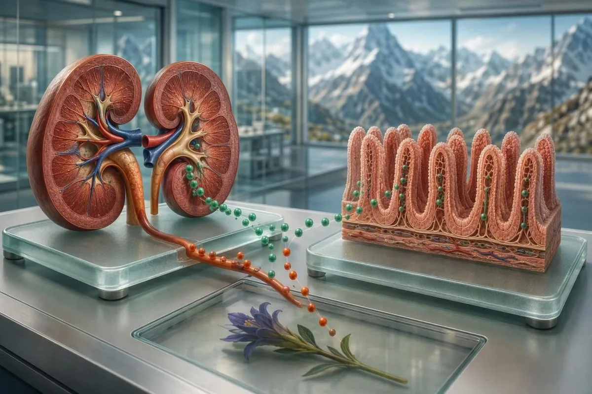
কম মেগনেছিয়াম সাধাৰণতে ঔষধৰ পৰা কিডনিৰ ক্ষতি, অন্ত্ৰৰ শোষণ কমি যোৱা, দীঘলীয়া ডায়েৰিয়া, বা বেছি পৰিমাণে এলক’হল সেৱনৰ ফলত হয়—কেৱল কম-মেগনেছিয়াম খাদ্যাভ্যাসৰ বাবে নহয়। পটাছিয়াম, কেলচিয়াম, কিডনিৰ কাৰ্যক্ষমতা, ঔষধ, আৰু পায়খানাৰ লক্ষণৰ ধৰণে সাধাৰণতে উৎসটোলৈ আঙুলিয়াই দিয়ে।.


- হাইপোমেগনেছেমিয়া মানে ছিৰাম মেগনেছিয়াম তলত প্রায়ে অথবা তকৈ কম মান অধিকাংশ প্ৰাপ্তবয়স্কৰ লেব’ৰেটৰিত।.
- লুপ আৰু থিয়াজাইড ডাইইউৰেটিক মূত্ৰৰ মাধ্যমে মেগনেছিয়ামৰ ক্ষতি বৃদ্ধি কৰিব পাৰে, বিশেষকৈ পটাছিয়ামো কম থাকিলে।.
- প্ৰ’টন-পাম্প ইনহিবিটৰ অন্ত্ৰৰ মেগনেছিয়াম শোষণ কমাব পাৰে, সাধাৰণতে কেইমাহমানৰ পৰা কেইবছৰলৈকে অবিৰত ব্যৱহাৰৰ পিছত।.
- এলক’হল-সম্পৰ্কীয় কম মেগনেছিয়াম প্ৰায়ে কম খাদ্য গ্ৰহণ, ডায়েৰিয়া, বমি, আৰু কিডনিৰ মেগনেছিয়াম অপচয় একেলগে দেখা যায়।.
- দীর্ঘস্থায়ী ডায়েৰিয়া ডাইজেষ্টিভ ফ্লুইডত ম্যাগনেছিয়াম থাকে আৰু অন্ত্ৰই তাক শোষণ কৰিবলৈ কম সময় পায় বাবে ই দ্ৰুতভাৱে মেগনেছিয়াম কমাই দিব পাৰে।.
- 2%ৰ ওপৰৰ ম্যাগনেছিয়ামৰ ফ্ৰেকশ্যনেল এক্সক্রিশন হাইপোমেগনেছিয়েমিয়াৰ সময়ত কিডনিৰ কাৰ্য্যক্ষমতা যথেষ্টভাৱে সংৰক্ষিত থাকিলে অনুপযুক্তভাৱে কিডনিয়ে ম্যাগনেছিয়াম হেৰুৱাই দিয়া (renal magnesium loss) বুলি সূচায়।.
- গুৰুতৰ লক্ষণ যেনে অজ্ঞান হোৱা, খিঁচুনি, স্থায়ী হৃদস্পন্দন অস্বাভাবিকতা, বা স্পষ্টভাৱে অধিক দুৰ্বলতা—ইবোৰে তৎক্ষণাৎ চিকিৎসাজনিত মূল্যায়ন (urgent clinical assessment)ৰ প্ৰয়োজন।.
- কম পটাছিয়াম বা কম কেলচিয়াম যি সংশোধন নহয় ম্যাগনেছিয়ামৰ ঘাটতি এৰি যোৱা হৈছে বুলি এটা সূত্ৰ হ’ব পাৰে।.
কম মেগনেছিয়াম সাধাৰণতে মানে কি—আৰু কিয় কাৰণটো প্ৰথমে আহে
কম মেগনেছিয়াম বেছিভাগ সময়ে কিডনিয়ে মেগনেছিয়াম নষ্ট কৰি দিয়া (waste) কিছুমান ঔষধ, অন্ত্ৰৰ শোষণ কমি যোৱা, চলি থকা গ্যাস্ট্ৰ’ইনটেষ্টাইনেল ফ্লুইড হেৰুৱা, বা অধিক পৰিমাণে মদ্যপান (heavy alcohol use)ৰ ফলত হয়।. ছিৰাম মেগনেছিয়াম 0.70 mmol/L (1.7 mg/dL)ৰ তলত বেছিভাগ প্ৰাপ্তবয়স্কৰ লেবত হাইপোমেগনেছিয়েমিয়া (hypomagnesemia) বুলি ধৰা হয়, কিন্তু কাৰণটোৱে নিৰ্ধাৰণ কৰে যে পৰৱৰ্তী সঠিক পদক্ষেপটো ঔষধ পৰ্যালোচনা কৰা, ডায়েৰিয়া চিকিৎসা কৰা, মদ্যপান সম্বোধন কৰা, নে কিডনিৰ পৰা হোৱা হেৰুৱা (renal losses) পৰীক্ষা কৰা—কোনটো হ’ব। ১ আগষ্ট, ২০২৬ অনুসৰি, এই পাৰ্থক্যটো স্বয়ংক্ৰিয়ভাৱে এটা সাপ্লিমেণ্ট খোৱাৰ ওপৰত অধিক গুৰুত্বপূৰ্ণ।.
মাত্ৰ প্ৰায় মুঠ শৰীৰৰ মেগনেছিয়ামৰ 1% ছিৰামত থাকে, সেয়ে স্বাভাৱিক ছিৰাম ফলাফলে সম্পূৰ্ণভাৱে ভিতৰৰ কোষীয় (intracellular) সঞ্চয় কমি যোৱা (depleted) বাদ দিব নোৱাৰে; বিপৰীতে, স্পষ্টভাৱে কম ছিৰাম মানে মনোযোগৰ যোগ্য। মোৰ ক্লিনিকেল অনুশীলনত, আটাইতকৈ তথ্যবহুল পেনেলসমূহে মেগনেছিয়ামক পটাছিয়ামৰ সৈতে, সংশোধিত কেলচিয়াম (corrected calcium), ক্ৰিয়েটিনিন (creatinine), বাইকাৰ্বনেট (bicarbonate), গ্লুক’জ (glucose), আৰু ঔষধৰ সময়ৰেখা (medication timeline)ৰ সৈতে মিলাই চায়—এটা এটা মাত্র সংখ্যা আলাদাভাৱে চিকিৎসা কৰাৰ পৰিৱৰ্তে।.
ড° থমাছ ক্লেইনে এটা পুনৰাবৃত্তিমূলক ধৰণ দেখা পায়: এজন ৰোগীয়ে ডাইইউৰেটিক আৰম্ভ কৰে বা প্ৰ’টন-পাম্প ইনহিবিটৰ (proton-pump inhibitor)ত থাকি যায়, কেইমাহমান পিছত কম পটাছিয়াম গঢ়ি উঠে, তাৰ পিছতহে অৱশেষত মেগনেছিয়াম পৰিমাপ কৰা হয়।. Kantesti হৈছে এটা AI ৰক্ত পৰীক্ষা বিশ্লেষক (blood test analyzer) যিয়ে মেগনেছিয়ামক কিডনিৰ কাৰ্য্যক্ষমতা, কেলচিয়াম, পটাছিয়াম, আৰু সময়ৰেখাভিত্তিক (longitudinal) ঔষধ-সম্পৰ্কীয় ধৰণৰ সৈতে একেলগে পঢ়ে—এটা একক “isolated” সংকেত (flag) দেখুৱাই নাথাকে।.
লক্ষণসমূহ সাধাৰণতে তলত অধিক সম্ভাৱনাময় হয় 0.50 mmol/L (1.2 mg/dL)ৰ, তলত, যদিও অৱনতিৰ গতি (rate of decline) আৰু একেলগে থকা হাইপ’কেলেমিয়া (coexisting hypokalemia) ফলাফলটোৰ নিজস্ব মানৰ সমানেই গুৰুত্বপূৰ্ণ। আমাৰ বায়'মাৰ্কাৰ ৰেফাৰেন্স গাইড পাঠকসকলক তেওঁলোকৰ নিজৰ লেবত ব্যৱহৃত একক (units) চিনাক্ত কৰাত সহায় কৰিব পাৰে, মানসমূহ তুলনা কৰাৰ আগতে।.
কোনবোৰ ঔষধে আটাইতকৈ বেছি কম মেগনেছিয়ামৰ কাৰণ হয়?
ডাইইউৰেটিক, প্ৰ’টন-পাম্প ইনহিবিটৰ, কিছুমান কেঞ্চাৰ ঔষধ, কেলচিনিউৰিন ইনহিবিটৰ (calcineurin inhibitors), এমিন’গ্লাইক’ছাইড এন্টিবায়’টিক (aminoglycoside antibiotics), এম্ফ’টেৰিচিন বি (amphotericin B), আৰু কিছুমান এন্টিভাইৰেল (antivirals) কম মেগনেছিয়ামৰ কাৰণ হ’ব পাৰে।. তেওঁলোকে হয় অন্ত্ৰৰ গ্ৰহণ (intestinal uptake) কমাই দিয়ে, নহ’লে শৰীৰে সংৰক্ষণ কৰা উচিত হ’লেও কিডনিয়ে মেগনেছিয়াম নিৰ্গত (excrete) কৰিবলৈ বাধ্য কৰে।.
লুপ ডাইইউৰেটিক (Loop diuretics) যেনে ফুৰ’ছেমাইড (furosemide) আৰু বুমেটানাইড (bumetanide) ডাঠ উঠা অংশত (thick ascending limb) মেগনেছিয়াম পুনঃশোষণ (reabsorption) কমাই দিয়ে, আনহাতে থাইয়াজাইড দূৰবর্তী টিউবুলত মেগনেছিয়াম হেৰাই যোৱাত সহায় কৰিব পাৰে। অধিক ড’জ, বয়স বঢ়া, খাদ্যত মেগনেছিয়াম কম খোৱা, ডায়েৰিয়া, আৰু আন এটা মেগনেছিয়াম-কমোৱা ঔষধৰ সৈতে একেলগে চিকিৎসা কৰিলে ঝুঁকি বাঢ়ে; সেইবাবেই ডাইইউৰেটিক আৰু কম মেগনেছিয়াম প্ৰায়ে পটাছিয়ামৰ অস্বাভাৱিকতাৰ সৈতে একেলগে দেখা যায়।.
প্ৰ’টন-পাম্প ইনহিবিটৰ, omeprazole আৰু pantoprazole-কে ধৰি, TRPM6 আৰু TRPM7 পথৰ জৰিয়তে সক্ৰিয় অন্ত্ৰীয় মেগনেছিয়াম পৰিবহণত বাধা দিব পাৰে যেন লাগে। FDA-এ অন্তত ৰ বাবে প্লেটলেট গণনা স্থায়ীভাৱে, পিছত হোৱা কিছুমান কেছ বৰ্ণনা কৰিছে, যদিও বহু কেছ; 1 বছৰৰ পাছত হয়; এটা উপগোটত, কেৱল মুখে খোৱা মেগনেছিয়ামে PPI বন্ধ নকৰালৈ বা চিকিৎসা তত্ত্বাৱধানত সলনি নকৰালৈকে স্তৰ স্বাভাৱিক নকৰিলে।.
Liamis et al. (2022)-এ cisplatin, anti-EGFR থেৰাপি, tacrolimus, cyclosporine, aminoglycosides, foscarnet, আৰু amphotericin B-ৰ পৰা কিডনিৰ মেগনেছিয়াম অপচয় (wasting) বিৱৰণ দিছে। নিৰ্ধাৰিত ঔষধ হঠাৎ বন্ধ নকৰিব: ক্লিনিচিয়ানলৈ এটা সম্পূৰ্ণ তালিকা লৈ যাওক—য’ত over-the-counter acid remedies-ও থাকিব—তাৰ পিছত দীৰ্ঘম্যাদী PPI নিৰীক্ষণ আৰু আপোনাৰ নিৰ্দিষ্ট প্ৰয়োগৰ বাবে অধিক সুৰক্ষিত বিকল্পসমূহ পৰ্যালোচনা কৰক।.
কিয় ডাইইউৰেটিকে একেলগে কম মেগনেছিয়াম আৰু কম পটাছিয়াম ঘটাব পাৰে
ডাইইউৰেটিকসমূহে কিডনীৰ সেই অংশসমূহত ছ’ডিয়াম ডেলিভাৰী আৰু মূত্ৰৰ প্ৰবাহ বৃদ্ধি কৰি মেগনেছিয়াম হ্ৰাস ঘটায়, য’ত সাধাৰণতে মেগনেছিয়াম পুনৰ আহৰণ হয়।. ডাইইউৰেটিক সলনি কৰাৰ পিছত কম পটাছিয়ামৰ সৈতে কম মেগনেছিয়াম ফলাফল কেৱল দুৰ্বল খাদ্যাভ্যাসৰ তুলনাত কিডনিৰ পৰা অপচয় (renal wasting) হোৱাৰ সম্ভাৱনা বেছি সূচায়।.
স্বাভাৱিক বা প্ৰায়-স্বাভাৱিক কিডনি কাৰ্য থকা ৰোগীসকলৰ ক্ষেত্ৰত, 2%-ৰ ওপৰত মেগনেছিয়ামৰ fractional excretion , কিন্তু ছিৰাম মেগনেছিয়াম কম থকাটো অনুপযুক্ত কিডনিৰ পৰা হেৰাই যোৱা (inappropriate renal loss) সমৰ্থন কৰে। হাইপোমেগনেছিয়েমিয়াৰ সময়ত প্ৰায় প্ৰতিদিন 24 mg-ৰ ওপৰত 24-ঘণ্টীয়া মূত্ৰৰ মেগনেছিয়ামেও একে ধৰণৰ কাহিনী ক’ব পাৰে, যদিও সংগ্ৰহজনিত ভুল আৰু কম eGFR দুয়োটা মাপ দুয়োটাকে কম বিশ্বাসযোগ্য কৰি তোলে।.
ক্লিনিকেল ফাঁদটো হ’ল কেৱল পটাছিয়ামৰ প্ৰতিস্থাপনে সমস্যাটো ঠিক কৰিব বুলি ধৰি লোৱা। কিডনিৰ পটাছিয়াম সংৰক্ষণৰ বাবে মেগনেছিয়ামৰ প্ৰয়োজন, সেয়ে বুদ্ধিসম্পন্ন প্ৰতিস্থাপন সত্ত্বেও 3.5 mmol/L তলত পটাছিয়াম অব্যাহত থাকিলে মেগনেছিয়াম পৰীক্ষা কৰাটো উচিত; মেগনেছিয়াম পুনৰ স্থাপন নোহোৱালৈকে পটাছিয়াম কেৱল মূত্ৰলৈ লিক হৈ থাকিব পাৰে।.
মই এইটো দেখিছোঁ এটা 68 বছৰীয়া ব্যক্তিত, য’ত মাত্ৰ তুলনামূলকভাৱে কম পৰিমাণে hydrochlorothiazide বৃদ্ধি কৰা হৈছিল, আৰু মেগনেছিয়াম আছিল 0.54 mmol/L আৰু পটাছিয়াম আছিল 3.0 mmol/L. । উপযোগী প্ৰশ্নটো “ঔষধটো বেয়া নে নহয়” সেইটো নাছিল, বৰং সংশোধিত ৰেজিমেন আৰু পুনৰ লেবৰেটৰী পৰীক্ষাৰে ৰক্তচাপ নিয়ন্ত্ৰণ বজাই ৰাখিব পাৰিনে—সেইটো আছিল; আমাৰ গাইড চাওক ৰক্তচাপৰ ঔষধৰ পৰিৱৰ্তনৰ পিছত পটাছিয়াম.
এলক’হলে কেনেকৈ মেগনেছিয়ামৰ ঘাটতি ঘটায়
অধিক পৰিমাণে মদ্যপান কৰিলে খাদ্য গ্ৰহণ কমি যোৱা, বমি, ডায়েৰিয়া, পেনক্ৰিয়াটাইটিছ, আৰু সৰাসৰি কিডনিৰ পৰা মেগনেছিয়াম অপচয়ৰ জৰিয়তে মেগনেছিয়াম কমি যাব পাৰে।. হঠাৎ মদ বন্ধ কৰা আৰু চিকিৎসালয়ত চিকিৎসা লোৱা এই ঘাটতি উন্মোচন কৰিব পাৰে বা বেয়া কৰিব পাৰে, কাৰণ কেটেক’লামিনৰ ঢৌ বৃদ্ধি পায় আৰু ৰিফিডিঙে ইলেক্ট্ৰ’লাইটসমূহ কোষলৈ স্থানান্তৰ কৰে।.
মদ-সম্পৰ্কীয় হাইপোমেগনেছিয়েমিয়া প্ৰায়ে এটা একক সমস্যা নহয়—ই এটা গোট (cluster) হ’ব পাৰে: মেগনেছিয়াম কম থাকিব পাৰে লগতে ০.৮০ mmol/L ৰ তলত ফছফেট, পটাছিয়াম তলত 3.5 mmol/L, আৰু কম বা কম-স্বাভাবিক কেলচিয়াম। অধিক পৰিমাণে গ্ৰহণ কৰা এজন ব্যক্তিৰ ক্ষেত্ৰত এই গোটটোৱে একেলগে বেয়া পুষ্টি, গ্যাস্ট্ৰ’ইনটেষ্টাইনেল ক্ষতি, আৰু কিডনিৰ টিউবুলাৰ বিকলতা সন্দেহজনক কৰি তুলিব লাগে।.
স্বাভাৱিক লিভাৰ পেনেলে এলক’হল-সম্পৰ্কীয় মেগনেছিয়াম হেৰুওৱা নাকচ নকৰে। GGT, AST, ALT, mean corpuscular volume, albumin, আৰু triglycerides এ কিছুমান প্ৰসংগ যোগ কৰিব পাৰে, কিন্তু কোনোটোৱেই এলক’হল এক্সপ’জাৰ নিশ্চিত নকৰে বা কেৱলেই কম মেগনেছিয়াম ফলাফল ব্যাখ্যা নকৰে; ড° থমাছ ক্লেইনে লেব’ৰেটৰী ট্ৰেণ্ড আৰু সৎ গ্ৰহণৰ ইতিহাস একেলগে ব্যৱহাৰ কৰিবলৈ পৰামৰ্শ দিয়ে।.
এলক’হল বন্ধ কৰিলে মূত্ৰত হোৱা অপচয় উন্নত হ’ব পাৰে, কিন্তু প্ৰথম ৭২ ঘণ্টা নিৰ্ভৰশীল লোকৰ বাবে চিকিৎসাগতভাৱে বিপদজনক হ’ব পাৰে, কিয়নো উইথড্ৰ’ৱালে খিঁচুনি, ডিলিৰিয়াম, ডিহাইড্ৰেচন, আৰু এৰিথমিয়া ঘটাব পাৰে। পুনৰুদ্ধাৰৰ সময়ত কি কি সলনি হ’ব পাৰে তাৰ বাস্তৱমুখী দৃষ্টিভংগী পাবলৈ পঢ়ক এলক’হল কমোৱাৰ পিছত ৰক্ত-মাৰ্কাৰ ট্ৰেণ্ডসমূহ; নিৰ্ভৰশীলতা সম্ভৱ হ’লে হঠাৎ উইথড্ৰ’ৱাল চেষ্টা কৰাৰ পৰিৱর্তে তত্ত্বাৱধানত চিকিৎসা বিচাৰক।.
কেতিয়া ডায়েৰিয়া আৰু বমি হৈছে মূল মেগনেছিয়ামৰ ক্ষতি
স্থায়ী ডায়েৰিয়া কম মেগনেছিয়ামৰ অন্যতম সৰ্বাধিক দ্ৰুত গ্যাস্ট্ৰ’ইনটেষ্টাইনেল কাৰণ, কিয়নো পায়খানা আৰু হজমজনিত নিঃসৰণত মেগনেছিয়াম থাকে আৰু শোষণৰ সময় কমি যায়।. বমি পৰোক্ষভাৱে কম গ্ৰহণ, ভলিউম কমি যোৱা, আৰু হৰম’ন-সম্পৰ্কীয় কিডনিৰ সঁহাৰিৰ জৰিয়তে অৰিহণা যোগায়, যদিও ডায়েৰিয়াই সাধাৰণতে অধিক পৰিমাণে সৰাসৰি মেগনেছিয়াম হেৰুওৱা কৰে।.
২ দিনতকৈ বেছি দিন ধৰি থকা ডায়েৰিয়া 14 দিন কাৰণ-কেন্দ্ৰিক পৰ্যালোচনাৰ যোগ্য, বিশেষকৈ যদি ই ওজন কমা, নিশাৰ পায়খানা, জ্বৰ, তেলীয়া/চিকচিকীয়া পায়খানা, বা প্রায়ে. তলৰ মেগনেছিয়াম ফলাফলৰ সৈতে ঘটে। ল্যাক্সেটিভ বা মেগনেছিয়াম-যুক্ত উৎপাদনৰ পৰা হোৱা অ’সম’টিক ডায়েৰিয়া বিভ্ৰান্তিকৰ হ’ব পাৰে: অতিমাত্ৰা সম্পূৰক মেগনেছিয়ামে ডায়েৰিয়া ঘটাব পাৰে, তাৰ পিছত পায়খানাৰ হেৰুওৱাই সামগ্ৰিক ইলেক্ট্ৰ’লাইট ছবিখন জটিল কৰি তুলিব পাৰে।.
পানীয়-ধৰণৰ ডায়েৰিয়া আৰু কম বাইকাৰ্বনেট সাধাৰণতে গ্যাস্ট্ৰ’ইনটেষ্টাইনেল বাইকাৰ্বনেট হেৰুওৱাৰ দিশে আঙুলিয়াই দিয়ে, আনহাতে বমিয়ে বেছিকৈ কম ক্ল’ৰাইড আৰু বঢ়া বাইকাৰ্বনেট সৃষ্টি কৰে। কোনো ধৰণেই নিখুঁত নহয়, কিন্তু এই লগত থকা ইলেক্ট্ৰ’লাইটসমূহে এজন চিকিৎসকক প্ৰতিটো কম মেগনেছিয়াম ফলাফলক কেৱল খাদ্যজনিত ঘাটতি বুলি ভুলকৈ লেবেল কৰাত বাধা দিব পাৰে; আমাৰ পায়খানাৰ-পেটাৰ্ণৰ ৰেড ফ্লেগ গাইড এ কিহৰ পৰিৱর্তনে তৎক্ষণাৎ পৰ্যালোচনাৰ যোগ্য তাক ব্যাখ্যা কৰে।.
সংক্রমণ, ইনফ্লেমেট’ৰি বাওয়েল ডিজিজ, মাইক্ৰ’স্ক’পিক ক’লাইটিছ, বাইল এচিড ডায়েৰিয়া, থাইৰয়েডৰ অতিমাত্ৰা, আৰু ঔষধৰ পাৰ্শ্বক্ৰিয়া—এই সকলোবোৰে ক্ষতি অব্যাহত ৰাখিব পাৰে। ৰিহাইড্ৰেচন ফ্লুইড সহায়ক, কিন্তু কেৱল সাধাৰণ পানীয়ে—বিশেষকৈ যথেষ্ট পৰিমাণে ফ্লুইড হেৰুওৱা হ’লে—ডাইলিউশ্বনজনিত লক্ষণ বঢ়াব পাৰে; সঠিক মুখে খোৱাৰ দ্ৰৱণটো সোডিয়াম, পটাছিয়াম, গ্লুক’জ, আৰু কিডনিৰ প্ৰসংগৰ ওপৰত নিৰ্ভৰ কৰে।.
কোনবোৰ অন্ত্ৰৰ অৱস্থাই মেগনেছিয়ামৰ শোষণ কমায়?
চিলিয়াক ডিজিজ, সৰু অন্ত্ৰত প্ৰভাৱ পেলোৱা ক্ৰ’ন ডিজিজ, পেনক্ৰিয়াটিক ইনছাফিচিয়েন্সি, শর্ট-বাওয়েল অৱস্থা, আৰু বেৰিয়াট্ৰিক ছাৰ্জাৰীয়ে মেগনেছিয়াম শোষণ কমাব পাৰে।. এই হাইপ’মেগনেছেমিয়া কাৰণসমূহ ডায়েৰিয়াৰ সৈতে কম আয়ৰণ, কম ভিটামিন D, কম albumin, বা অনিচ্ছাকৃতভাৱে ওজন কমাৰ সহ-অৱস্থান থাকিলে বেছি সম্ভাৱনা থাকে।.
ডিষ্টেল সৰু অন্ত্ৰ আৰু ক’লনে মেগনেছিয়াম শোষণত সহায় কৰে, নিয়ন্ত্ৰিত TRPM6 পৰিবহণৰ জৰিয়তেও; এই অঞ্চলসমূহত হোৱা ৰোগ বা ৰিসেকচনে অসমানুপাতিকভাৱে গুৰুত্ব ধাৰণ কৰিব পাৰে। পূৰ্বৰ অন্ত্ৰীয় ছাৰ্জাৰীৰ ইতিহাস থকা ৰোগীৰ ক্ষেত্ৰত 0.62 mmol/L ৰ এটা ছিৰাম মেগনেছিয়াম মানে, আহাৰ পুষ্টিগতভাৱে যথেষ্ট যেন লাগিলেও, ক্ৰমাগত হেৰুওৱা প্ৰতিফলিত কৰিব পাৰে।.
তেলীয়া, ভাঁহি থকা, আৰু ধুবলৈ কঠিন পায়খানাই চৰ্বি শোষণৰ সমস্যা (ফেট মেলএবজৰ্পচন) সূচায় আৰু কেৱল সম্পূৰক বঢ়োৱাৰ পৰিৱর্তে কাম-কাজ পেনক্ৰিয়াটিক, বাইলি, বা সৰু অন্ত্ৰজনিত কাৰণৰ দিশে ঘূৰাই দিব লাগে। এটা ফিকেল ফেট ফলাফল ইয়া এটা মেগনেছিয়াম পৰীক্ষা নহয়, কিন্তু কম মেগনেছিয়ামৰ সৈতে ওজন কমা থাকিলে ডায়াগন’ষ্টিক পথত ই এক উপযোগী মোড় ঘূৰাই দিব পাৰে।.
চেলিয়াক পৰীক্ষাৰ এটা সূক্ষ্মতা আছে যিটো বহু ৰোগীয়ে বুজি নাপায়: টিছ্যু-ট্ৰান্সগ্লুটামিনেজ IgA ৰ সৈতে মুঠ IgA থাকিব লাগে, কিয়নো নিৰ্বাচিত IgA ঘাটতিয়ে এটা মিছা-ভাল ফলাফল দেখুৱাব পাৰে। যদি ৰক্তহীনতা, কম ফেৰিটিন, বা পুনৰাবৃত্ত ইলেক্ট্ৰ’লাইট হেৰুওৱা থাকিও লক্ষণবোৰ টিকি থাকে, তেন্তে এখন নেগেটিভ স্ক্ৰিনিং পেনেলৰ পিছতো গেষ্ট্ৰ’এণ্টেৰ’ল’জি মতামত লোৱা যুক্তিসংগত।.
চিকিৎসকে কেনেকৈ কিডনিৰ মেগনেছিয়াম অপচয়ক অন্ত্ৰৰ ক্ষতিৰ পৰা পৃথক কৰে
অনুপযুক্তভাৱে উচ্চ ইউৰিন মেগনেছিয়াম স্তৰৰ সৈতে কম মেগনেছিয়াম থাকিলে কিডনিৰ “wasting” (কিডনি-জনিত হেৰুওৱা) সূচায়, আনহাতে কম ইউৰিন মেগনেছিয়াম দেখালে বুজায় যে কিডনিয়ে কম খাদ্য গ্ৰহণ বা অন্ত্ৰৰ পৰা হেৰুওৱাৰ সময়ত মেগনেছিয়াম ঠিকমতে সংৰক্ষণ কৰি আছে।. তুলনাটো সৰ্বাধিক উপযোগী হয় যেতিয়া ডাঙৰ পৰিমাণৰ ৰিপ্লেচমেণ্ট ড’জ দিয়াৰ আগতে লেবৰেটৰী নমুনা আৰু ইউৰিন সংগ্ৰহ কৰা হয়।.
এজন চিকিৎসকে একেলগে ছিৰাম মেগনেছিয়াম, ক্ৰিয়েটিনিন, পটাছিয়াম, কেলচিয়াম, ফ’ছফেট, ইউৰিন মেগনেছিয়াম, আৰু ইউৰিন ক্ৰিয়েটিনিন নিৰ্দেশ দিব পাৰে।. Kantesti হৈছে এটা AI blood test interpretation প্লেটফৰ্ম যিয়ে সময়ৰ লগে লগে এই সম্পৰ্কীয় ফলাফলবোৰ সংগঠিত কৰিব পাৰে, কিন্তু urinary indices আৰু চিকিৎসা সিদ্ধান্তসমূহ এতিয়াও clinician ৰ পৰ্যালোচনা লাগিব, কিয়নো eGFR আৰু শেহতীয়া ছাপ্লিমেণ্টে ব্যাখ্যা সলনি কৰিব পাৰে।.
কিডনিৰ কাৰ্যক্ষমতা সাধাৰণতে মেগনেছিয়াম ধৰি ৰখাৰ (retention) কাৰণ হয়, হেৰুওৱাৰ নহয়, কিন্তু ব্যতিক্ৰম আছে: টিউবুল’ইণ্টাৰ্ষ্টিটিয়েল আঘাত, acute kidney injury ৰ পিছৰ আৰোগ্য, osmotic diuresis সহ uncontrolled diabetes, আৰু কিছুমান ঔষধে সকলোবোৰে মেগনেছিয়াম নষ্ট কৰিব পাৰে। সেয়ে নতুন glycosuria, উচ্চ গ্লুক’জ, বা ক্ৰিয়েটিনিনৰ পৰিৱর্তন—এইবোৰে ব্যাখ্যাৰ অংশ হ’ব পাৰে, অচিনাকি ফলাফল নহয়।.
ইউৰিন albumin-creatinine ratio এ মেগনেছিয়াম wasting জুখিব নোৱাৰে, কিন্তু albuminuria ৰ বাবে কিডনি-জনিত ঝুঁকিৰ আলোচনা সলনি হয় আৰু ছাপ্লিমেণ্টৰ সুৰক্ষাও প্ৰভাৱিত হয়। ডায়েবেটিছ বা হাইপাৰটেনচন থকা পাঠকে review কৰিব পাৰে ইউৰিন ACR preparation এটা একক ইউৰিন ফলাফলে সকলো কিডনি প্ৰশ্নৰ উত্তৰ দিয়ে বুলি ধৰি লোৱাৰ আগতে।.
ডায়েবেটিছ, হৰম’ন, আৰু কম মেগনেছিয়ামৰ অন্যান্য বিপাকীয় কাৰণ
বেয়াকৈ নিয়ন্ত্ৰিত ডায়েবেটিছে osmotic diuresis ৰ জৰিয়তে কম মেগনেছিয়াম ঘটাব পাৰে, আৰু hyperaldosteronism এ renal electrolyte wasting ৰ জৰিয়তে সহায় কৰিব পাৰে।. এই কাৰণবোৰ অধিক সম্ভাৱ্য হয় যেতিয়া মেগনেছিয়াম কম থাকে আৰু একেলগে উচ্চ গ্লুক’জ, ঘন ঘন মূত্ৰত্যাগ, হাইপাৰটেনচন, বা স্থায়ীভাৱে কম পটাছিয়াম থাকে।.
গ্লুক’জ কিডনিৰ reabsorptive capacity অতিক্ৰম কৰিলে, সাধাৰণতে প্ৰায় 10 mmol/L (180 mg/dL) যদিও ব্যক্তিভেদে ভিন্ন হ’ব পাৰে, গ্লুক’জ ইউৰিনলৈ সোমায় আৰু লগে লগে পানী আৰু ইলেক্ট্ৰ’লাইট কঢ়িয়াই আনে। মেগনেছিয়ামো সমান্তৰালভাৱে কমিব পাৰে—বিশেষকৈ dehydration বা তীব্ৰ hyperglycemia ৰ চিকিৎসাৰ সময়ত—স’ডিয়াম, পটাছিয়াম, আৰু ফ’ছফেটৰ সৈতে।.
Primary aldosteronism এ সাধাৰণতে কম পটাছিয়ামৰ সৈতে হাইপাৰটেনচন সৃষ্টি কৰে, কিন্তু distal sodium transport বৃদ্ধি পালে urinary losses বাঢ়ি মেগনেছিয়ামো কম হ’ব পাৰে। এজন ব্যক্তিৰ ক্ষেত্ৰত যিয়ে কোনো diuretic গ্ৰহণ কৰা নাই আৰু যাৰ ৰক্তচাপ নিয়ন্ত্ৰণ কৰাত কঠিন, তেন্তে supplement-only পদ্ধতিৰ পৰিৱর্তে এটা directed endocrine review উপযুক্ত। 3.2 mmol/L কোনো ডাইইউৰেটিক নলোৱা আৰু নিয়ন্ত্ৰণ কৰিবলৈ কঠিন হোৱা ৰক্তচাপ থকা ব্যক্তিৰ ক্ষেত্ৰত পৰিপূৰক-ভিত্তিক পদ্ধতিৰ সলনি এটা নিৰ্দেশিত অন্তঃস্ৰাৱী পৰ্যালোচনাৰ প্ৰয়োজন।.
Gitelman syndrome দৰে inherited renal tubulopathies বিৰল কিন্তু মনত ৰখা যোগ্য: কম মেগনেছিয়াম, কম পটাছিয়াম, metabolic alkalosis, আৰু কম ইউৰিন কেলচিয়াম—এইবোৰ এটা ক্লাছিক সংমিশ্ৰণ। যদি এই অস্বাভাৱিকতাবোৰ সৰুতে আৰম্ভ হৈছিল, চিকিৎসাৰ পিছতো পুনৰাবৃত্ত হয়, বা আত্মীয়সকলকো প্ৰভাৱিত কৰে, তেন্তে chronic kidney disease গাইড কিয় renal assessment আৰু specialist input গুৰুত্বপূৰ্ণ সেইটো বুজাবলৈ সহায় কৰে।.
কিয় কম মেগনেছিয়াম চিকিৎসালয়ৰ যত্নৰ সময়ত বা ৰিফিডিংৰ সময়ত দেখা দিব পাৰে
refeeding ৰ সময়ত, diabetic ketoacidosis ৰ চিকিৎসাৰ সময়ত, দীঘলীয়া hospitalization ৰ সময়ত, আৰু critical illness ৰ পৰা আৰোগ্য লাভৰ সময়ত কম মেগনেছিয়াম দেখা দিব পাৰে, কিয়নো মেগনেছিয়াম কোষলৈ সলনি হয় বা ইউৰিনত হেৰাই যায়।. পূৰ্বতে স্বাভাৱিক ফলাফল থকাটোৱে কেলৰি, ইনচুলিন, ফ্লুইড, বা diuretics যোগ কৰাৰ পিছত সুৰক্ষাৰ নিশ্চয়তা নিদিয়ে।.
refeeding syndrome বেছি চিন্তাজনক হয় দীঘলীয়া সময়ৰ বাবে অপৰ্যাপ্ত খাদ্য গ্ৰহণ, উল্লেখযোগ্য ওজন কমা, অ্যালক’হল নিৰ্ভৰশীলতা, বা তীব্ৰ অসুস্থতাৰ পিছত। ইনচুলিন-চালিত cellular uptake এ phosphate, potassium, আৰু magnesium কমাব পাৰে within ২৪ৰ পৰা ৭২ ঘণ্টা কেলৰি পুনৰ আৰম্ভ কৰাৰ; ফছফেট প্ৰায়ে সৰ্বপ্ৰথমেই নাটকীয়ভাৱে দেখা দিয়া অস্বাভাৱিকতা, কিন্তু মেগনেছিয়ামো সমানে ক্লিনিকেলভাৱে গুৰুত্বপূৰ্ণ হ’ব পাৰে।.
ডায়াবেটিক কেট’এচিড’ছিছত, চিকিৎসাৰ আগতে সম্পূৰ্ণ-শৰীৰৰ ঘাটিৰ সত্ত্বেও পৰিমাপ কৰা ৰক্তৰ মেগনেছিয়াম স্বাভাবিক থাকিব পাৰে, কাৰণ ডিহাইড্ৰেচনে ৰক্তৰ মান ঘনীভূত কৰে। এবাৰ ইনচুলিন আৰু ফ্লুইড আৰম্ভ হ’লে, কেৱল ভাৰ্তিৰ পেনেল নহয়—পুনৰ ইলেক্ট্ৰ’লাইটসমূহে প্ৰতিস্থাপন নিৰ্দেশ দিব; মাংসপেশীৰ টুইচিং বা ৰিদমৰ পৰিৱর্তনৰ সৈতে মেগনেছিয়াম কমি যোৱাটো সক্ৰিয় ব্যৱস্থাপনা দাবী কৰে।.
এইটো এটা এনে পৰিস্থিতি য’ত “অধিক মেগনেছিয়াম খাওক” বুলি কোৱা যথেষ্ট উত্তৰ নহয়। চিকিৎসালয়ৰ টিমে ক্ৰমাগতভাৱে ইলেক্ট্ৰ’লাইট পৰীক্ষা কৰে, প্ৰয়োজন হ’লে হৃদযন্ত্ৰৰ মনিটৰিং কৰে, থায়ামিন দিয়ে, আৰু পৰিমাপভিত্তিক প্ৰতিস্থাপন কৰে; আমাৰ refeeding laboratory guide-এ কিয় phosphate, potassium, আৰু magnesium-ৰ অধিক তৎক্ষণাৎ monitoring প্ৰয়োজন হয়—সেই কথা ব্যাখ্যা কৰে। কিয় তিনিটা খনিজ একেলগে পৰীক্ষা কৰা হয়—সেয়া ব্যাখ্যা কৰে।.
কম মেগনেছিয়ামৰ লক্ষণ আৰু গুৰুত্বপূৰ্ণ ইলেক্ট্ৰ’লাইটৰ ধৰণ
কম মেগনেছিয়ামৰ লক্ষণসমূহৰ ভিতৰত কঁপনি, মাংসপেশীৰ খিঁচ, দুৰ্বলতা, টিংগলিং, ভাগৰুৱা, খিঁচুনি, আৰু হৃদযন্ত্ৰৰ ৰিদম-সম্পৰ্কীয় লক্ষণ থাকে, কিন্তু মৃদু ঘাটিত বহু সময়ে নীৰৱেই থাকে।. আটাইতকৈ উপযোগী সতৰ্ক সংকেতৰ ধৰণটো হ’ল মেগনেছিয়াম 0.50 mmol/L তকৈ তলত থাকিলে, লগতে পটাছিয়াম কম, কেলচিয়াম কম, বা ECG-ৰ লক্ষণ থাকিলে।.
মেগনেছিয়ামৰ ঘাটিয়ে নিউৰোমাস্কুলাৰ উত্তেজনাশীলতা বৃদ্ধি কৰে, যিটো চকুৰ পাতাৰ টুইচিং, কঁপনি, কারপ’পেডাল স্পাজম, বা সাধাৰণ দুৰ্বলতা হিচাপে দেখা দিব পাৰে। এই লক্ষণসমূহ বিশেষ নহয়—উদ্বেগ, থাইৰয়েড ৰোগ, ষ্টিমুলেণ্ট ব্যৱহাৰ, কেলচিয়াম কম, আৰু অতিরিক্ত পৰিশ্ৰম একে ধৰণে দেখা দিব পাৰে—সেয়েহে লক্ষণে পৰীক্ষা নিৰ্দেশ দিব লাগে, স্ব-নিদান নহয়।.
কম মেগনেছিয়ামে পেৰাথাইৰয়েড হৰম’নৰ মুক্তি দমন কৰিব পাৰে আৰু ইয়াৰ কাৰ্যৰ প্ৰতি ৰেজিষ্টেন্স সৃষ্টি কৰিব পাৰে, যাৰ ফলত কেলচিয়াম কমে; মেগনেছিয়াম প্ৰতিস্থাপন নোহোৱালৈকে এই কেলচিয়াম কমা স্বাভাৱিক নাহ’বও পাৰে। এইটো এটা মূল্যবান ক্লিনিকেল সংকেত: হাইপ’কেলচেমিয়া + হাইপ’মেগনেছেমিয়া—দুয়োটাৰ ভিতৰত যিকোনো এটা মৃদুভাৱে অস্বাভাৱিক ফলৰ তুলনাত—অধিক চিন্তাজনক, বিশেষকৈ টিংগলিং বা স্পাজম থাকিলে।.
তীব্ৰ হাইপ’মেগনেছেমিয়াই ভেণ্ট্ৰিকুলাৰ এৰিথমিয়া (হৃদস্পন্দনৰ অনিয়ম)ৰ ঝুঁকি বৃদ্ধি কৰে, বিশেষকৈ পটাছিয়াম কম থাকিলে বা QT ইণ্টাৰভেল দীঘলীয়া কৰা ঔষধ ব্যৱহাৰ কৰিলে। নতুনকৈ ধপধপনি, প্ৰায় অজ্ঞান হোৱা, বা দ্ৰুত অনিয়মীয়া হৃদস্পন্দন—এইবোৰক ঘৰত সাপ্লিমেণ্টেৰে ব্যৱস্থাপনা কৰা উচিত নহয়; আমাৰ পেলিটেশ্বন পৰীক্ষাৰ সহায়িকা ব্যৱহাৰ কৰি ক্লিনিকেল মূল্যায়নৰ বাবে সাজু হওক—কিন্তু দেৰি নকৰিব।.
এটা ফলক অতিমাত্ৰা ব্যাখ্যা নকৰাকৈ মেগনেছিয়াম কেনেকৈ পৰীক্ষা কৰিব
চিৰাম মেগনেছিয়াম হৈছে মানক প্ৰথম পৰীক্ষা; অধিকাংশ প্ৰাপ্তবয়স্কৰ ৰেফাৰেন্স ৰেঞ্জ প্ৰায় 0.70 ৰ পৰা 0.95 mmol/L বা 1.7 ৰ পৰা 2.3 mg/dL।. তলৰ মানসমূহ 0.50 mmol/L (1.2 mg/dL)ৰ সাধাৰণতে লক্ষণ, ECG-ৰ ঝুঁকি, আৰু ক্ষতি হোৱাৰ কাৰণৰ বাবে সময়মতে মূল্যায়ন দাবী কৰে।.
ৰেফাৰেন্স ইণ্টাৰভেল ভিন্ন হয়: কিছুমান ইউৰোপীয় লেব’ৰেটৰীয়ে তলৰ সীমা প্ৰায় 0.66 mmol/L, ব্যৱহাৰ কৰে, আনহাতে বহু চিকিৎসকে উচ্চ-ঝুঁকিৰ ৰোগীত 0.75 mmol/L ক অধিক শাৰীৰবৃত্তীয়ভাৱে আৰামদায়ক সীমা বুলি দেখে। সংখ্যাটোৱে নিজেই এটা নিৰ্ণয় নহয়; লক্ষণ নথকা এজন ব্যক্তিত 0.69 mmol/L ৰ স্থিৰ ফল 0.86 ৰ পৰা 0.69 mmol/L লৈ কেম’থেৰাপি বা ডায়েৰিয়াৰ পিছত হোৱা পতনৰ পৰা বেলেগ।.
কিছুমান পৰিস্থিতিত RBC মেগনেছিয়াম আৰু ionized magnesium বিক্ৰী কৰা হয়, কিন্তু কোনোটোৱেই সৰ্বজনীনভাৱে মানকীকৃত ক্লিনিকেল কাটঅফ বা নিয়মিত নিৰ্ণয়ৰ বাবে বিস্তৃত গাইডলাইন সমৰ্থন নাই। মই সাধাৰণতে পুনৰ চিৰাম মেগনেছিয়াম, ঔষধৰ পৰ্যালোচনা, আৰু জোড়া ইলেক্ট্ৰ’লাইটৰ সৈতে আৰম্ভ কৰোঁ; কাৰণটো স্পষ্ট নাথাকিলে তেতিয়াহে মূত্ৰ পৰীক্ষা যোগ কৰোঁ।.
Kantesti AI এ মেগনেছিয়ামৰ ধাৰা ব্যাখ্যা কৰে লেব’ৰেটৰীয়ে নিজৰ ৰেঞ্জ, ফলাফলৰ গতিপথ, আৰু সহ-অৱস্থিত ইলেক্ট্ৰ’লাইটৰ পৰিৱর্তন তুলনা কৰি—কোনো “red flag”ক নিৰ্ণয় বুলি ধৰি চিকিৎসা কৰাৰ পৰিৱর্তে। এই পদ্ধতিৰ কাৰিকৰী সুৰক্ষা-সীমাৰ বাবে চাওক— চিকিৎসাগত বৈধতা মানদণ্ড.
কষ্টকৰ ড্ৰ’ৰ পিছত কম ফলাফল সাধাৰণতে বাস্তৱ হয়, হিম’লাইছিছৰ ফলত হোৱা মেগনেছিয়ামৰ মিছা উচ্চতা নহয়, কাৰণ কোষীয় মেগনেছিয়াম নমুনালৈ সৰি যাব পাৰে। পৰীক্ষা পুনৰ কৰাৰ আগতে শেহতীয়া শিৰাৰ ভিতৰত (intravenous) মেগনেছিয়াম, লেক্সেটিভ ব্যৱহাৰ, তীব্ৰ ব্যায়াম, ডায়েৰিয়া, আৰু শেষ ড’জৰ সঠিক সময়ৰ বিষয়ে লেব’ৰেটৰী বা চিকিৎসকক জনাওক।.
কেতিয়া কম মেগনেছিয়ামে নিয়মীয়া ফলো-আপৰ বদলে তৎক্ষণাৎ চিকিৎসা লাগে
মেগনেছিয়াম অতি কম হ’লে, খিঁচুনি বা স্থায়ী ধপধপনি (palpitations) সৃষ্টি কৰিলে, অজ্ঞান হোৱাৰ লগত থাকিলে, বা পটাছিয়াম বা কেলচিয়ামৰ গুৰুতৰ অস্বাভাৱিকতাৰ সৈতে থাকিলে মেগনেছিয়াম কমক তৎক্ষণাৎ মূল্যায়ন কৰিব লাগে।. এটা ফলাফল তলত 0.40 mmol/L (1.0 mg/dL) লক্ষণসমূহ সামান্য যেন লাগিলেও একে দিনাই ক্লিনিকেল পৰামৰ্শৰ যোগ্য।.
খিঁচুনি, ধপধপকৈ পৰি যোৱা (collapse), বুকৰ বিষ (chest pain), তীব্ৰ শ্বাসকষ্ট (severe breathlessness), বা স্থায়ীভাৱে দ্ৰুত বা অনিয়মীয়া হৃদস্পন্দন থাকিলে emergency services-লৈ ফোন কৰক। emergency দলসমূহে ECG পৰীক্ষা কৰে, মেগনেছিয়াম আৰু পটাছিয়াম পুনৰ মাপে, গ্লুক’জ আৰু কিডনিৰ কাৰ্যক্ষমতা (renal function) পৰীক্ষা কৰে, আৰু শিৰাৰ ভিতৰত মেগনেছিয়াম মৌখিক (oral) প্ৰতিস্থাপনৰ তুলনাত সুৰক্ষিত নে নহয় নিৰ্ণয় কৰে।.
অধিক ঝুঁকিত থকা লোকসকলৰ ভিতৰত QT-prolonging ঔষধ গ্ৰহণ কৰা লোক, digoxin, loop diuretics, cisplatin, tacrolimus, বা একাধিক লেক্সেটিভ গ্ৰহণ কৰা লোক অন্তৰ্ভুক্ত; তীব্ৰ ডায়েৰিয়া বা এলক’হল ত্যাগ (alcohol withdrawal)ৰ পিছতো ঝুঁকি বৃদ্ধি পায়। মেগনেছিয়ামৰ ফলাফল যদি 0.47 mmol/L QT prolongation আৰু পটাছিয়ামৰ (potassium) 2.9 mmol/L.
কিডনিৰ ৰোগ থাকিলে যত্ন নোপোৱালৈকে পুনৰ পুনৰ ডাঙৰ ড’জ নল’ব, কাৰণ কম নিৰ্গমন (excretion) হ’লে প্ৰতিস্থাপন উচ্চ মেগনেছিয়ামলৈ পৰিণত কৰিব পাৰে। কম ফ’ছফেটৰ (phosphate) সতৰ্ক-চিহ্নৰ গাইড অপুষ্টি (malnutrition), এলক’হল সেৱন, বা refeedingৰ পিছত দুৰ্বলতা হ’লে এইটোও প্ৰাসংগিক।.
কাৰণটো স্পষ্ট হোৱাৰ পিছত চিকিৎসা কেনেকৈ সলনি হয়
কম মেগনেছিয়ামৰ বাবে সৰ্বাধিক সুৰক্ষিত চিকিৎসাই হেৰুৱাৰ উৎস (source of loss) শুধৰে আৰু লক্ষণ, কিডনিৰ কাৰ্যক্ষমতা, আৰু ঘাটতিৰ তীব্ৰতা অনুযায়ী ড’জত মেগনেছিয়াম পুনৰ যোগান ধৰে।. মৃদু আৰু স্থিতিশীল ঘাটতি সাধাৰণতে মুখেৰে (oral) পৰিচালনা কৰা হয়, আনহাতে গুৰুতৰ লক্ষণযুক্ত ঘাটতি বা শোষণ (absorption) কম হ’লে পৰ্যবেক্ষণসহ শিৰাৰ ভিতৰত (monitored intravenous) প্ৰতিস্থাপন লাগিব পাৰে।.
জটিলতা নথকা আউটপেশ্যেণ্ট ঘাটতিত চিকিৎসকসকলে প্ৰায়েই ভাগ ভাগ ড’জত মুখেৰে মেগনেছিয়াম ব্যৱহাৰ কৰে, কাৰণ ডাঙৰ একক ড’জত শোষণ কমে আৰু ডায়েৰিয়া বাঢ়ে। magnesium citrate, chloride, lactate, আৰু glycinate সাধাৰণতে oxide-তকৈ ভালকৈ সহ্য হয়; কিন্তু oxide-ত প্ৰতি টেবলেটে অধিক elemental magnesium থাকে যদিও bioavailability কম হ’ব পাৰে—ব্যৱহাৰিক সমঝোতা (trade-off) ব্যক্তিভেদে।.
এটা এটা এটা এটা এটা এটা এটা এটা এটা এটা এটা এটা এটা এটা এটা এটা এটা এটা এটা এটা এটা এটা এটা এটা এটা এটা এটা এটা এটা এটা এটা এটা এটা এটা এটা এটা এটা এটা এটা এটা এটা এটা এটা এটা এটা এটা এটা এটা এটা এটা এটা এটা এটা এটা এটা এটা এটা এটা এটা এটা এটা এটা এটা এটা এটা এটা এটা এটা এটা এটা এটা এটা এটা এটা এটা এটা এটা এটা এটা এটা এটা এটা এটা এটা এটা এটা এটা এটা এটা এটা এটা এটা এটা এটা এটা এটা এটা এটা এটা এটা এটা এটা এটা এটা এটা এটা এটা এটা এটা এটা এটা এটা এটা এটা এটা এটা এটা এটা এটা এটা এটা এটা এটা এটা এটা এটা এটা এটা এটা এটা এটা এটা এটা এটা এটা এটা এটা এটা এটা এটা এটা এটা এটা এটা এটা এটা এটা এটা এটা এটা এটা এটা এটা এটা এটা এটা এটা এটা এটা এটা এটা এটা এটা এটা এটা এটা এটা এটা এটা এটা এটা এটা এটা এটা এটা এটা এটা এটা এটা এটা এটা এটা এটা এটা এটা এটা এটা এটা এটা এটা এটা এটা এটা এটা এটা এটা এটা এটা এটা এটা এটা এটা এটা এটা এটা এটা এটা এটা এটা এটা এটা এটা এটা এটা এটা এটা এটা এটা এটা এটা এটা এটা এটা এটা এটা এটা এটা এটা এটা এটা এটা এটা এটা এটা এটা এটা এটা এটা এটা এটা এটা এটা এটা এটা এটা এটা এটা এটা এটা এটা এটা এটা এটা এটা এটা এটা এটা এটা এটা এটা এটা এটা এটা এটা এটা এটা এটা এটা এটা এটা এটা এটা এটা এটা এটা এটা এটা এটা এটা এটা এটা এটা এটা এটা এটা এটা এটা এটা এটা এটা এটা এটা এটা এটা এটা এটা এটা এটা এটা এটা এটা এটা এটা এটা এটা এটা এটা এটা এটা এটা এটা এটা এটা এটা এটা এটা এটা এটা এটা এটা এটা এটা এটা এটা এটা এটা এটা এটা এটা এটা এটা এটা এটা এটা এটা এটা এটা এটা এটা এটা এটা এটা এটা এটা এটা এটা এটা এটা এটা এটা এটা এটা এটা এটা এটা এটা এটা এটা এটা এটা এটা এটা এটা এটা এটা এটা এটা এটা এটা এটা এটা এটা এটা এটা এটা এটা এটা এটা এটা এটা এটা এটা এটা এটা এটা এটা এটা এটা এটা এটা এটা এটা এটা এটা এটা এটা এটা এটা এটা এটা এটা এটা এটা এটা এটা এটা এটা এটা এটা এটা এটা এটা এটা এটা এটা এটা এটা এটা এটা এটা এটা এটা এটা এটা এটা এটা এটা এটা এটা এটা এটা এটা এটা এটা এটা এটা এটা এটা এটা এটা এটা এটা এটা এটা এটা এটা এটা এটা এটা এটা এটা এটা এটা এটা এটা এটা এটা এটা এটা এটা এটা এটা এটা এটা এটা এটা এটা এটা এটা এটা এটা এটা এটা এটা এটা এটা এটা এটা এটা এটা এটা এটা এটা এটা এটা এটা এটা এটা এটা এটা এটা এটা এটা এটা এটা এটা এটা এটা এটা এটা এটা এটা এটা এটা এটা এটা এটা এটা এটা এটা এটা এটা এটা এটা এটা এটা এটা এটা এটা এটা এটা এটা এটা এটা এটা এটা এটা এটা এটা এটা এটা এটা এটা এটা এটা এটা এটা এটা এটা এটা এটা এটা এটা এটা এটা এটা এটা এটা এটা এটা এটা এটা এটা এটা এটা এটা এটা এটা এটা এটা এটা এটা এটা এটা এটা এটা এটা এটা এটা এটা এটা এটা এটা এটা এটা এটা এটা এটা এটা এটা এটা এটা এটা এটা এটা এটা এটা এটা এটা এটা এটা এটা এটা এটা এটা এটা এটা এটা এটা এটা এটা এটা এটা এটা এটা এটা এটা এটা এটা এটা এটা এটা এটা এটা এটা এটা এটা এটা এটা এটা এটা এটা এটা এটা এটা এটা এটা এটা এটা এটা এটা এটা এটা এটা এটা এটা এটা এটা এটা এটা এটা এটা এটা এটা এটা এটা এটা এটা এটা এটা এটা এটা এটা এটা এটা এটা এটা এটা এটা এটা এটা এটা এটা এটা এটা এটা এটা এটা এটা এটা এটা এটা এটা এটা এটা এটা এটা এটা এটা এটা এটা এটা এটা এটা এটা এটা এটা এটা এটা এটা এটা এটা এটা এটা এটা এটা এটা এটা এটা এটা এটা এটা এটা এটা এটা এটা এটা এটা এটা এটা এটা এটা এটা এটা এটা এটা এটা এটা এটা এটা এটা এটা এটা এটা এটা এটা এটা এটা এটা এটা এটা এটা এটা এটা এটা এটা এটা এটা এটা এটা এটা এটা এটা এটা এটা এটা এটা এটা এটা এটা এটা এটা এটা এটা এটা এটা এটা এটা এটা এটা এটা এটা এটা এটা এটা এটা এটা এটা এটা এটা এটা এটা এটা এটা এটা এটা এটা এটা এটা এটা এটা এটা এটা এটা এটা এটা এটা এটা এটা এটা এটা এটা এটা এটা এটা এটা এটা এটা এটা এটা এটা এটা এটা এটা এটা এটা এটা এটা এটা এটা এটা এটা এটা এটা এটা এটা এটা এটা এটা এটা এটা এটা এটা এটা এটা এটা এটা এটা এটা এটা এটা এটা এটা এটা এটা এটা এটা এটা এটা এটা এটা এটা এটা এটা এটা এটা এটা এটা এটা এটা এটা এটা এটা এটা এটা এটা এটা এটা এটা এটা এটা এটা এটা এটা এটা এটা এটা এটা এটা এটা এটা এটা এটা এটা এটা এটা এটা এটা এটা এটা এটা এটা এটা এটা এটা এটা এটা এটা এটা এটা এটা এটা এটা এটা এটা এটা এটা এটা এটা এটা এটা এটা এটা এটা এটা এটা এটা এটা এটা এটা এটা এটা এটা এটা এটা এটা এটা এটা এটা এটা এটা এটা এটা এটা এটা এটা এটা এটা এটা এটা এটা এটা এটা এটা এটা এটা এটা এটা এটা এটা এটা এটা এটা এটা এটা এটা এটা এটা এটা এটা এটা এটা এটা এটা এটা এটা এটা এটা এটা এটা এটা এটা এটা এটা এটা এটা এটা এটা এটা এটা এটা এটা এটা এটা এটা এটা এটা এটা এটা এটা এটা এটা এটা এটা এটা এটা এটা এটা এটা এটা এটা এটা এটা এটা এটা এটা এটা এটা এটা এটা এটা এটা এটা এটা এটা এটা এটা এটা এটা এটা এটা এটা এটা এটা এটা এটা এটা এটা এটা এটা এটা এটা এটা এটা এটা এটা এটা এটা এটা এটা এটা এটা এটা এটা এটা এটা এটা এটা এটা এটা এটা এটা এটা এটা এটা এটা এটা এটা এটা এটা এটা এটা এটা এটা এটা এটা এটা এটা এটা এটা এটা এটা এটা এটা এটা এটা এটা এটা এটা এটা এটা এটা এটা এটা এটা এটা এটা এটা এটা এটা এটা এটা এটা এটা এটা এটা এটা এটা এটা এটা এটা এটা এটা এটা এটা এটা এটা এটা এটা এটা এটা এটা এটা এটা এটা এটা এটা এটা এটা এটা এটা এটা এটা এটা এটা এটা এটা এটা এটা এটা এটা এটা এটা এটা এটা এটা এটা এটা এটা এটা এটা এটা এটা এটা এটা এটা এটা এটা এটা এটা এটা এটা এটা এটা এটা এটা এটা এটা এটা এটা এটা এটা এটা এটা এটা এটা এটা এটা এটা এটা এটা এটা এটা এটা এটা এটা এটা এটা এটা এটা এটা এটা এটা এটা এটা এটা এটা এটা এটা এটা এটা এটা এটা এটা এটা এটা এটা এটা এটা এটা এটা এটা এটা এটা এটা এটা এটা এটা এটা এটা এটা এটা এটা এটা এটা এটা এটা এটা এটা এটা এটা এটা এটা এটা এটা এটা এটা এটা এটা এটা এটা এটা এটা এটা এটা এটা এটা এটা এটা এটা এটা এটা এটা এটা এটা এটা এটা এটা এটা এটা এটা এটা এটা এটা এটা এটা এটা এটা এটা এটা এটা এটা এটা এটা এটা এটা এটা এটা এটা এটা এটা এটা এটা এটা এটা এটা এটা এটা এটা এটা এটা এটা এটা এটা এটা এটা এটা এটা এটা এটা এটা এটা এটা এটা এটা এটা এটা এটা এটা এটা এটা এটা এটা এটা এটা এটা এটা এটা এটা এটা এটা এটা এটা এটা এটা এটা এটা এটা এটা এটা এটা এটা এটা এটা এটা এটা এটা এটা এটা এটা এটা এটা এটা এটা এটা এটা এটা এটা এটা এটা এটা এটা এটা এটা এটা এটা এটা এটা এটা এটা এটা এটা এটা এটা এটা এটা এটা এটা এটা এটা এটা এটা এটা এটা এটা এটা এটা এটা এটা এটা এটা এটা এটা এটা এটা এটা এটা এটা এটা এটা এটা এটা এটা এটা এটা এটা এটা এটা এটা এটা এটা এটা এটা এটা এটা এটা এটা এটা এটা এটা এটা এটা এটা এটা এটা এটা এটা এটা এটা এটা এটা এটা এটা এটা এটা এটা এটা এটা এটা এটা এটা এটা এটা এটা এটা এটা এটা এটা এটা এটা এটা এটা এটা এটা এটা এটা এটা এটা এটা এটা এটা এটা এটা এটা এটা এটা এটা এটা এটা এটা এটা এটা এটা এটা এটা এটা এটা এটা এটা এটা এটা এটা এটা এটা এটা এটা এটা এটা এটা এটা এটা এটা এটা এটা এটা এটা এটা এটা এটা এটা এটা এটা এটা এটা এটা এটা এটা এটা এটা এটা এটা এটা এটা এটা এটা এটা এটা এটা এটা এটা এটা এটা এটা এটা এটা এটা এটা এটা এটা এটা এটা এটা এটা এটা এটা এটা এটা এটা এটা এটা এটা এটা এটা এটা এটা এটা এটা এটা এটা এটা এটা এটা এটা এটা এটা এটা এটা এটা এটা এটা এটা এটা এটা এটা এটা এটা এটা এটা এটা এটা এটা এটা এটা এটা এটা এটা এটা এটা এটা এটা এটা এটা এটা এটা এটা এটা এটা এটা এটা এটা এটা এটা এটা এটা এটা এটা এটা এটা এটা এটা এটা এটা এটা এটা এটা এটা এটা এটা এটা এটা এটা এটা এটা এটা এটা এটা এটা এটা এটা এটা এটা এটা এটা এটা এটা এটা এটা এটা এটা এটা এটা এটা এটা এটা এটা এটা এটা এটা এটা এটা এটা এটা এটা এটা এটা এটা এটা এটা এটা এটা এটা এটা এটা এটা এটা এটা এটা এটা এটা এটা এটা এটা এটা এটা এটা এটা এটা এটা এটা এটা এটা এটা এটা এটা এটা এটা এটা এটা এটা এটা এটা এটা এটা এটা এটা এটা এটা এটা এটা এটা এটা এটা এটা এটা এটা এটা এটা এটা এটা এটা এটা এটা এটা এটা এটা এটা এটা এটা এটা এটা এটা এটা এটা এটা এটা এটা এটা এটা এটা এটা এটা এটা এটা এটা এটা এটা এটা এটা এটা এটা এটা এটা এটা এটা এটা এটা এটা এটা এটা এটা এটা এটা এটা এটা এটা এটা এটা এটা এটা এটা এটা এটা এটা এটা এটা এটা এটা এটা এটা এটা এটা এটা এটা এটা এটা এটা এটা এটা এটা এটা এটা এটা এটা এটা এটা এটা এটা এটা এটা এটা এটা এটা এটা এটা এটা এটা এটা এটা এটা এটা এটা এটা এটা এটা এটা এটা এটা এটা এটা এটা এটা এটা এটা এটা এটা এটা এটা এটা এটা এটা এটা এটা এটা এটা এটা এটা এটা এটা এটা এটা এটা এটা এটা এটা এটা এটা এটা এটা এটা এটা এটা এটা এটা এটা এটা এটা এটা এটা এটা এটা এটা এটা এটা এটা এটা এটা এটা এটা এটা এটা এটা এটা এটা এটা এটা এটা এটা এটা এটা এটা এটা এটা এটা এটা এটা এটা এটা এটা এটা এটা এটা এটা এটা এটা এটা এটা এটা এটা এটা এটা এটা এটা এটা এটা এটা এটা এটা এটা এটা এটা এটা এটা এটা এটা এটা এটা এটা এটা এটা এটা এটা এটা এটা এটা এটা এটা এটা এটা এটা এটা এটা এটা এটা এটা এটা এটা এটা এটা এটা এটা এটা এটা এটা এটা এটা এটা এটা এটা এটা এটা এটা এটা এটা এটা এটা এটা এটা এটা এটা এটা এটা এটা এটা এটা এটা এটা এটা এটা এটা এটা এটা এটা এটা এটা এটা এটা এটা এটা এটা এটা এটা এটা এটা এটা এটা এটা এটা এটা এটা এটা এটা এটা এটা এটা এটা এটা এটা এটা এটা এটা এটা এটা এটা এটা এটা এটা এটা এটা এটা এটা এটা এটা এটা এটা এটা এটা এটা এটা এটা এটা এটা এটা এটা এটা এটা এটা এটা এটা এটা এটা এটা এটা এটা এটা এটা এটা এটা এটা এটা এটা এটা এটা এটা এটা এটা এটা এটা এটা এটা এটা এটা এটা এটা এটা এটা এটা এটা এটা এটা এটা এটা এটা এটা এটা এটা এটা এটা এটা এটা এটা এটা এটা এটা এটা এটা এটা এটা এটা এটা এটা এটা এটা এটা এটা এটা এটা এটা এটা এটা এটা এটা এটা এটা এটা এটা এটা এটা এটা এটা এটা এটা এটা এটা এটা এটা এটা এটা এটা এটা এটা এটা এটা এটা এটা এটা এটা এটা এটা এটা এটা এটা এটা এটা এটা এটা এটা এটা এটা এটা এটা এটা এটা এটা এটা এটা এটা এটা এটা এটা এটা এটা এটা এটা এটা এটা এটা এটা এটা এটা এটা এটা এটা এটা এটা এটা এটা এটা এটা এটা এটা এটা এটা এটা এটা এটা এটা এটা এটা এটা এটা এটা এটা এটা এটা এটা এটা এটা এটা এটা এটা এটা এটা এটা এটা এটা এটা এটা এটা এটা এটা এটা এটা এটা এটা এটা এটা এটা এটা এটা এটা এটা এটা এটা এটা এটা এটা এটা এটা এটা এটা এটা এটা এটা এটা এটা এটা এটা এটা এটা এটা এটা এটা এটা এটা এটা এটা এটা এটা এটা এটা এটা এটা এটা এটা এটা এটা এটা এটা এটা এটা এটা এটা এটা এটা এটা এটা এটা এটা এটা এটা এটা এটা এটা এটা এটা এটা এটা এটা এটা এটা এটা এটা এটা এটা এটা এটা এটা এটা এটা এটা এটা এটা এটা এটা এটা এটা এটা এটা এটা এটা এটা এটা এটা এটা এটা এটা এটা এটা এটা এটা এটা এটা এটা এটা এটা এটা এটা এটা এটা এটা এটা এটা এটা এটা এটা এটা এটা এটা এটা এটা এটা এটা এটা এটা এটা এটা এটা এটা এটা এটা এটা এটা এটা এটা এটা এটা এটা এটা এটা এটা এটা এটা এটা এটা এটা এটা এটা এটা এটা এটা এটা এটা এটা এটা এটা এটা এটা এটা এটা এটা এটা এটা এটা এটা এটা এটা এটা এটা এটা এটা এটা এটা এটা এটা এটা এটা এটা এটা এটা এটা এটা এটা এটা এটা এটা এটা এটা এটা এটা এটা এটা এটা এটা এটা এটা এটা এটা এটা এটা এটা এটা এটা এটা এটা এটা এটা এটা এটা এটা এটা এটা এটা এটা এটা এটা এটা এটা এটা এটা এটা এটা এটা এটা এটা এটা এটা এটা এটা এটা এটা এটা এটা এটা এটা এটা এটা এটা এটা এটা এটা এটা এটা এটা এটা এটা এটা এটা এটা এটা এটা এটা এটা এটা এটা এটা এটা এটা এটা এটা এটা এটা এটা এটা এটা এটা এটা এটা এটা এটা এটা এটা এটা এটা এটা এটা এটা এটা এটা এটা এটা এটা এটা এটা এটা এটা এটা এটা এটা এটা এটা এটা এটা এটা এটা এটা এটা এটা এটা এটা এটা এটা এটা এটা এটা এটা এটা এটা এটা এটা এটা এটা এটা এটা এটা এটা এটা এটা এটা এটা এটা এটা এটা এটা এটা এটা এটা এটা এটা এটা এটা এটা এটা এটা এটা এটা এটা এটা এটা এটা এটা এটা এটা এটা এটা এটা এটা এটা এটা এটা এটা এটা এটা এটা এটা এটা এটা এটা এটা এটা এটা এটা এটা এটা এটা এটা এটা এটা এটা এটা এটা এটা এটা এটা এটা এটা এটা এটা এটা এটা এটা এটা এটা এটা এটা এটা এটা এটা এটা এটা এটা এটা এটা এটা এটা এটা এটা এটা এটা এটা এটা এটা এটা এটা এটা এটা এটা এটা এটা এটা এটা এটা এটা এটা এটা এটা এটা এটা এটা এটা এটা এটা এটা এটা এটা এটা এটা এটা এটা এটা এটা এটা এটা এটা এটা এটা এটা এটা এটা এটা এটা এটা এটা এটা এটা এটা এটা এটা এটা এটা এটা এটা এটা এটা এটা এটা এটা এটা এটা এটা এটা এটা এটা এটা এটা এটা এটা এটা এটা এটা এটা এটা এটা এটা এটা এটা এটা এটা এটা এটা এটা এটা এটা এটা এটা এটা এটা এটা এটা এটা এটা এটা এটা এটা এটা এটা এটা এটা এটা এটা এটা এটা এটা এটা এটা এটা এটা এটা এটা এটা এটা এটা এটা এটা এটা এটা এটা এটা এটা এটা এটা এটা এটা এটা এটা এটা এটা এটা এটা এটা এটা এটা এটা এটা এটা এটা এটা এটা এটা এটা এটা এটা এটা এটা এটা এটা এটা এটা এটা এটা এটা এটা এটা এটা এটা এটা এটা এটা এটা এটা এটা এটা এটা এটা এটা এটা এটা এটা এটা এটা এটা এটা এটা এটা এটা এটা এটা এটা এটা এটা এটা এটা এটা এটা এটা এটা এটা এটা এটা এটা এটা এটা এটা এটা এটা এটা এটা এটা এটা এটা এটা এটা এটা এটা এটা এটা এটা এটা এটা এটা এটা এটা এটা এটা এটা এটা এটা এটা এটা এটা এটা এটা এটা এটা এটা এটা এটা এটা এটা এটা এটা এটা এটা এটা এটা এটা এটা এটা এটা এটা এটা এটা এটা এটা এটা এটা এটা এটা এটা এটা এটা এটা এটা এটা এটা এটা এটা এটা এটা এটা এটা এটা এটা এটা এটা এটা এটা এটা এটা এটা এটা এটা এটা এটা এটা এটা এটা এটা এটা এটা এটা এটা এটা এটা এটা এটা এটা এটা এটা এটা এটা এটা এটা এটা এটা এটা এটা এটা এটা এটা এটা এটা এটা এটা এটা এটা এটা এটা এটা এটা এটা এটা এটা এটা এটা এটা এটা এটা এটা এটা এটা এটা এটা এটা এটা এটা এটা এটা এটা এটা এটা এটা এটা এটা এটা এটা এটা এটা এটা এটা এটা এটা এটা এটা এটা এটা এটা এটা এটা এটা এটা এটা এটা এটা এটা এটা এটা এটা এটা এটা এটা এটা এটা এটা এটা এটা এটা এটা এটা এটা এটা এটা এটা এটা এটা এটা এটা এটা এটা এটা এটা এটা এটা এটা এটা এটা এটা এটা এটা এটা এটা এটা এটা এটা এটা এটা এটা এটা এটা এটা এটা এটা এটা এটা এটা এটা এটা এটা এটা এটা এটা এটা এটা এটা এটা এটা এটা এটা এটা এটা এটা এটা এটা এটা এটা এটা এটা এটা এটা এটা এটা এটা এটা এটা এটা এটা এটা এটা এটা এটা এটা এটা এটা এটা এটা এটা এটা এটা এটা এটা এটা এটা এটা এটা এটা এটা এটা এটা এটা এটা এটা এটা এটা এটা এটা এটা এটা এটা এটা এটা এটা এটা এটা এটা এটা এটা এটা এটা এটা এটা এটা এটা এটা এটা এটা এটা এটা এটা এটা এটা এটা এটা এটা এটা এটা এটা এটা এটা এটা এটা এটা এটা এটা এটা এটা এটা এটা এটা এটা এটা এটা এটা এটা এটা এটা এটা এটা এটা এটা এটা এটা এটা এটা এটা এটা এটা এটা এটা এটা এটা এটা এটা এটা এটা এটা এটা এটা এটা এটা এটা এটা এটা এটা এটা এটা এটা এটা এটা এটা এটা এটা এটা এটা এটা এটা এটা এটা এটা এটা এটা এটা এটা এটা এটা এটা এটা এটা এটা এটা এটা এটা এটা এটা এটা এটা এটা এটা এটা এটা এটা এটা এটা এটা এটা এটা এটা এটা এটা এটা এটা এটা এটা এটা এটা এটা এটা এটা এটা এটা এটা এটা এটা এটা এটা এটা এটা এটা এটা এটা এটা এটা এটা এটা এটা এটা এটা এটা এটা এটা এটা এটা এটা এটা এটা এটা এটা এটা এটা এটা এটা এটা এটা এটা এটা এটা এটা এটা এটা এটা এটা এটা এটা এটা এটা এটা এটা এটা এটা এটা এটা এটা এটা এটা এটা এটা এটা এটা এটা এটা এটা এটা এটা এটা এটা এটা এটা এটা এটা এটা এটা এটা এটা এটা এটা এটা এটা এটা এটা এটা এটা এটা এটা এটা এটা এটা এটা এটা এটা এটা এটা এটা এটা এটা এটা এটা এটা এটা এটা এটা এটা এটা এটা এটা এটা এটা এটা এটা এটা এটা এটা এটা এটা এটা এটা এটা এটা এটা এটা এটা এটা এটা এটা এটা এটা এটা এটা এটা এটা এটা এটা এটা এটা এটা এটা এটা এটা এটা এটা এটা এটা এটা এটা এটা এটা এটা এটা এটা এটা এটা এটা এটা এটা এটা এটা এটা এটা এটা এটা এটা এটা এটা এটা এটা এটা এটা এটা এটা এটা এটা এটা এটা এটা এটা এটা এটা এটা এটা এটা এটা এটা এটা এটা এটা এটা এটা এটা এটা এটা এটা এটা এটা এটা এটা এটা এটা এটা এটা এটা এটা এটা এটা এটা এটা এটা এটা এটা এটা এটা এটা এটা এটা এটা এটা এটা এটা এটা এটা এটা এটা এটা এটা এটা এটা এটা এটা এটা এটা এটা এটা এটা এটা এটা এটা এটা এটা এটা এটা এটা এটা এটা এটা এটা এটা এটা এটা এটা এটা এটা এটা এটা এটা এটা এটা এটা এটা এটা এটা এটা এটা এটা এটা এটা এটা এটা এটা এটা এটা এটা এটা এটা এটা এটা এটা এটা এটা এটা এটা এটা এটা এটা এটা এটা এটা এটা এটা এটা এটা এটা এটা এটা এটা এটা এটা এটা এটা এটা এটা এটা এটা এটা এটা এটা এটা এটা এটা এটা এটা এটা এটা এটা এটা এটা এটা এটা এটা এটা এটা এটা এটা এটা এটা এটা এটা এটা এটা এটা এটা এটা এটা এটা এটা এটা এটা এটা এটা এটা এটা এটা এটা এটা এটা এটা এটা এটা এটা এটা এটা এটা এটা এটা এটা এটা এটা এটা এটা এটা এটা এটা এটা এটা এটা এটা এটা এটা এটা এটা এটা এটা এটা এটা এটা এটা এটা এটা এটা এটা এটা এটা এটা এটা এটা এটা এটা এটা এটা এটা এটা এটা এটা এটা এটা এটা এটা এটা এটা এটা এটা এটা এটা এটা এটা এটা এটা এটা এটা এটা এটা এটা এটা এটা এটা এটা এটা এটা এটা এটা এটা এটা এটা এটা এটা এটা এটা এটা এটা এটা এটা এটা এটা এটা এটা এটা এটা এটা এটা এটা এটা এটা এটা এটা এটা এটা এটা এটা এটা এটা এটা এটা এটা এটা এটা এটা এটা এটা এটা এটা এটা এটা এটা এটা এটা এটা এটা এটা এটা এটা এটা এটা এটা এটা এটা এটা এটা এটা এটা এটা এটা এটা এটা এটা এটা এটা এটা এটা এটা এটা এটা এটা এটা এটা এটা এটা এটা এটা এটা এটা এটা এটা এটা এটা এটা এটা এটা এটা এটা এটা এটা এটা এটা এটা এটা এটা এটা এটা এটা এটা এটা এটা এটা এটা এটা এটা এটা এটা এটা এটা এটা এটা এটা এটা এটা এটা এটা এটা এটা এটা এটা এটা এটা এটা এটা এটা এটা এটা এটা এটা এটা এটা এটা এটা এটা এটা এটা এটা এটা এটা এটা এটা এটা এটা এটা এটা এটা এটা এটা এটা এটা এটা এটা এটা এটা এটা এটা এটা এটা এটা এটা এটা এটা এটা এটা এটা এটা এটা এটা এটা এটা এটা এটা এটা এটা এটা এটা এটা এটা এটা এটা এটা এটা এটা এটা এটা এটা এটা এটা এটা এটা এটা এটা এটা এটা এটা এটা এটা এটা এটা এটা এটা এটা এটা এটা এটা এটা এটা এটা এটা এটা এটা এটা এটা এটা এটা এটা এটা এটা এটা এটা এটা এটা এটা এটা এটা এটা এটা এটা এটা এটা এটা এটা এটা এটা এটা এটা এটা এটা এটা এটা এটা এটা এটা এটা এটা এটা এটা এটা এটা এটা এটা এটা এটা এটা এটা এটা এটা এটা এটা এটা এটা এটা এটা এটা এটা এটা এটা এটা এটা এটা এটা এটা এটা এটা এটা এটা এটা এটা এটা এটা এটা এটা এটা এটা এটা এটা এটা এটা এটা এটা এটা এটা এটা এটা এটা এটা এটা এটা এটা এটা এটা এটা এটা এটা এটা এটা এটা এটা এটা এটা এটা এটা এটা এটা এটা এটা এটা এটা এটা এটা এটা এটা এটা এটা এটা এটা এটা এটা এটা এটা এটা এটা এটা এটা এটা এটা এটা এটা এটা এটা এটা এটা এটা এটা এটা এটা এটা এটা এটা এটা এটা এটা এটা এটা এটা এটা এটা এটা এটা এটা এটা এটা এটা এটা এটা এটা এটা এটা এটা এটা এটা এটা এটা এটা এটা এটা এটা এটা এটা এটা এটা এটা এটা এটা এটা এটা এটা এটা এটা এটা এটা এটা এটা এটা এটা এটা এটা এটা এটা এটা এটা এটা এটা এটা এটা এটা এটা এটা এটা এটা এটা এটা এটা এটা এটা এটা এটা এটা এটা এটা এটা এটা এটা এটা এটা এটা এটা এটা এটা এটা এটা এটা এটা এটা এটা এটা এটা এটা এটা এটা এটা এটা এটা এটা এটা এটা এটা এটা এটা এটা এটা এটা এটা এটা এটা এটা এটা এটা এটা এটা এটা এটা এটা এটা এটা এটা এটা এটা এটা এটা এটা এটা এটা এটা এটা এটা এটা এটা এটা এটা এটা এটা এটা এটা এটা এটা এটা এটা এটা এটা এটা এটা এটা এটা এটা এটা এটা এটা এটা এটা এটা এটা এটা এটা এটা এটা এটা এটা এটা এটা এটা এটা এটা এটা এটা এটা এটা এটা এটা এটা এটা এটা এটা এটা এটা এটা এটা এটা এটা এটা এটা এটা এটা এটা এটা এটা এটা এটা এটা এটা এটা এটা এটা এটা এটা এটা এটা এটা এটা এটা এটা এটা এটা এটা এটা এটা এটা এটা এটা এটা এটা এটা এটা এটা এটা এটা এটা এটা এটা এটা এটা এটা এটা এটা এটা এটা এটা এটা এটা এটা এটা এটা এটা এটা এটা এটা এটা এটা এটা এটা এটা এটা এটা এটা এটা এটা এটা এটা এটা এটা এটা এটা এটা এটা এটা এটা এটা এটা এটা এটা এটা এটা এটা এটা এটা এটা এটা এটা এটা এটা এটা এটা এটা এটা এটা এটা এটা এটা এটা এটা এটা এটা এটা এটা এটা এটা এটা এটা এটা এটা এটা এটা এটা এটা এটা এটা এটা এটা এটা এটা এটা এটা এটা এটা এটা এটা এটা এটা এটা এটা এটা এটা এটা এটা এটা এটা এটা এটা এটা এটা এটা এটা এটা এটা এটা এটা এটা এটা এটা এটা এটা এটা এটা এটা এটা এটা এটা এটা এটা এটা এটা এটা এটা এটা এটা এটা এটা এটা এটা এটা এটা এটা এটা এটা এটা এটা এটা এটা এটা এটা এটা এটা এটা এটা এটা এটা এটা এটা এটা এটা এটা এটা এটা এটা এটা এটা এটা এটা এটা এটা এটা এটা এটা এটা এটা এটা এটা এটা এটা এটা এটা এটা এটা এটা এটা এটা এটা এটা এটা এটা এটা এটা এটা এটা এটা এটা এটা এটা এটা এটা এটা এটা এটা এটা এটা এটা এটা এটা এটা এটা এটা এটা এটা এটা এটা এটা এটা এটা এটা এটা এটা এটা এটা এটা এটা এটা এটা এটা এটা এটা এটা এটা এটা এটা এটা এটা এটা এটা এটা এটা এটা এটা এটা এটা এটা এটা এটা এটা এটা এটা এটা এটা এটা এটা এটা এটা এটা এটা এটা এটা এটা এটা এটা এটা এটা এটা এটা এটা এটা এটা এটা এটা এটা এটা এটা এটা এটা এটা এটা এটা এটা এটা এটা এটা এটা এটা এটা এটা এটা এটা এটা এটা এটা এটা এটা এটা এটা এটা এটা এটা এটা এটা এটা এটা এটা এটা এটা এটা এটা এটা এটা এটা এটা এটা এটা এটা এটা এটা এটা এটা এটা এটা এটা এটা এটা এটা এটা এটা এটা এটা এটা এটা এটা এটা এটা এটা এটা এটা এটা এটা এটা এটা এটা এটা এটা এটা এটা এটা এটা এটা এটা এটা এটা এটা এটা এটা এটা এটা এটা এটা এটা এটা এটা এটা এটা এটা এটা এটা এটা এটা এটা এটা এটা এটা এটা এটা এটা এটা এটা এটা এটা এটা এটা এটা এটা এটা এটা এটা এটা এটা এটা এটা এটা এটা এটা এটা এটা এটা এটা এটা এটা এটা এটা এটা এটা এটা এটা এটা এটা এটা এটা এটা এটা এটা এটা এটা এটা এটা এটা এটা এটা এটা এটা এটা এটা এটা এটা এটা এটা এটা এটা এটা এটা এটা এটা এটা এটা এটা এটা এটা এটা এটা এটা এটা এটা এটা এটা এটা এটা এটা এটা এটা এটা এটা এটা এটা এটা এটা এটা এটা এটা এটা এটা এটা এটা এটা এটা এটা এটা এটা এটা এটা এটা এটা এটা এটা এটা এটা এটা এটা এটা এটা এটা এটা এটা এটা এটা এটা এটা এটা এটা এটা এটা এটা এটা এটা এটা এটা এটা এটা এটা এটা এটা এটা এটা এটা এটা এটা এটা এটা এটা এটা এটা এটা এটা এটা এটা এটা এটা এটা এটা এটা এটা এটা এটা এটা এটা এটা এটা এটা এটা এটা এটা এটা এটা এটা এটা এটা এটা এটা এটা এটা এটা এটা এটা এটা এটা এটা এটা এটা এটা এটা এটা এটা এটা এটা এটা এটা এটা এটা এটা এটা এটা এটা এটা এটা এটা এটা এটা এটা এটা এটা এটা এটা এটা এটা এটা এটা এটা এটা এটা এটা এটা এটা এটা এটা এটা এটা এটা এটা এটা এটা এটা এটা এটা এটা এটা এটা এটা এটা এটা এটা এটা এটা এটা এটা এটা এটা এটা এটা এটা এটা এটা এটা এটা এটা এটা এটা এটা এটা এটা এটা এটা এটা এটা এটা এটা এটা এটা এটা এটা এটা এটা এটা এটা এটা এটা এটা এটা এটা এটা এটা এটা এটা এটা এটা এটা এটা এটা এটা এটা এটা এটা এটা এটা এটা এটা এটা এটা এটা এটা এটা এটা এটা এটা এটা এটা এটা এটা এটা এটা এটা এটা এটা এটা এটা এটা এটা এটা এটা এটা এটা এটা এটা এটা এটা এটা এটা এটা এটা এটা এটা এটা এটা এটা এটা এটা এটা এটা এটা এটা এটা এটা এটা এটা এটা এটা এটা এটা এটা এটা এটা এটা এটা এটা এটা এটা এটা এটা এটা এটা এটা এটা এটা এটা এটা এটা এটা এটা এটা এটা এটা এটা এটা এটা এটা এটা এটা এটা এটা এটা এটা এটা এটা এটা এটা এটা এটা এটা এটা এটা এটা এটা এটা এটা এটা এটা এটা এটা এটা এটা এটা এটা এটা এটা এটা এটা এটা এটা এটা এটা এটা এটা এটা এটা এটা এটা এটা এটা এটা এটা এটা এটা এটা এটা এটা এটা এটা এটা এটা এটা এটা এটা এটা এটা এটা এটা এটা এটা এটা এটা এটা এটা এটা এটা এটা এটা এটা এটা এটা এটা এটা এটা এটা এটা এটা এটা এটা এটা এটা এটা এটা এটা এটা এটা এটা এটা এটা এটা এটা এটা এটা এটা এটা এটা এটা এটা এটা এটা এটা এটা এটা এটা এটা এটা এটা এটা এটা এটা এটা এটা এটা এটা এটা এটা এটা এটা এটা এটা এটা এটা এটা এটা এটা এটা এটা এটা এটা এটা এটা এটা এটা এটা এটা এটা এটা এটা এটা এটা এটা এটা এটা এটা এটা এটা এটা এটা এটা এটা এটা এটা এটা এটা এটা এটা এটা এটা এটা এটা এটা এটা এটা এটা এটা এটা এটা এটা এটা এটা এটা এটা এটা এটা এটা এটা এটা এটা এটা এটা এটা এটা এটা এটা এটা এটা এটা এটা এটা এটা এটা এটা এটা এটা এটা এটা এটা এটা এটা এটা এটা এটা এটা এটা এটা এটা এটা এটা এটা এটা এটা এটা এটা এটা এটা এটা এটা এটা এটা এটা এটা এটা এটা এটা এটা এটা এটা এটা এটা এটা এটা এটা এটা এটা এটা এটা এটা এটা এটা এটা এটা এটা এটা এটা এটা এটা এটা এটা এটা এটা এটা এটা এটা এটা এটা এটা এটা এটা এটা এটা এটা এটা এটা এটা এটা এটা এটা এটা এটা এটা এটা এটা এটা এটা এটা এটা এটা এটা এটা এটা এটা এটা এটা এটা এটা এটা এটা এটা এটা এটা এটা এটা এটা এটা এটা এটা এটা এটা এটা এটা এটা এটা এটা এটা এটা এটা এটা এটা এটা এটা এটা এটা এটা এটা এটা এটা এটা এটা এটা এটা এটা এটা এটা এটা এটা এটা এটা এটা এটা এটা এটা এটা এটা এটা এটা এটা এটা এটা এটা এটা এটা এটা এটা এটা এটা এটা এটা এটা এটা এটা এটা এটা এটা এটা এটা এটা এটা এটা এটা এটা এটা এটা এটা এটা এটা এটা এটা এটা এটা এটা এটা এটা এটা এটা এটা এটা এটা এটা এটা এটা এটা এটা এটা এটা এটা এটা এটা এটা এটা এটা এটা এটা এটা এটা এটা এটা এটা এটা এটা এটা এটা এটা এটা এটা এটা এটা এটা এটা এটা এটা এটা এটা এটা এটা এটা এটা এটা এটা এটা এটা এটা এটা এটা এটা এটা এটা এটা এটা এটা এটা এটা এটা এটা এটা এটা এটা এটা এটা এটা এটা এটা এটা এটা এটা এটা এটা এটা এটা এটা এটা এটা এটা এটা এটা এটা এটা এটা এটা এটা এটা এটা এটা এটা এটা এটা এটা এটা এটা এটা এটা এটা এটা এটা এটা এটা এটা এটা এটা এটা এটা এটা এটা এটা এটা এটা এটা এটা এটা এটা এটা এটা এটা এটা এটা এটা এটা এটা এটা এটা এটা এটা এটা এটা এটা এটা এটা এটা এটা এটা এটা এটা এটা এটা এটা এটা এটা এটা এটা এটা এটা এটা এটা এটা এটা এটা এটা এটা এটা এটা এটা এটা এটা এটা এটা এটা এটা এটা এটা এটা এটা এটা এটা এটা এটা এটা এটা এটা এটা এটা এটা এটা এটা এটা এটা এটা এটা এটা এটা এটা এটা এটা এটা এটা এটা এটা এটা এটা এটা এটা এটা এটা এটা এটা এটা এটা এটা এটা এটা এটা এটা এটা এটা এটা এটা এটা এটা এটা এটা এটা এটা এটা এটা এটা এটা এটা এটা এটা এটা এটা এটা এটা এটা এটা এটা এটা এটা এটা এটা এটা এটা এটা এটা এটা এটা এটা এটা এটা এটা এটা এটা এটা এটা এটা এটা এটা এটা এটা এটা এটা এটা এটা এটা এটা এটা এটা এটা এটা এটা এটা এটা এটা এটা এটা এটা এটা এটা এটা এটা এটা এটা এটা এটা এটা এটা এটা এটা এটা এটা এটা এটা এটা এটা এটা এটা এটা এটা এটা এটা এটা এটা এটা এটা এটা এটা এটা এটা এটা এটা এটা এটা এটা এটা এটা এটা এটা এটা এটা এটা এটা এটা এটা এটা এটা এটা এটা এটা এটা এটা এটা এটা এটা এটা এটা এটা এটা এটা এটা এটা এটা এটা এটা এটা এটা এটা এটা এটা এটা এটা এটা এটা এটা এটা এটা এটা এটা এটা এটা এটা এটা এটা এটা এটা এটা এটা এটা এটা এটা এটা এটা এটা এটা এটা এটা এটা এটা এটা এটা এটা এটা এটা এটা এটা এটা এটা এটা এটা এটা এটা এটা এটা এটা এটা এটা এটা এটা এটা এটা এটা এটা এটা এটা এটা এটা এটা এটা এটা এটা এটা এটা এটা এটা এটা এটা এটা এটা এটা এটা এটা এটা এটা এটা এটা এটা এটা এটা এটা এটা এটা এটা এটা এটা এটা এটা এটা এটা এটা এটা এটা এটা এটা এটা এটা এটা এটা এটা এটা এটা এটা এটা এটা এটা এটা এটা এটা এটা এটা এটা এটা এটা এটা এটা এটা এটা এটা এটা এটা এটা এটা এটা এটা এটা এটা এটা এটা এটা এটা এটা এটা এটা এটা এটা এটা এটা এটা এটা এটা এটা এটা এটা এটা এটা এটা এটা এটা এটা এটা এটা এটা এটা এটা এটা এটা এটা এটা এটা এটা এটা এটা এটা এটা এটা এটা এটা এটা এটা এটা এটা এটা এটা এটা এটা এটা এটা এটা এটা এটা এটা এটা এটা এটা এটা এটা এটা এটা এটা এটা এটা এটা এটা এটা এটা এটা এটা এটা এটা এটা এটা এটা এটা এটা এটা এটা এটা এটা এটা এটা এটা এটা এটা এটা এটা এটা এটা এটা এটা এটা এটা এটা এটা এটা এটা এটা এটা এটা এটা এটা এটা এটা এটা এটা এটা এটা এটা এটা এটা এটা এটা এটা এটা এটা এটা এটা এটা এটা এটা এটা এটা এটা এটা এটা এটা এটা এটা এটা এটা এটা এটা এটা এটা এটা এটা এটা এটা এটা এটা এটা এটা এটা এটা এটা এটা এটা এটা এটা এটা এটা এটা এটা এটা এটা এটা এটা এটা এটা এটা এটা এটা এটা এটা এটা এটা এটা এটা এটা এটা এটা এটা এটা এটা এটা এটা এটা এটা এটা এটা এটা এটা এটা এটা এটা এটা এটা এটা এটা এটা এটা এটা এটা এটা এটা এটা এটা এটা এটা এটা এটা এটা এটা এটা এটা এটা এটা এটা এটা এটা এটা এটা এটা এটা এটা এটা এটা এটা এটা এটা এটা এটা এটা এটা এটা এটা এটা এটা এটা এটা এটা এটা এটা এটা এটা এটা এটা এটা এটা এটা এটা এটা এটা এটা এটা এটা এটা এটা এটা এটা এটা এটা এটা এটা এটা এটা এটা এটা এটা এটা এটা এটা এটা এটা এটা এটা এটা এটা এটা এটা এটা এটা এটা এটা এটা এটা এটা এটা এটা এটা এটা এটা এটা এটা এটা এটা এটা এটা এটা এটা এটা এটা এটা এটা এটা এটা এটা এটা এটা এটা এটা এটা এটা এটা এটা এটা এটা এটা এটা এটা এটা এটা এটা এটা এটা এটা এটা এটা এটা এটা এটা এটা এটা এটা এটা এটা এটা এটা এটা এটা এটা এটা এটা এটা এটা এটা এটা এটা এটা এটা এটা এটা এটা এটা এটা এটা এটা এটা এটা এটা এটা এটা এটা এটা এটা এটা এটা এটা এটা এটা এটা এটা এটা এটা এটা এটা এটা এটা এটা এটা এটা এটা এটা এটা এটা এটা এটা এটা এটা এটা এটা এটা এটা এটা এটা এটা এটা এটা এটা এটা এটা এটা এটা এটা এটা এটা এটা এটা এটা এটা এটা এটা এটা এটা এটা এটা এটা এটা এটা এটা এটা এটা এটা এটা এটা এটা এটা এটা এটা এটা এটা এটা এটা এটা এটা এটা এটা এটা এটা এটা এটা এটা এটা এটা এটা এটা এটা এটা এটা এটা এটা এটা এটা এটা এটা এটা এটা এটা এটা এটা এটা এটা এটা এটা এটা এটা এটা এটা এটা এটা এটা এটা এটা এটা এটা এটা এটা এটা এটা এটা এটা এটা এটা এটা এটা এটা এটা এটা এটা এটা এটা এটা এটা এটা এটা এটা এটা এটা এটা এটা এটা এটা এটা এটা এটা এটা এটা এটা এটা এটা এটা এটা এটা এটা এটা এটা এটা এটা এটা এটা এটা এটা এটা এটা এটা এটা এটা এটা এটা এটা এটা এটা এটা এটা এটা এটা এটা এটা এটা এটা এটা এটা এটা এটা এটা এটা এটা এটা এটা এটা এটা এটা এটা এটা এটা এটা এটা এটা এটা এটা এটা এটা এটা এটা এটা এটা এটা এটা এটা এটা এটা এটা এটা এটা এটা এটা এটা এটা এটা এটা এটা এটা এটা এটা এটা এটা এটা এটা এটা এটা এটা এটা এটা এটা এটা এটা এটা এটা এটা এটা এটা এটা এটা এটা এটা এটা এটা এটা এটা এটা এটা এটা এটা এটা এটা এটা এটা এটা এটা এটা এটা এটা এটা এটা এটা এটা এটা এটা এটা এটা এটা এটা এটা এটা এটা এটা এটা এটা এটা এটা এটা এটা এটা এটা এটা এটা এটা এটা এটা এটা এটা এটা এটা এটা এটা এটা এটা এটা এটা এটা এটা এটা এটা এটা এটা এটা এটা এটা এটা এটা এটা এটা এটা এটা এটা এটা এটা এটা এটা এটা এটা এটা এটা এটা এটা এটা এটা এটা এটা এটা এটা এটা এটা এটা এটা এটা এটা এটা এটা এটা এটা এটা এটা এটা এটা এটা এটা এটা এটা এটা এটা এটা এটা এটা এটা এটা এটা এটা এটা এটা এটা এটা এটা এটা এটা এটা এটা এটা এটা এটা এটা এটা এটা এটা এটা এটা এটা এটা এটা এটা এটা এটা এটা এটা এটা এটা এটা এটা এটা এটা এটা এটা এটা এটা এটা এটা এটা এটা এটা এটা এটা এটা এটা এটা এটা এটা এটা এটা এটা এটা এটা এটা এটা এটা এটা এটা এটা এটা এটা এটা এটা এটা এটা এটা এটা এটা এটা এটা এটা এটা এটা এটা এটা এটা এটা এটা এটা এটা এটা এটা এটা এটা এটা এটা এটা এটা এটা এটা এটা এটা এটা এটা এটা এটা এটা এটা এটা এটা এটা এটা এটা এটা এটা এটা এটা এটা এটা এটা এটা এটা এটা এটা এটা এটা এটা এটা এটা এটা এটা এটা এটা এটা এটা এটা এটা এটা এটা এটা এটা এটা এটা এটা এটা এটা এটা এটা এটা এটা এটা এটা এটা এটা এটা এটা এটা এটা এটা এটা এটা এটা এটা এটা এটা এটা এটা এটা এটা এটা এটা এটা এটা এটা এটা এটা এটা এটা এটা এটা এটা এটা এটা এটা এটা এটা এটা এটা এটা এটা এটা এটা এটা এটা এটা এটা এটা এটা এটা এটা এটা এটা এটা এটা এটা এটা এটা এটা এটা এটা এটা এটা এটা এটা এটা এটা এটা এটা এটা এটা এটা এটা এটা এটা এটা এটা এটা এটা এটা এটা এটা এটা এটা এটা এটা এটা এটা এটা এটা এটা এটা এটা এটা এটা এটা এটা এটা এটা এটা এটা এটা এটা এটা এটা এটা এটা এটা এটা এটা এটা এটা এটা এটা এটা এটা এটা এটা এটা এটা এটা এটা এটা এটা এটা এটা এটা এটা এটা এটা এটা এটা এটা এটা এটা এটা এটা এটা এটা এটা এটা এটা এটা এটা এটা এটা এটা এটা এটা এটা এটা এটা এটা এটা এটা এটা এটা এটা এটা এটা এটা এটা এটা এটা এটা এটা এটা এটা এটা এটা এটা এটা এটা এটা এটা এটা এটা এটা এটা এটা এটা এটা এটা এটা এটা এটা এটা এটা এটা এটা এটা এটা এটা এটা এটা এটা এটা এটা এটা এটা এটা এটা এটা এটা এটা এটা এটা এটা এটা এটা এটা এটা এটা এটা এটা এটা এটা এটা এটা এটা এটা এটা এটা এটা এটা এটা এটা এটা এটা এটা এটা এটা এটা এটা এটা এটা এটা এটা এটা এটা এটা এটা এটা এটা এটা এটা এটা এটা এটা এটা এটা এটা এটা এটা এটা এটা এটা এটা এটা এটা এটা এটা এটা এটা এটা এটা এটা এটা এটা এটা এটা এটা এটা এটা এটা এটা এটা এটা এটা এটা এটা এটা এটা এটা এটা এটা এটা এটা এটা এটা এটা এটা এটা এটা এটা এটা এটা এটা এটা এটা এটা এটা এটা এটা এটা এটা এটা এটা এটা এটা এটা এটা এটা এটা এটা এটা এটা এটা এটা এটা এটা এটা এটা এটা এটা এটা এটা এটা এটা এটা এটা এটা এটা এটা এটা এটা এটা এটা এটা এটা এটা এটা এটা এটা এটা এটা এটা এটা এটা এটা এটা এটা এটা এটা এটা এটা এটা এটা এটা এটা এটা এটা এটা এটা এটা এটা এটা এটা এটা এটা এটা এটা এটা এটা এটা এটা এটা এটা এটা এটা এটা এটা এটা এটা এটা এটা এটা এটা এটা এটা এটা এটা এটা এটা এটা এটা এটা এটা এটা এটা এটা এটা এটা এটা এটা এটা এটা এটা এটা এটা এটা এটা এটা এটা এটা এটা এটা এটা এটা এটা এটা এটা এটা এটা এটা এটা এটা এটা এটা এটা এটা এটা এটা এটা এটা এটা এটা এটা এটা এটা এটা এটা এটা এটা এটা এটা এটা এটা এটা এটা এটা এটা এটা এটা এটা এটা এটা এটা এটা এটা এটা এটা এটা এটা এটা এটা এটা এটা এটা এটা এটা এটা এটা এটা এটা এটা এটা এটা এটা এটা এটা এটা এটা এটা এটা এটা এটা এটা এটা এটা এটা এটা এটা এটা এটা এটা এটা এটা এটা এটা এটা এটা এটা এটা এটা এটা এটা এটা এটা এটা এটা এটা এটা এটা এটা এটা এটা এটা এটা এটা এটা এটা এটা এটা এটা এটা এটা এটা এটা এটা এটা এটা এটা এটা এটা এটা এটা এটা এটা এটা এটা এটা এটা এটা এটা এটা এটা এটা এটা এটা এটা এটা এটা এটা এটা এটা এটা এটা এটা এটা এটা এটা এটা এটা এটা এটা এটা এটা এটা এটা এটা এটা এটা এটা এটা এটা এটা এটা এটা এটা এটা এটা এটা এটা এটা এটা এটা এটা এটা এটা এটা এটা এটা এটা এটা এটা এটা এটা এটা এটা এটা এটা এটা এটা এটা এটা এটা এটা এটা এটা এটা এটা এটা এটা এটা এটা এটা এটা এটা এটা এটা এটা এটা এটা এটা এটা এটা এটা এটা এটা এটা এটা এটা এটা এটা এটা এটা এটা এটা এটা এটা এটা এটা এটা এটা এটা এটা এটা এটা এটা এটা এটা এটা এটা এটা এটা এটা এটা এটা এটা এটা এটা এটা এটা এটা এটা এটা এটা এটা এটা এটা এটা এটা এটা এটা এটা এটা এটা এটা এটা এটা এটা এটা এটা এটা এটা এটা এটা এটা এটা এটা এটা এটা এটা এটা এটা এটা এটা এটা এটা এটা এটা এটা এটা এটা এটা এটা এটা এটা এটা এটা এটা এটা এটা এটা এটা এটা এটা এটা এটা এটা এটা এটা এটা এটা এটা এটা এটা এটা এটা এটা এটা এটা এটা এটা এটা এটা এটা এটা এটা এটা এটা এটা এটা এটা এটা এটা এটা এটা এটা এটা এটা এটা এটা এটা এটা এটা এটা এটা এটা এটা এটা এটা এটা এটা এটা এটা এটা এটা এটা এটা এটা এটা এটা এটা এটা এটা এটা এটা এটা এটা এটা এটা এটা এটা এটা এটা এটা এটা এটা এটা এটা এটা এটা এটা এটা এটা এটা এটা এটা এটা এটা এটা এটা এটা এটা এটা এটা এটা এটা এটা এটা এটা এটা এটা এটা এটা এটা এটা এটা এটা এটা এটা এটা এটা এটা এটা এটা এটা এটা এটা এটা এটা এটা এটা এটা এটা এটা এটা এটা এটা এটা এটা এটা এটা এটা এটা এটা এটা এটা এটা এটা এটা এটা এটা এটা এটা এটা এটা এটা এটা এটা এটা এটা এটা এটা এটা এটা এটা এটা এটা এটা এটা এটা এটা এটা এটা এটা এটা এটা এটা এটা এটা এটা এটা এটা এটা এটা এটা এটা এটা এটা এটা এটা এটা এটা এটা এটা এটা এটা এটা এটা এটা এটা এটা এটা এটা এটা এটা এটা এটা এটা এটা এটা এটা এটা এটা এটা এটা এটা এটা এটা এটা এটা এটা এটা এটা এটা এটা এটা এটা এটা এটা এটা এটা এটা এটা এটা এটা এটা এটা এটা এটা এটা এটা এটা এটা এটা এটা এটা এটা এটা এটা এটা এটা এটা এটা এটা এটা এটা এটা এটা এটা এটা এটা এটা এটা এটা এটা এটা এটা এটা এটা এটা এটা এটা এটা এটা এটা এটা এটা এটা এটা এটা এটা এটা এটা এটা এটা এটা এটা এটা এটা এটা এটা এটা এটা এটা এটা এটা এটা এটা এটা এটা এটা এটা এটা এটা এটা এটা এটা এটা এটা এটা এটা এটা এটা এটা এটা এটা এটা এটা এটা এটা এটা এটা এটা এটা এটা এটা এটা এটা এটা এটা এটা এটা এটা এটা এটা এটা এটা এটা এটা এটা এটা এটা এটা এটা এটা এটা এটা এটা এটা এটা এটা এটা এটা এটা এটা এটা এটা এটা এটা এটা এটা এটা এটা এটা এটা এটা এটা এটা এটা এটা এটা এটা এটা এটা এটা এটা এটা এটা এটা এটা এটা এটা এটা এটা এটা এটা এটা এটা এটা এটা এটা এটা এটা এটা এটা এটা এটা এটা এটা এটা এটা এটা এটা এটা এটা এটা এটা এটা এটা এটা এটা এটা এটা এটা এটা এটা এটা এটা এটা এটা এটা এটা এটা এটা এটা এটা এটা এটা এটা এটা এটা এটা এটা এটা এটা এটা এটা এটা এটা এটা এটা এটা এটা এটা এটা এটা এটা এটা এটা এটা এটা এটা এটা এটা এটা এটা এটা এটা এটা এটা এটা এটা এটা এটা এটা এটা এটা এটা এটা এটা এটা এটা এটা এটা এটা এটা এটা এটা এটা এটা এটা এটা এটা এটা এটা এটা এটা এটা এটা এটা এটা এটা এটা এটা এটা এটা এটা এটা এটা এটা এটা এটা এটা এটা এটা এটা এটা এটা এটা এটা এটা এটা এটা এটা এটা এটা এটা এটা এটা এটা এটা এটা এটা এটা এটা এটা এটা এটা এটা এটা এটা এটা এটা এটা এটা এটা এটা এটা এটা এটা এটা এটা এটা এটা এটা এটা এটা এটা এটা এটা এটা এটা এটা এটা এটা এটা এটা এটা এটা এটা এটা এটা এটা এটা এটা এটা এটা এটা এটা এটা এটা এটা এটা এটা এটা এটা এটা এটা এটা এটা এটা এটা এটা এটা এটা এটা এটা এটা এটা এটা এটা এটা এটা এটা এটা এটা এটা এটা এটা এটা এটা এটা এটা এটা এটা এটা এটা এটা এটা এটা এটা এটা এটা এটা এটা এটা এটা এটা এটা এটা এটা এটা এটা এটা এটা এটা এটা এটা এটা এটা এটা এটা এটা এটা এটা এটা এটা এটা এটা এটা এটা এটা এটা এটা এটা এটা এটা এটা এটা এটা এটা এটা এটা এটা এটা এটা এটা এটা এটা এটা এটা এটা এটা এটা এটা এটা এটা এটা এটা এটা এটা এটা এটা এটা এটা এটা এটা এটা এটা এটা এটা এটা এটা এটা এটা এটা এটা এটা এটা এটা এটা এটা এটা এটা এটা এটা এটা এটা এটা এটা এটা এটা এটা এটা এটা এটা এটা এটা এটা এটা এটা এটা এটা এটা এটা এটা এটা এটা এটা এটা এটা এটা এটা এটা এটা এটা এটা এটা এটা এটা এটা এটা এটা এটা এটা এটা এটা এটা এটা এটা এটা এটা এটা এটা এটা এটা এটা এটা এটা এটা এটা এটা এটা এটা এটা এটা এটা এটা এটা এটা এটা এটা এটা এটা এটা এটা এটা এটা এটা এটা এটা এটা এটা এটা এটা এটা এটা এটা এটা এটা এটা এটা এটা এটা এটা এটা এটা এটা এটা এটা এটা এটা এটা এটা এটা এটা এটা এটা এটা এটা এটা এটা এটা এটা এটা এটা এটা এটা এটা এটা এটা এটা এটা এটা এটা এটা এটা এটা এটা এটা এটা এটা এটা এটা এটা এটা এটা এটা এটা এটা এটা এটা এটা এটা এটা এটা এটা এটা এটা এটা এটা এটা এটা এটা এটা এটা এটা এটা এটা এটা এটা এটা এটা এটা এটা এটা এটা এটা এটা এটা এটা এটা এটা এটা এটা এটা এটা এটা এটা এটা এটা এটা এটা এটা এটা এটা এটা এটা এটা এটা এটা এটা এটা এটা এটা এটা এটা এটা এটা এটা এটা এটা এটা এটা এটা এটা এটা এটা এটা এটা এটা এটা এটা এটা এটা এটা এটা এটা এটা এটা এটা এটা এটা এটা এটা এটা এটা এটা এটা এটা এটা এটা এটা এটা এটা এটা এটা এটা এটা এটা এটা এটা এটা এটা এটা এটা এটা এটা এটা এটা এটা এটা এটা এটা এটা এটা এটা এটা এটা এটা এটা এটা এটা এটা এটা এটা এটা এটা এটা এটা এটা এটা এটা এটা এটা এটা এটা এটা এটা এটা এটা এটা এটা এটা এটা এটা এটা এটা এটা এটা এটা এটা এটা এটা এটা এটা এটা এটা এটা এটা এটা এটা এটা এটা এটা এটা এটা এটা এটা এটা এটা এটা এটা এটা এটা এটা এটা এটা এটা এটা এটা এটা এটা এটা এটা এটা এটা এটা এটা এটা এটা এটা এটা এটা এটা এটা এটা এটা এটা এটা এটা এটা এটা এটা এটা এটা এটা এটা এটা এটা এটা এটা এটা এটা এটা এটা এটা এটা এটা এটা এটা এটা এটা এটা এটা এটা এটা এটা এটা এটা এটা এটা এটা এটা এটা এটা এটা এটা এটা এটা এটা এটা এটা এটা এটা এটা এটা এটা এটা এটা এটা এটা এটা এটা এটা এটা এটা এটা এটা এটা এটা এটা এটা এটা এটা এটা এটা এটা এটা এটা এটা এটা এটা এটা এটা এটা এটা এটা এটা এটা এটা এটা এটা এটা এটা এটা এটা এটা এটা এটা এটা এটা এটা এটা এটা এটা এটা এটা এটা এটা এটা এটা এটা এটা এটা এটা এটা এটা এটা এটা এটা এটা এটা এটা এটা এটা এটা এটা এটা এটা এটা এটা এটা এটা এটা এটা এটা এটা এটা এটা এটা এটা এটা এটা এটা এটা এটা এটা এটা এটা এটা এটা এটা এটা এটা এটা এটা এটা এটা এটা এটা এটা এটা এটা এটা এটা এটা এটা এটা এটা এটা এটা এটা এটা এটা এটা এটা এটা এটা এটা এটা এটা এটা এটা এটা এটা এটা এটা এটা এটা এটা এটা এটা এটা এটা এটা এটা এটা এটা এটা এটা এটা এটা এটা এটা এটা এটা এটা এটা এটা এটা এটা এটা এটা এটা এটা এটা এটা এটা এটা এটা এটা এটা এটা এটা এটা এটা এটা এটা এটা এটা এটা এটা এটা এটা এটা এটা এটা এটা এটা এটা এটা এটা এটা এটা এটা এটা এটা এটা এটা এটা এটা এটা এটা এটা এটা এটা এটা এটা এটা এটা এটা এটা এটা এটা এটা এটা এটা এটা এটা এটা এটা এটা এটা এটা এটা এটা এটা এটা এটা এটা এটা এটা এটা এটা এটা এটা এটা এটা এটা এটা এটা এটা এটা এটা এটা এটা এটা এটা এটা এটা এটা এটা এটা এটা এটা এটা এটা এটা এটা এটা এটা এটা এটা এটা এটা এটা এটা এটা এটা এটা এটা এটা এটা এটা এটা এটা এটা এটা এটা এটা এটা এটা এটা এটা এটা এটা এটা এটা এটা এটা এটা এটা এটা এটা এটা এটা এটা এটা এটা এটা এটা এটা এটা এটা এটা এটা এটা এটা এটা এটা এটা এটা এটা এটা এটা এটা এটা এটা এটা এটা এটা এটা এটা এটা এটা এটা এটা এটা এটা এটা এটা এটা এটা এটা এটা এটা এটা এটা এটা এটা এটা এটা এটা এটা এটা এটা এটা এটা এটা এটা এটা এটা এটা এটা এটা এটা এটা এটা এটা এটা এটা এটা এটা এটা এটা এটা এটা এটা এটা এটা এটা এটা এটা এটা এটা এটা এটা এটা এটা এটা এটা এটা এটা এটা এটা এটা এটা এটা এটা এটা এটা এটা এটা এটা এটা এটা এটা এটা এটা এটা এটা এটা এটা এটা এটা এটা এটা এটা এটা এটা এটা এটা এটা এটা এটা এটা এটা এটা এটা এটা এটা এটা এটা এটা এটা এটা এটা এটা এটা এটা এটা এটা এটা এটা এটা এটা এটা এটা এটা এটা এটা এটা এটা এটা এটা এটা এটা এটা এটা এটা এটা এটা এটা এটা এটা এটা এটা এটা এটা এটা এটা এটা এটা এটা এটা এটা এটা এটা এটা এটা এটা এটা এটা এটা এটা এটা এটা এটা এটা এটা এটা এটা এটা এটা এটা এটা এটা এটা এটা এটা এটা এটা এটা এটা এটা এটা এটা এটা এটা এটা এটা এটা এটা এটা এটা এটা এটা এটা এটা এটা এটা এটা এটা এটা এটা এটা এটা এটা এটা এটা এটা এটা এটা এটা এটা এটা এটা এটা এটা এটা এটা এটা এটা এটা এটা এটা এটা এটা এটা এটা এটা এটা এটা এটা এটা এটা এটা এটা এটা এটা এটা এটা এটা এটা এটা এটা এটা এটা এটা এটা এটা এটা এটা এটা এটা এটা এটা এটা এটা এটা এটা এটা এটা এটা এটা এটা এটা এটা এটা এটা এটা এটা এটা এটা এটা এটা এটা এটা এটা এটা এটা এটা এটা এটা এটা এটা এটা এটা এটা এটা এটা এটা এটা এটা এটা এটা এটা এটা এটা এটা এটা এটা এটা এটা এটা এটা এটা এটা এটা এটা এটা এটা এটা এটা এটা এটা এটা এটা এটা এটা এটা এটা এটা এটা এটা এটা এটা এটা এটা এটা এটা এটা এটা এটা এটা এটা এটা এটা এটা এটা এটা এটা এটা এটা এটা এটা এটা এটা এটা এটা এটা এটা এটা এটা এটা এটা এটা এটা এটা এটা এটা এটা এটা এটা এটা এটা এটা এটা এটা এটা এটা এটা এটা এটা এটা এটা এটা এটা এটা এটা এটা এটা এটা এটা এটা এটা এটা এটা এটা এটা এটা এটা এটা এটা এটা এটা এটা এটা এটা এটা এটা এটা এটা এটা এটা এটা এটা এটা এটা এটা এটা এটা এটা এটা এটা এটা এটা এটা এটা এটা এটা এটা এটা এটা এটা এটা এটা এটা এটা এটা এটা এটা এটা এটা এটা এটা এটা এটা এটা এটা এটা এটা এটা এটা এটা এটা এটা এটা এটা এটা এটা এটা এটা এটা এটা এটা এটা এটা এটা এটা এটা এটা এটা এটা এটা এটা এটা এটা এটা এটা এটা এটা এটা এটা এটা এটা এটা এটা এটা এটা এটা এটা এটা এটা এটা এটা এটা এটা এটা এটা এটা এটা এটা এটা এটা এটা এটা এটা এটা এটা এটা এটা এটা এটা এটা এটা এটা এটা এটা এটা এটা এটা এটা এটা এটা এটা এটা এটা এটা এটা এটা এটা এটা এটা এটা এটা এটা এটা এটা এটা এটা এটা এটা এটা এটা এটা এটা এটা এটা এটা এটা এটা এটা এটা এটা এটা এটা এটা এটা এটা এটা এটা এটা এটা এটা এটা এটা এটা এটা এটা এটা এটা এটা এটা এটা এটা এটা এটা এটা এটা এটা এটা এটা এটা এটা এটা এটা এটা এটা এটা এটা এটা এটা এটা এটা এটা এটা এটা এটা এটা এটা এটা এটা এটা এটা এটা এটা এটা এটা এটা এটা এটা এটা এটা এটা এটা এটা এটা এটা এটা এটা এটা এটা এটা এটা এটা এটা এটা এটা এটা এটা এটা এটা এটা এটা এটা এটা এটা এটা এটা এটা এটা এটা এটা এটা এটা এটা এটা এটা এটা এটা এটা এটা এটা এটা এটা এটা এটা এটা এটা এটা এটা এটা এটা এটা এটা এটা এটা এটা এটা এটা এটা এটা এটা এটা এটা এটা এটা এটা এটা এটা এটা এটা এটা এটা এটা এটা এটা এটা এটা এটা এটা এটা এটা এটা এটা এটা এটা এটা এটা এটা এটা এটা এটা এটা এটা এটা এটা এটা এটা এটা এটা এটা এটা এটা এটা এটা এটা এটা এটা এটা এটা এটা এটা এটা এটা এটা এটা এটা এটা এটা এটা এটা এটা এটা এটা এটা এটা এটা এটা এটা এটা এটা এটা এটা এটা এটা এটা এটা এটা এটা এটা এটা এটা এটা এটা এটা এটা এটা এটা এটা এটা এটা এটা এটা এটা এটা এটা এটা এটা এটা এটা এটা এটা এটা এটা এটা এটা এটা এটা এটা এটা এটা এটা এটা এটা এটা এটা এটা এটা এটা এটা এটা এটা এটা এটা এটা এটা এটা এটা এটা এটা এটা এটা এটা এটা এটা এটা এটা এটা এটা এটা এটা এটা এটা এটা এটা এটা এটা এটা এটা এটা এটা এটা এটা এটা এটা এটা এটা এটা এটা এটা এটা এটা এটা এটা এটা এটা এটা এটা এটা এটা এটা এটা এটা এটা এটা এটা এটা এটা এটা এটা এটা এটা এটা এটা এটা এটা এটা এটা এটা এটা এটা এটা এটা এটা এটা এটা এটা এটা এটা এটা এটা এটা এটা এটা এটা এটা এটা এটা এটা এটা এটা এটা এটা এটা এটা এটা এটা এটা এটা এটা এটা এটা এটা এটা এটা এটা এটা এটা এটা এটা এটা এটা এটা এটা এটা এটা এটা এটা এটা এটা এটা এটা এটা এটা এটা এটা এটা এটা এটা এটা এটা এটা এটা এটা এটা এটা এটা এটা এটা এটা এটা এটা এটা এটা এটা এটা এটা এটা এটা এটা এটা এটা এটা এটা এটা এটা এটা এটা এটা এটা এটা এটা এটা এটা এটা এটা এটা এটা এটা এটা এটা এটা এটা এটা এটা এটা এটা এটা এটা এটা এটা এটা এটা এটা এটা এটা এটা এটা এটা এটা এটা এটা এটা এটা এটা এটা এটা এটা এটা এটা এটা এটা এটা এটা এটা এটা এটা এটা এটা এটা এটা এটা এটা এটা এটা এটা এটা এটা এটা এটা এটা এটা এটা এটা এটা এটা এটা এটা এটা এটা এটা এটা এটা এটা এটা এটা এটা এটা এটা এটা এটা এটা এটা এটা এটা এটা এটা এটা এটা এটা এটা এটা এটা এটা এটা এটা এটা এটা এটা এটা এটা এটা এটা এটা এটা এটা এটা এটা এটা এটা এটা এটা এটা এটা এটা এটা এটা এটা এটা এটা এটা এটা এটা এটা এটা এটা এটা এটা এটা এটা এটা এটা এটা এটা এটা এটা এটা এটা এটা এটা এটা এটা এটা এটা এটা এটা এটা এটা এটা এটা এটা এটা এটা এটা এটা এটা এটা এটা এটা এটা এটা এটা এটা এটা এটা এটা এটা এটা এটা এটা এটা এটা এটা এটা এটা এটা এটা এটা এটা এটা এটা এটা এটা এটা এটা এটা এটা এটা এটা এটা এটা এটা এটা এটা এটা এটা এটা এটা এটা এটা এটা এটা এটা এটা এটা এটা এটা এটা এটা এটা এটা এটা এটা এটা এটা এটা এটা এটা এটা এটা এটা এটা এটা এটা এটা এটা এটা এটা এটা এটা এটা এটা এটা এটা এটা এটা এটা এটা এটা এটা এটা এটা এটা এটা এটা এটা এটা এটা এটা এটা এটা এটা এটা এটা এটা এটা এটা এটা এটা এটা এটা এটা এটা এটা এটা এটা এটা এটা এটা এটা এটা এটা এটা এটা এটা এটা এটা এটা এটা এটা এটা এটা এটা এটা এটা এটা এটা এটা এটা এটা এটা এটা এটা এটা এটা এটা এটা এটা এটা এটা এটা এটা এটা এটা এটা এটা এটা এটা এটা এটা এটা এটা এটা এটা এটা এটা এটা এটা এটা এটা এটা এটা এটা এটা এটা এটা এটা এটা এটা এটা এটা এটা এটা এটা এটা এটা এটা এটা এটা এটা এটা এটা এটা এটা এটা এটা এটা এটা এটা এটা এটা এটা এটা এটা এটা এটা এটা এটা এটা এটা এটা এটা এটা এটা এটা এটা এটা এটা এটা এটা এটা এটা এটা এটা এটা এটা এটা এটা এটা এটা এটা এটা এটা এটা এটা এটা এটা এটা এটা এটা এটা এটা এটা এটা এটা এটা এটা এটা এটা এটা এটা এটা এটা এটা এটা এটা এটা এটা এটা এটা এটা এটা এটা এটা এটা এটা এটা এটা এটা এটা এটা এটা এটা এটা এটা এটা এটা এটা এটা এটা এটা এটা এটা এটা এটা এটা এটা এটা এটা এটা এটা এটা এটা এটা এটা এটা এটা এটা এটা এটা এটা এটা এটা এটা এটা এটা এটা এটা এটা এটা এটা এটা এটা এটা এটা এটা এটা এটা এটা এটা এটা এটা এটা এটা এটা এটা এটা এটা এটা এটা এটা এটা এটা এটা এটা এটা এটা এটা এটা এটা এটা এটা এটা এটা এটা এটা এটা এটা এটা এটা এটা এটা এটা এটা এটা এটা এটা এটা এটা এটা এটা এটা এটা এটা এটা এটা এটা এটা এটা এটা এটা এটা এটা এটা এটা এটা এটা এটা এটা এটা এটা এটা এটা এটা এটা এটা এটা এটা এটা এটা এটা এটা এটা এটা এটা এটা এটা এটা এটা এটা এটা এটা এটা এটা এটা এটা এটা এটা এটা এটা এটা এটা এটা এটা এটা এটা এটা এটা এটা এটা এটা এটা এটা এটা এটা এটা এটা এটা এটা এটা এটা এটা এটা এটা এটা এটা এটা এটা এটা এটা এটা এটা এটা এটা এটা এটা এটা এটা এটা এটা এটা এটা এটা এটা এটা এটা এটা এটা এটা এটা এটা এটা এটা এটা এটা এটা এটা এটা এটা এটা এটা এটা এটা এটা এটা এটা এটা এটা এটা এটা এটা এটা এটা এটা এটা এটা এটা এটা এটা এটা এটা এটা এটা এটা এটা এটা এটা এটা এটা এটা এটা এটা এটা এটা এটা এটা এটা এটা এটা এটা এটা এটা এটা এটা এটা এটা এটা এটা এটা এটা এটা এটা এটা এটা এটা এটা এটা এটা এটা এটা এটা এটা এটা এটা এটা এটা এটা এটা এটা এটা এটা এটা এটা এটা এটা এটা এটা এটা এটা এটা এটা এটা এটা এটা এটা এটা এটা এটা এটা এটা এটা এটা এটা এটা এটা এটা এটা এটা এটা এটা এটা এটা এটা এটা এটা এটা এটা এটা এটা এটা এটা এটা এটা এটা এটা এটা এটা এটা এটা এটা এটা এটা এটা এটা এটা এটা এটা এটা এটা এটা এটা এটা এটা এটা এটা এটা এটা এটা এটা এটা এটা এটা এটা এটা এটা এটা এটা এটা এটা এটা এটা এটা এটা এটা এটা এটা এটা এটা এটা এটা এটা এটা এটা এটা এটা এটা এটা এটা এটা এটা এটা এটা এটা এটা এটা এটা এটা এটা এটা এটা এটা এটা এটা এটা এটা এটা এটা এটা এটা এটা এটা এটা এটা এটা এটা এটা এটা এটা এটা এটা এটা এটা এটা এটা এটা এটা এটা এটা এটা এটা এটা এটা এটা এটা এটা এটা এটা এটা এটা এটা এটা এটা এটা এটা এটা এটা এটা এটা এটা এটা এটা এটা এটা এটা এটা এটা এটা এটা এটা এটা এটা এটা এটা এটা এটা এটা এটা এটা এটা এটা এটা এটা এটা এটা এটা এটা এটা এটা এটা এটা এটা এটা এটা এটা এটা এটা এটা এটা এটা এটা এটা এটা এটা এটা এটা এটা এটা এটা এটা এটা এটা এটা এটা এটা এটা এটা এটা এটা এটা এটা এটা এটা এটা এটা এটা এটা এটা এটা এটা এটা এটা এটা এটা এটা এটা এটা এটা এটা এটা এটা এটা এটা এটা এটা এটা এটা এটা এটা এটা এটা এটা এটা এটা এটা এটা এটা এটা এটা এটা এটা এটা এটা এটা এটা এটা এটা এটা এটা এটা এটা এটা এটা এটা এটা এটা এটা এটা এটা এটা এটা এটা এটা এটা এটা এটা এটা এটা এটা এটা এটা এটা এটা এটা এটা এটা এটা এটা এটা এটা এটা এটা এটা এটা এটা এটা এটা এটা এটা এটা এটা এটা এটা এটা এটা এটা এটা এটা এটা এটা এটা এটা এটা এটা এটা এটা এটা এটা এটা এটা এটা এটা এটা এটা এটা এটা এটা এটা এটা এটা এটা এটা এটা এটা এটা এটা এটা এটা এটা এটা এটা এটা এটা এটা এটা এটা এটা এটা এটা এটা এটা এটা এটা এটা এটা এটা এটা এটা এটা এটা এটা এটা এটা এটা এটা এটা এটা এটা এটা এটা এটা এটা এটা এটা এটা এটা এটা এটা এটা এটা এটা এটা এটা এটা এটা এটা এটা এটা এটা এটা এটা এটা এটা এটা এটা এটা এটা এটা এটা এটা এটা এটা এটা এটা এটা এটা এটা এটা এটা এটা এটা এটা এটা এটা এটা এটা এটা এটা এটা এটা এটা এটা এটা এটা এটা এটা এটা এটা এটা এটা এটা এটা এটা এটা এটা এটা এটা এটা এটা এটা এটা এটা এটা এটা এটা এটা এটা এটা এটা এটা এটা এটা এটা এটা এটা এটা এটা এটা এটা এটা এটা এটা এটা এটা এটা এটা এটা এটা এটা এটা এটা এটা এটা এটা এটা এটা এটা এটা এটা এটা এটা এটা এটা এটা এটা এটা এটা এটা এটা এটা এটা এটা এটা এটা এটা এটা এটা এটা এটা এটা এটা এটা এটা এটা এটা এটা এটা এটা এটা এটা এটা এটা এটা এটা এটা এটা এটা এটা এটা এটা এটা এটা এটা এটা এটা এটা এটা এটা এটা এটা এটা এটা এটা এটা এটা এটা এটা এটা এটা এটা এটা এটা এটা এটা এটা এটা এটা এটা এটা এটা এটা এটা এটা এটা এটা এটা এটা এটা এটা এটা এটা এটা এটা এটা এটা এটা এটা এটা এটা এটা এটা এটা এটা এটা এটা এটা এটা এটা এটা এটা এটা এটা এটা এটা এটা এটা এটা এটা এটা এটা এটা এটা এটা এটা এটা এটা এটা এটা এটা এটা এটা এটা এটা এটা এটা এটা এটা এটা এটা এটা এটা এটা এটা এটা এটা এটা এটা এটা এটা এটা এটা এটা এটা এটা এটা এটা এটা এটা এটা এটা এটা এটা এটা এটা এটা এটা এটা এটা এটা এটা এটা এটা এটা এটা এটা এটা এটা এটা এটা এটা এটা এটা এটা এটা এটা এটা এটা এটা এটা এটা এটা এটা এটা এটা এটা এটা এটা এটা এটা এটা এটা এটা এটা এটা এটা এটা এটা এটা এটা এটা এটা এটা এটা এটা এটা এটা এটা এটা এটা এটা এটা এটা এটা এটা এটা এটা এটা এটা এটা এটা এটা এটা এটা এটা এটা এটা এটা এটা এটা এটা এটা এটা এটা এটা এটা এটা এটা এটা এটা এটা এটা এটা এটা এটা এটা এটা এটা এটা এটা এটা এটা এটা এটা এটা এটা এটা এটা এটা এটা এটা এটা এটা এটা এটা এটা এটা এটা এটা এটা এটা এটা এটা এটা এটা এটা এটা এটা এটা এটা এটা এটা এটা এটা এটা এটা এটা এটা এটা এটা এটা এটা এটা এটা এটা এটা এটা এটা এটা এটা এটা এটা এটা এটা এটা এটা এটা এটা এটা এটা এটা এটা এটা এটা এটা এটা এটা এটা এটা এটা এটা এটা এটা এটা এটা এটা এটা এটা এটা এটা এটা এটা এটা এটা এটা এটা এটা এটা এটা এটা এটা এটা এটা এটা এটা এটা এটা এটা এটা এটা এটা এটা এটা এটা এটা এটা এটা এটা এটা এটা এটা এটা এটা এটা এটা এটা এটা এটা এটা এটা এটা এটা এটা এটা এটা এটা এটা এটা এটা এটা এটা এটা এটা এটা এটা এটা এটা এটা এটা এটা এটা এটা এটা এটা এটা এটা এটা এটা এটা এটা এটা এটা এটা এটা এটা এটা এটা এটা এটা এটা এটা এটা এটা এটা এটা এটা এটা এটা এটা এটা এটা এটা এটা এটা এটা এটা এটা এটা এটা এটা এটা এটা এটা এটা এটা এটা এটা এটা এটা এটা এটা এটা এটা এটা এটা এটা এটা এটা এটা এটা এটা এটা এটা এটা এটা এটা এটা এটা এটা এটা এটা এটা এটা এটা এটা এটা এটা এটা এটা এটা এটা এটা এটা এটা এটা এটা এটা এটা এটা এটা এটা এটা এটা এটা এটা এটা এটা এটা এটা এটা এটা এটা এটা এটা এটা এটা এটা এটা এটা এটা এটা এটা এটা এটা এটা এটা এটা এটা এটা এটা এটা এটা এটা এটা এটা এটা এটা এটা এটা এটা এটা এটা এটা এটা এটা এটা এটা এটা এটা এটা এটা এটা এটা এটা এটা এটা এটা এটা এটা এটা এটা এটা এটা এটা এটা এটা এটা এটা এটা এটা এটা এটা এটা এটা এটা এটা এটা এটা এটা এটা এটা এটা এটা এটা এটা এটা এটা এটা এটা এটা এটা এটা এটা এটা এটা এটা এটা এটা এটা এটা এটা এটা এটা এটা এটা এটা এটা এটা এটা এটা এটা এটা এটা এটা এটা এটা এটা এটা এটা এটা এটা এটা এটা এটা এটা এটা এটা এটা এটা এটা এটা এটা এটা এটা এটা এটা এটা এটা এটা এটা এটা এটা এটা এটা এটা এটা এটা এটা এটা এটা এটা এটা এটা এটা এটা এটা এটা এটা এটা এটা এটা এটা এটা এটা এটা এটা এটা এটা এটা এটা এটা এটা এটা এটা এটা এটা এটা এটা এটা এটা এটা এটা এটা এটা এটা এটা এটা এটা এটা এটা এটা এটা এটা এটা এটা এটা এটা এটা এটা এটা এটা এটা এটা এটা এটা এটা এটা এটা এটা এটা এটা এটা এটা এটা এটা এটা এটা এটা এটা এটা এটা এটা এটা এটা এটা এটা এটা এটা এটা এটা এটা এটা এটা এটা এটা এটা এটা এটা এটা এটা এটা এটা এটা এটা এটা এটা এটা এটা এটা এটা এটা এটা এটা এটা এটা এটা এটা এটা এটা এটা এটা এটা এটা এটা এটা এটা এটা এটা এটা এটা এটা এটা এটা এটা এটা এটা এটা এটা এটা এটা এটা এটা এটা এটা এটা এটা এটা এটা এটা এটা এটা এটা এটা এটা এটা এটা এটা এটা এটা এটা এটা এটা এটা এটা এটা এটা এটা এটা এটা এটা এটা এটা এটা এটা এটা এটা এটা এটা এটা এটা এটা এটা এটা এটা এটা এটা এটা এটা এটা এটা এটা এটা এটা এটা এটা এটা এটা এটা এটা এটা এটা এটা এটা এটা এটা এটা এটা এটা এটা এটা এটা এটা এটা এটা এটা এটা এটা এটা এটা এটা এটা এটা এটা এটা এটা এটা এটা এটা এটা এটা এটা এটা এটা এটা এটা এটা এটা এটা এটা এটা এটা এটা এটা এটা এটা এটা এটা এটা এটা এটা এটা এটা এটা এটা এটা এটা এটা এটা এটা এটা এটা এটা এটা এটা এটা এটা এটা এটা এটা এটা এটা এটা এটা এটা এটা এটা এটা এটা এটা এটা এটা এটা এটা এটা এটা এটা এটা এটা এটা এটা এটা এটা এটা এটা এটা এটা এটা এটা এটা এটা এটা এটা এটা এটা এটা এটা এটা এটা এটা এটা এটা এটা এটা এটা এটা এটা এটা এটা এটা এটা এটা এটা এটা এটা এটা এটা এটা এটা এটা এটা এটা এটা এটা এটা এটা এটা এটা এটা এটা এটা এটা এটা এটা এটা এটা এটা এটা এটা এটা এটা এটা এটা এটা এটা এটা এটা এটা এটা এটা এটা এটা এটা এটা এটা এটা এটা এটা এটা এটা এটা এটা এটা এটা এটা এটা এটা এটা এটা এটা এটা এটা এটা এটা এটা এটা এটা এটা এটা এটা এটা এটা এটা এটা এটা এটা এটা এটা এটা এটা এটা এটা এটা এটা এটা এটা এটা এটা এটা এটা এটা এটা এটা এটা এটা এটা এটা এটা এটা এটা এটা এটা এটা এটা এটা এটা এটা এটা এটা এটা এটা এটা এটা এটা এটা এটা এটা এটা এটা এটা এটা এটা এটা এটা এটা এটা এটা এটা এটা এটা এটা এটা এটা এটা এটা এটা এটা এটা এটা এটা এটা এটা এটা এটা এটা এটা এটা এটা এটা এটা এটা এটা এটা এটা এটা এটা এটা এটা এটা এটা এটা এটা এটা এটা এটা এটা এটা এটা এটা এটা এটা এটা এটা এটা এটা এটা এটা এটা এটা এটা এটা এটা এটা এটা এটা এটা এটা এটা এটা এটা এটা এটা এটা এটা এটা এটা এটা এটা এটা এটা এটা এটা এটা এটা এটা এটা এটা এটা এটা এটা এটা এটা এটা এটা এটা এটা এটা এটা এটা এটা এটা এটা এটা এটা এটা এটা এটা এটা এটা এটা এটা এটা এটা এটা এটা এটা এটা এটা এটা এটা এটা এটা এটা এটা এটা এটা এটা এটা এটা এটা এটা এটা এটা এটা এটা এটা এটা এটা এটা এটা এটা এটা এটা এটা এটা এটা এটা এটা এটা এটা এটা এটা এটা এটা এটা এটা এটা এটা এটা এটা এটা এটা এটা এটা এটা এটা এটা এটা এটা এটা এটা এটা এটা এটা এটা এটা এটা এটা এটা এটা এটা এটা এটা এটা এটা এটা এটা এটা এটা এটা এটা এটা এটা এটা এটা এটা এটা এটা এটা এটা এটা এটা এটা এটা এটা এটা এটা এটা এটা এটা এটা এটা এটা এটা এটা এটা এটা এটা এটা এটা এটা এটা এটা এটা এটা এটা এটা এটা এটা এটা এটা এটা এটা এটা এটা এটা এটা এটা এটা এটা এটা এটা এটা এটা এটা এটা এটা এটা এটা এটা এটা এটা এটা এটা এটা এটা এটা এটা এটা এটা এটা এটা এটা এটা এটা এটা এটা এটা এটা এটা এটা এটা এটা এটা এটা এটা এটা এটা এটা এটা এটা এটা এটা এটা এটা এটা এটা এটা এটা এটা এটা এটা এটা এটা এটা এটা এটা এটা এটা এটা এটা এটা এটা এটা এটা এটা এটা এটা এটা এটা এটা এটা এটা এটা এটা এটা এটা এটা এটা এটা এটা এটা এটা এটা এটা এটা এটা এটা এটা এটা এটা এটা এটা এটা এটা এটা এটা এটা এটা এটা এটা এটা এটা এটা এটা এটা এটা এটা এটা এটা এটা এটা এটা এটা এটা এটা এটা এটা এটা এটা এটা এটা এটা এটা এটা এটা এটা এটা এটা এটা এটা এটা এটা এটা এটা এটা এটা এটা এটা এটা এটা এটা এটা এটা এটা এটা এটা এটা এটা এটা এটা এটা এটা এটা এটা এটা এটা এটা এটা এটা এটা এটা এটা এটা এটা এটা এটা এটা এটা এটা এটা এটা এটা এটা এটা এটা এটা এটা এটা এটা এটা এটা এটা এটা এটা এটা এটা এটা এটা এটা এটা এটা এটা এটা এটা এটা এটা এটা এটা এটা এটা এটা এটা এটা এটা এটা এটা এটা এটা এটা এটা এটা এটা এটা এটা এটা এটা এটা এটা এটা এটা এটা এটা এটা এটা এটা এটা এটা এটা এটা এটা এটা এটা এটা এটা এটা এটা এটা এটা এটা এটা এটা এটা এটা এটা এটা এটা এটা এটা এটা এটা এটা এটা এটা এটা এটা এটা এটা এটা এটা এটা এটা এটা এটা এটা এটা এটা এটা এটা এটা এটা এটা এটা এটা এটা এটা এটা এটা এটা এটা এটা এটা এটা এটা এটা এটা এটা এটা এটা এটা এটা এটা এটা এটা এটা এটা এটা এটা এটা এটা এটা এটা এটা এটা এটা এটা এটা এটা এটা এটা এটা এটা এটা এটা এটা এটা এটা এটা এটা এটা এটা এটা এটা এটা এটা এটা এটা এটা এটা এটা এটা এটা এটা এটা এটা এটা এটা এটা এটা এটা এটা এটা এটা এটা এটা এটা এটা এটা এটা এটা এটা এটা এটা এটা এটা এটা এটা এটা এটা এটা এটা এটা এটা এটা এটা এটা এটা এটা এটা এটা এটা এটা এটা এটা এটা এটা এটা এটা এটা এটা এটা এটা এটা এটা এটা এটা এটা এটা এটা এটা এটা এটা এটা এটা এটা এটা এটা এটা এটা এটা এটা এটা এটা এটা এটা এটা এটা এটা এটা এটা এটা এটা এটা এটা এটা এটা এটা এটা এটা এটা এটা এটা এটা এটা এটা এটা এটা এটা এটা এটা এটা এটা এটা এটা এটা এটা এটা এটা এটা এটা এটা এটা এটা এটা এটা এটা এটা এটা এটা এটা এটা এটা এটা এটা এটা এটা এটা এটা এটা এটা এটা এটা এটা এটা এটা এটা এটা এটা এটা এটা এটা এটা এটা এটা এটা এটা এটা এটা এটা এটা এটা এটা এটা এটা এটা এটা এটা এটা এটা এটা এটা এটা এটা এটা এটা এটা এটা এটা এটা এটা এটা এটা এটা এটা এটা এটা এটা এটা এটা এটা এটা এটা এটা এটা এটা এটা এটা এটা এটা এটা এটা এটা এটা এটা এটা এটা এটা এটা এটা এটা এটা এটা এটা এটা এটা এটা এটা এটা এটা এটা এটা এটা এটা এটা এটা এটা এটা এটা এটা এটা এটা এটা এটা এটা এটা এটা এটা এটা এটা এটা এটা এটা এটা এটা এটা এটা এটা এটা এটা এটা এটা এটা এটা এটা এটা এটা এটা এটা এটা এটা এটা এটা এটা এটা এটা এটা এটা এটা এটা এটা এটা এটা এটা এটা এটা এটা এটা এটা এটা এটা এটা এটা এটা এটা এটা এটা এটা এটা এটা এটা এটা এটা এটা এটা এটা এটা এটা এটা এটা এটা এটা এটা এটা এটা এটা এটা এটা এটা এটা এটা এটা এটা এটা এটা এটা এটা এটা এটা এটা এটা এটা এটা এটা এটা এটা এটা এটা এটা এটা এটা এটা এটা এটা এটা এটা এটা এটা এটা এটা এটা এটা এটা এটা এটা এটা এটা এটা এটা এটা এটা এটা এটা এটা এটা এটা এটা এটা এটা এটা এটা এটা এটা এটা এটা এটা এটা এটা এটা এটা এটা এটা এটা এটা এটা এটা এটা এটা এটা এটা এটা এটা এটা এটা এটা এটা এটা এটা এটা এটা এটা এটা এটা এটা এটা এটা এটা এটা এটা এটা এটা এটা এটা এটা এটা এটা এটা এটা এটা এটা এটা এটা এটা এটা এটা এটা এটা এটা এটা এটা এটা এটা এটা এটা এটা এটা এটা এটা এটা এটা এটা এটা এটা এটা এটা এটা এটা এটা এটা এটা এটা এটা এটা এটা এটা এটা এটা এটা এটা এটা এটা এটা এটা এটা এটা এটা এটা এটা এটা এটা এটা এটা এটা এটা এটা এটা এটা এটা এটা এটা এটা এটা এটা এটা এটা এটা এটা এটা এটা এটা এটা এটা এটা এটা এটা এটা এটা এটা এটা এটা এটা এটা এটা এটা এটা এটা এটা এটা এটা এটা এটা এটা এটা এটা এটা এটা এটা এটা এটা এটা এটা এটা এটা এটা এটা এটা এটা এটা এটা এটা এটা এটা এটা এটা এটা এটা এটা এটা এটা এটা এটা এটা এটা এটা এটা এটা এটা এটা এটা এটা এটা এটা এটা এটা এটা এটা এটা এটা এটা এটা এটা এটা এটা এটা এটা এটা এটা এটা এটা এটা এটা এটা এটা এটা এটা এটা এটা এটা এটা এটা এটা এটা এটা এটা এটা এটা এটা এটা এটা এটা এটা এটা এটা এটা এটা এটা এটা এটা এটা এটা এটা এটা এটা এটা এটা এটা এটা এটা এটা এটা এটা এটা এটা এটা এটা এটা এটা এটা এটা এটা এটা এটা এটা এটা এটা এটা এটা এটা এটা এটা এটা এটা এটা এটা এটা এটা এটা এটা এটা এটা এটা এটা এটা এটা এটা এটা এটা এটা এটা এটা এটা এটা এটা এটা এটা এটা এটা এটা এটা এটা এটা এটা এটা এটা এটা এটা এটা এটা এটা এটা এটা এটা এটা এটা এটা এটা এটা এটা এটা এটা এটা এটা এটা এটা এটা এটা এটা এটা এটা এটা এটা এটা এটা এটা এটা এটা এটা এটা এটা এটা এটা এটা এটা এটা এটা এটা এটা এটা এটা এটা এটা এটা এটা এটা এটা এটা এটা এটা এটা এটা এটা এটা এটা এটা এটা এটা এটা এটা এটা এটা এটা এটা এটা এটা এটা এটা এটা এটা এটা এটা এটা এটা এটা এটা এটা এটা এটা এটা এটা এটা এটা এটা এটা এটা এটা এটা এটা এটা এটা এটা এটা এটা এটা এটা এটা এটা এটা এটা এটা এটা এটা এটা এটা এটা এটা এটা এটা এটা এটা এটা এটা এটা এটা এটা এটা এটা এটা এটা এটা এটা এটা এটা এটা এটা এটা এটা এটা এটা এটা এটা এটা এটা এটা এটা এটা এটা এটা এটা এটা এটা এটা এটা এটা এটা এটা এটা এটা এটা এটা এটা এটা এটা এটা এটা এটা এটা এটা এটা এটা এটা এটা এটা এটা এটা এটা এটা এটা এটা এটা এটা এটা এটা এটা এটা এটা এটা এটা এটা এটা এটা এটা এটা এটা এটা এটা এটা এটা এটা এটা এটা এটা এটা এটা এটা এটা এটা এটা এটা এটা এটা এটা এটা এটা এটা এটা এটা এটা এটা এটা এটা এটা এটা এটা এটা এটা এটা এটা এটা এটা এটা এটা এটা এটা এটা এটা এটা এটা এটা এটা এটা এটা এটা এটা এটা এটা এটা এটা এটা এটা এটা এটা এটা এটা এটা এটা এটা এটা এটা এটা এটা এটা এটা এটা এটা এটা এটা এটা এটা এটা এটা এটা এটা এটা এটা এটা এটা এটা এটা এটা এটা এটা এটা এটা এটা এটা এটা এটা এটা এটা এটা এটা এটা এটা এটা এটা এটা এটা এটা এটা এটা এটা এটা এটা এটা এটা এটা এটা এটা এটা এটা এটা এটা এটা এটা এটা এটা এটা এটা এটা এটা এটা এটা এটা এটা এটা এটা এটা এটা এটা এটা এটা এটা এটা এটা এটা এটা এটা এটা এটা এটা এটা এটা এটা এটা এটা এটা এটা এটা এটা এটা এটা এটা এটা এটা এটা এটা এটা এটা এটা এটা এটা এটা এটা এটা এটা এটা এটা এটা এটা এটা এটা এটা এটা এটা এটা এটা এটা এটা এটা এটা এটা এটা এটা এটা এটা এটা এটা এটা এটা এটা এটা এটা এটা এটা এটা এটা এটা এটা এটা এটা এটা এটা এটা এটা এটা এটা এটা এটা এটা এটা এটা এটা এটা এটা এটা এটা এটা এটা এটা এটা এটা এটা এটা এটা এটা এটা এটা এটা এটা এটা এটা এটা এটা এটা এটা এটা এটা এটা এটা এটা এটা এটা এটা এটা এটা এটা এটা এটা এটা এটা এটা এটা এটা এটা এটা এটা এটা এটা এটা এটা এটা এটা এটা এটা এটা এটা এটা এটা এটা এটা এটা এটা এটা এটা এটা এটা এটা এটা এটা এটা এটা এটা এটা এটা এটা এটা এটা এটা এটা এটা এটা এটা এটা এটা এটা এটা এটা এটা এটা এটা এটা এটা এটা এটা এটা এটা এটা এটা এটা এটা এটা এটা এটা এটা এটা এটা এটা এটা এটা এটা এটা এটা এটা এটা এটা এটা এটা এটা এটা এটা এটা এটা এটা এটা এটা এটা এটা এটা এটা এটা এটা এটা এটা এটা এটা এটা এটা এটা এটা এটা এটা এটা এটা এটা এটা এটা এটা এটা এটা এটা এটা এটা এটা এটা এটা এটা এটা এটা এটা এটা এটা এটা এটা এটা এটা এটা এটা এটা এটা এটা এটা এটা এটা এটা এটা এটা এটা এটা এটা এটা এটা এটা এটা এটা এটা এটা এটা এটা এটা এটা এটা এটা এটা এটা এটা এটা এটা এটা এটা এটা এটা এটা এটা এটা এটা এটা এটা এটা এটা এটা এটা এটা এটা এটা এটা এটা এটা এটা এটা এটা এটা এটা এটা এটা এটা এটা এটা এটা এটা এটা এটা এটা এটা এটা এটা এটা এটা এটা এটা এটা এটা এটা এটা এটা এটা এটা এটা এটা এটা এটা এটা এটা এটা এটা এটা এটা এটা এটা এটা এটা এটা এটা এটা এটা এটা এটা এটা এটা এটা এটা এটা এটা এটা এটা এটা এটা এটা এটা এটা এটা এটা এটা এটা এটা এটা এটা এটা এটা এটা এটা এটা এটা এটা এটা এটা এটা এটা এটা এটা এটা এটা এটা এটা এটা এটা এটা এটা এটা এটা এটা এটা এটা এটা এটা এটা এটা এটা এটা এটা এটা এটা এটা এটা এটা এটা এটা এটা এটা এটা এটা এটা এটা এটা এটা এটা এটা এটা এটা এটা এটা এটা এটা এটা এটা এটা এটা এটা এটা এটা এটা এটা এটা এটা এটা এটা এটা এটা এটা এটা এটা এটা এটা এটা এটা এটা এটা এটা এটা এটা এটা এটা এটা এটা এটা এটা এটা এটা এটা এটা এটা এটা এটা এটা এটা এটা এটা এটা এটা এটা এটা এটা এটা এটা এটা এটা এটা এটা এটা এটা এটা এটা এটা এটা এটা এটা এটা এটা এটা এটা এটা এটা এটা এটা এটা এটা এটা এটা এটা এটা এটা এটা এটা এটা এটা এটা এটা এটা এটা এটা এটা এটা এটা এটা এটা এটা এটা এটা এটা এটা এটা এটা এটা এটা এটা এটা এটা এটা এটা এটা এটা এটা এটা এটা এটা এটা এটা এটা এটা এটা এটা এটা এটা এটা এটা এটা এটা এটা এটা এটা এটা এটা এটা এটা এটা এটা এটা এটা এটা এটা এটা এটা এটা এটা এটা এটা এটা এটা এটা এটা এটা এটা এটা এটা এটা এটা এটা এটা এটা এটা এটা এটা এটা এটা এটা এটা এটা এটা এটা এটা এটা এটা এটা এটা এটা এটা এটা এটা এটা এটা এটা এটা এটা এটা এটা এটা এটা এটা এটা এটা এটা এটা এটা এটা এটা এটা এটা এটা এটা এটা এটা এটা এটা এটা এটা এটা এটা এটা এটা এটা এটা এটা এটা এটা এটা এটা এটা এটা এটা এটা এটা এটা এটা এটা এটা এটা এটা এটা এটা এটা এটা এটা এটা এটা এটা এটা এটা এটা এটা এটা এটা এটা এটা এটা এটা এটা এটা এটা এটা এটা এটা এটা এটা এটা এটা এটা এটা এটা এটা এটা এটা এটা এটা এটা এটা এটা এটা এটা এটা এটা এটা এটা এটা এটা এটা এটা এটা এটা এটা এটা এটা এটা এটা এটা এটা এটা এটা এটা এটা এটা এটা এটা এটা এটা এটা এটা এটা এটা এটা এটা এটা এটা এটা এটা এটা এটা এটা এটা এটা এটা এটা এটা এটা এটা এটা এটা এটা এটা এটা এটা এটা এটা এটা এটা এটা এটা এটা এটা এটা এটা এটা এটা এটা এটা এটা এটা এটা এটা এটা এটা এটা এটা এটা এটা এটা এটা এটা এটা এটা এটা এটা এটা এটা এটা এটা এটা এটা এটা এটা এটা এটা এটা এটা এটা এটা এটা এটা এটা এটা এটা এটা এটা এটা এটা এটা এটা এটা এটা এটা এটা এটা এটা এটা এটা এটা এটা এটা এটা এটা এটা এটা এটা এটা এটা এটা এটা এটা এটা এটা এটা এটা এটা এটা এটা এটা এটা এটা এটা এটা এটা এটা এটা এটা এটা এটা এটা এটা এটা এটা এটা এটা এটা এটা এটা এটা এটা এটা এটা এটা এটা এটা এটা এটা এটা এটা এটা এটা এটা এটা এটা এটা এটা এটা এটা এটা এটা এটা এটা এটা এটা এটা এটা এটা এটা এটা এটা এটা এটা এটা এটা এটা এটা এটা এটা এটা এটা এটা এটা এটা এটা এটা এটা এটা এটা এটা এটা এটা এটা এটা এটা এটা এটা এটা এটা এটা এটা এটা এটা এটা এটা এটা এটা এটা এটা এটা এটা এটা এটা এটা এটা এটা এটা এটা এটা এটা এটা এটা এটা এটা এটা এটা এটা এটা এটা এটা এটা এটা এটা এটা এটা এটা এটা এটা এটা এটা এটা এটা এটা এটা এটা এটা এটা এটা এটা এটা এটা এটা এটা এটা এটা এটা এটা এটা এটা এটা এটা এটা এটা এটা এটা এটা এটা এটা এটা এটা এটা এটা এটা এটা এটা এটা এটা এটা এটা এটা এটা এটা এটা এটা এটা এটা এটা এটা এটা এটা এটা এটা এটা এটা এটা এটা এটা এটা এটা এটা এটা এটা এটা এটা এটা এটা এটা এটা এটা এটা এটা এটা এটা এটা এটা এটা এটা এটা এটা এটা এটা এটা এটা এটা এটা এটা এটা এটা এটা এটা এটা এটা এটা এটা এটা এটা এটা এটা এটা এটা এটা এটা এটা এটা এটা এটা এটা এটা এটা এটা এটা এটা এটা এটা এটা এটা এটা এটা এটা এটা এটা এটা এটা এটা এটা এটা এটা এটা এটা এটা এটা এটা এটা এটা এটা এটা এটা এটা এটা এটা এটা এটা এটা এটা এটা এটা এটা এটা এটা এটা এটা এটা এটা এটা এটা এটা এটা এটা এটা এটা এটা এটা এটা এটা এটা এটা এটা এটা এটা এটা এটা এটা এটা এটা এটা এটা এটা এটা এটা এটা এটা এটা এটা এটা এটা এটা এটা এটা এটা এটা এটা এটা এটা এটা এটা এটা এটা এটা এটা এটা এটা এটা এটা এটা এটা এটা এটা এটা এটা এটা এটা এটা এটা এটা এটা এটা এটা এটা এটা এটা এটা এটা এটা এটা এটা এটা এটা এটা এটা এটা এটা এটা এটা এটা এটা এটা এটা এটা এটা এটা এটা এটা এটা এটা এটা এটা এটা এটা এটা এটা এটা এটা এটা এটা এটা এটা এটা এটা এটা এটা এটা এটা এটা এটা এটা এটা এটা এটা এটা এটা এটা এটা এটা এটা এটা এটা এটা এটা এটা এটা এটা এটা এটা এটা এটা এটা এটা এটা এটা এটা এটা এটা এটা এটা এটা এটা এটা এটা এটা এটা এটা এটা এটা এটা এটা এটা এটা এটা এটা এটা এটা এটা এটা এটা এটা এটা এটা এটা এটা এটা এটা এটা এটা এটা এটা এটা এটা এটা এটা এটা এটা এটা এটা এটা এটা এটা এটা এটা এটা এটা এটা এটা এটা এটা এটা এটা এটা এটা এটা এটা এটা এটা এটা এটা এটা এটা এটা এটা এটা এটা এটা এটা এটা এটা এটা এটা এটা এটা এটা এটা এটা এটা এটা এটা এটা এটা এটা এটা এটা এটা এটা এটা এটা এটা এটা এটা এটা এটা এটা এটা এটা এটা এটা এটা এটা এটা এটা এটা এটা এটা এটা এটা এটা এটা এটা এটা এটা এটা এটা এটা এটা এটা এটা এটা এটা এটা এটা এটা এটা এটা এটা এটা এটা এটা এটা এটা এটা এটা এটা এটা এটা এটা এটা এটা এটা এটা এটা এটা এটা এটা এটা এটা এটা এটা এটা এটা এটা এটা এটা এটা এটা এটা এটা এটা এটা এটা এটা এটা এটা এটা এটা এটা এটা এটা এটা এটা এটা এটা এটা এটা এটা এটা এটা এটা এটা এটা এটা এটা এটা এটা এটা এটা এটা এটা এটা এটা এটা এটা এটা এটা এটা এটা এটা এটা এটা এটা এটা এটা এটা এটা এটা এটা এটা এটা এটা এটা এটা এটা এটা এটা এটা এটা এটা এটা এটা এটা এটা এটা এটা এটা এটা এটা এটা এটা এটা এটা এটা এটা এটা এটা এটা এটা এটা এটা এটা এটা এটা এটা এটা এটা এটা এটা এটা এটা এটা এটা এটা এটা এটা এটা এটা এটা এটা এটা এটা এটা এটা এটা এটা এটা এটা এটা এটা এটা এটা এটা এটা এটা এটা এটা এটা এটা এটা এটা এটা এটা এটা এটা এটা এটা এটা এটা এটা এটা এটা এটা এটা এটা এটা এটা এটা এটা এটা এটা এটা এটা এটা এটা এটা এটা এটা এটা এটা এটা এটা এটা এটা এটা এটা এটা এটা এটা এটা এটা এটা এটা এটা এটা এটা এটা এটা এটা এটা এটা এটা এটা এটা এটা এটা এটা এটা এটা এটা এটা এটা এটা এটা এটা এটা এটা এটা এটা এটা এটা এটা এটা এটা এটা এটা এটা এটা এটা এটা এটা এটা এটা এটা এটা এটা এটা এটা এটা এটা এটা এটা এটা এটা এটা এটা এটা এটা এটা এটা এটা এটা এটা এটা এটা এটা এটা এটা এটা এটা এটা এটা এটা এটা এটা এটা এটা এটা এটা এটা এটা এটা এটা এটা এটা এটা এটা এটা এটা এটা এটা এটা এটা এটা এটা এটা এটা এটা এটা এটা এটা এটা এটা এটা এটা এটা এটা এটা এটা এটা এটা এটা এটা এটা এটা এটা এটা এটা এটা এটা এটা এটা এটা এটা এটা এটা এটা এটা এটা এটা এটা এটা এটা এটা এটা এটা এটা এটা এটা এটা এটা এটা এটা এটা এটা এটা এটা এটা এটা এটা এটা এটা এটা এটা এটা এটা এটা এটা এটা এটা এটা এটা এটা এটা এটা এটা এটা এটা এটা এটা এটা এটা এটা এটা এটা এটা এটা এটা এটা এটা এটা এটা এটা এটা এটা এটা এটা এটা এটা এটা এটা এটা এটা এটা এটা এটা এটা এটা এটা এটা এটা এটা এটা এটা এটা এটা এটা এটা এটা এটা এটা এটা এটা এটা এটা এটা এটা এটা এটা এটা এটা এটা এটা এটা এটা এটা এটা এটা এটা এটা এটা এটা এটা এটা এটা এটা এটা এটা এটা এটা এটা এটা এটা এটা এটা এটা এটা এটা এটা এটা এটা এটা এটা এটা এটা এটা এটা এটা এটা এটা এটা এটা এটা এটা এটা এটা এটা এটা এটা এটা এটা এটা এটা এটা এটা এটা এটা এটা এটা এটা এটা এটা এটা এটা এটা এটা এটা এটা এটা এটা এটা এটা এটা এটা এটা এটা এটা এটা এটা এটা এটা এটা এটা এটা এটা এটা এটা এটা এটা এটা এটা এটা এটা এটা এটা এটা এটা এটা এটা এটা এটা এটা এটা এটা এটা এটা এটা এটা এটা এটা এটা এটা এটা এটা এটা এটা এটা এটা এটা এটা এটা এটা এটা এটা এটা এটা এটা এটা এটা এটা এটা এটা এটা এটা এটা এটা এটা এটা এটা এটা এটা এটা এটা এটা এটা এটা এটা এটা এটা এটা এটা এটা এটা এটা এটা এটা এটা এটা এটা এটা এটা এটা এটা এটা এটা এটা এটা এটা এটা এটা এটা এটা এটা এটা এটা এটা এটা এটা এটা এটা এটা এটা এটা এটা এটা এটা এটা এটা এটা এটা এটা এটা এটা এটা এটা এটা এটা এটা এটা এটা এটা এটা এটা এটা এটা এটা এটা এটা এটা এটা এটা এটা এটা এটা এটা এটা এটা এটা এটা এটা এটা এটা এটা এটা এটা এটা এটা এটা এটা এটা এটা এটা এটা এটা এটা এটা এটা এটা এটা এটা এটা এটা এটা এটা এটা এটা এটা এটা এটা এটা এটা এটা এটা এটা এটা এটা এটা এটা এটা এটা এটা এটা এটা এটা এটা এটা এটা এটা এটা এটা এটা এটা এটা এটা এটা এটা এটা এটা এটা এটা এটা এটা এটা এটা এটা এটা এটা এটা এটা এটা এটা এটা এটা এটা এটা এটা এটা এটা এটা এটা এটা এটা এটা এটা এটা এটা এটা এটা এটা এটা এটা এটা এটা এটা এটা এটা এটা এটা এটা এটা এটা এটা এটা এটা এটা এটা এটা এটা এটা এটা এটা এটা এটা এটা এটা এটা এটা এটা এটা এটা এটা এটা এটা এটা এটা এটা এটা এটা এটা এটা এটা এটা এটা এটা এটা এটা এটা এটা এটা এটা এটা এটা এটা এটা এটা এটা এটা এটা এটা এটা এটা এটা এটা এটা এটা এটা এটা এটা এটা এটা এটা এটা এটা এটা এটা এটা এটা এটা এটা এটা এটা এটা এটা এটা এটা এটা এটা এটা এটা এটা এটা এটা এটা এটা এটা এটা এটা এটা এটা এটা এটা এটা এটা এটা এটা এটা এটা এটা এটা এটা এটা এটা এটা এটা এটা এটা এটা এটা এটা এটা এটা এটা এটা এটা এটা এটা এটা এটা এটা এটা এটা এটা এটা এটা এটা এটা এটা এটা এটা এটা এটা এটা এটা এটা এটা এটা এটা এটা এটা এটা এটা এটা এটা এটা এটা এটা এটা এটা এটা এটা এটা এটা এটা এটা এটা এটা এটা এটা এটা এটা এটা এটা এটা এটা এটা এটা এটা এটা এটা এটা এটা এটা এটা এটা এটা এটা এটা এটা এটা এটা এটা এটা এটা এটা এটা এটা এটা এটা এটা এটা এটা এটা এটা এটা এটা এটা এটা এটা এটা এটা এটা এটা এটা এটা এটা এটা এটা এটা এটা এটা এটা এটা এটা এটা এটা এটা এটা এটা এটা এটা এটা এটা এটা এটা এটা এটা এটা এটা এটা এটা এটা এটা এটা এটা এটা এটা এটা এটা এটা এটা এটা এটা এটা এটা এটা এটা এটা এটা এটা এটা এটা এটা এটা এটা এটা এটা এটা এটা এটা এটা এটা এটা এটা এটা এটা এটা এটা এটা এটা এটা এটা এটা এটা এটা এটা এটা এটা এটা এটা এটা এটা এটা এটা এটা এটা এটা এটা এটা এটা এটা এটা এটা এটা এটা এটা এটা এটা এটা এটা এটা এটা এটা এটা এটা এটা এটা এটা এটা এটা এটা এটা এটা এটা এটা এটা এটা এটা এটা এটা এটা এটা এটা এটা এটা এটা এটা এটা এটা এটা এটা এটা এটা এটা এটা এটা এটা এটা এটা এটা এটা এটা এটা এটা এটা এটা এটা এটা এটা এটা এটা এটা এটা এটা এটা এটা এটা এটা এটা এটা এটা এটা এটা এটা এটা এটা এটা এটা এটা এটা এটা এটা এটা এটা এটা এটা এটা এটা এটা এটা এটা এটা এটা এটা এটা এটা এটা এটা এটা এটা এটা এটা এটা এটা এটা এটা এটা এটা এটা এটা এটা এটা এটা এটা এটা এটা এটা এটা এটা এটা এটা এটা এটা এটা এটা এটা এটা এটা এটা এটা এটা এটা এটা এটা এটা এটা এটা এটা এটা এটা এটা এটা এটা এটা এটা এটা এটা এটা এটা এটা এটা এটা এটা এটা এটা এটা এটা এটা এটা এটা এটা এটা এটা এটা এটা এটা এটা এটা এটা এটা এটা এটা এটা এটা এটা এটা এটা এটা এটা এটা এটা এটা এটা এটা এটা এটা এটা এটা এটা এটা এটা এটা এটা এটা এটা এটা এটা এটা এটা এটা এটা এটা এটা এটা এটা এটা এটা এটা এটা এটা এটা এটা এটা এটা এটা এটা এটা এটা এটা এটা এটা এটা এটা এটা এটা এটা এটা এটা এটা এটা এটা এটা এটা এটা এটা এটা এটা এটা এটা এটা এটা এটা এটা এটা এটা এটা এটা এটা এটা এটা এটা এটা এটা এটা এটা এটা এটা এটা এটা এটা এটা এটা এটা এটা এটা এটা এটা এটা এটা এটা এটা এটা এটা এটা এটা এটা এটা এটা এটা এটা এটা এটা এটা এটা এটা এটা এটা এটা এটা এটা এটা এটা এটা এটা এটা এটা এটা এটা এটা এটা এটা এটা এটা এটা এটা এটা এটা এটা এটা এটা এটা এটা এটা এটা এটা এটা এটা এটা এটা এটা এটা এটা এটা এটা এটা এটা এটা এটা এটা এটা এটা এটা এটা এটা এটা এটা এটা এটা এটা এটা এটা এটা এটা এটা এটা এটা এটা এটা এটা এটা এটা এটা এটা এটা এটা এটা এটা এটা এটা এটা এটা এটা এটা এটা এটা এটা এটা এটা এটা এটা এটা এটা এটা এটা এটা এটা এটা এটা এটা এটা এটা এটা এটা এটা এটা এটা এটা এটা এটা এটা এটা এটা এটা এটা এটা এটা এটা এটা এটা এটা এটা এটা এটা এটা এটা এটা এটা এটা এটা এটা এটা এটা এটা এটা এটা এটা এটা এটা এটা এটা এটা এটা এটা এটা এটা এটা এটা এটা এটা এটা এটা এটা এটা এটা এটা এটা এটা এটা এটা এটা এটা এটা এটা এটা এটা এটা এটা এটা এটা এটা এটা এটা এটা এটা এটা এটা এটা এটা এটা এটা এটা এটা এটা এটা এটা এটা এটা এটা এটা এটা এটা এটা এটা এটা এটা এটা এটা এটা এটা এটা এটা এটা এটা এটা এটা এটা এটা এটা এটা এটা এটা এটা এটা এটা এটা এটা এটা এটা এটা এটা এটা এটা এটা এটা এটা এটা এটা এটা এটা এটা এটা এটা এটা এটা এটা এটা এটা এটা এটা এটা এটা এটা এটা এটা এটা এটা এটা এটা এটা এটা এটা এটা এটা এটা এটা এটা এটা এটা এটা এটা এটা এটা এটা এটা এটা এটা এটা এটা এটা এটা এটা এটা এটা এটা এটা এটা এটা এটা এটা এটা এটা এটা এটা এটা এটা এটা এটা এটা এটা এটা এটা এটা এটা এটা এটা এটা এটা এটা এটা এটা এটা এটা এটা এটা এটা এটা এটা এটা এটা এটা এটা এটা এটা এটা এটা এটা এটা এটা এটা এটা এটা এটা এটা এটা এটা এটা এটা এটা এটা এটা এটা এটা এটা এটা এটা এটা এটা এটা এটা এটা এটা এটা এটা এটা এটা এটা এটা এটা এটা এটা এটা এটা এটা এটা এটা এটা এটা এটা এটা এটা এটা এটা দৈনিক 100 ৰ পৰা 300 mg, কিন্তু ই কোনো সাৰ্বজনীন বিধান নহয়। eGFR তলৰ লোকসকল 30 mL/min/1.73 m², হৃদ-ব্লাইক, মাইস্থেনিয়া গ্ৰেভিছ, বা সক্ৰিয় বৃক্কৰ আঘাতৰ ক্ষেত্ৰত পৰিপূৰকতাৰ পূৰ্বে এজন চিকিৎসকক সুধিব লাগে, আৰু কোনো ব্যক্তি যাৰ ডায়েৰিয়া অব্যাহত আছে তেওঁক প্ৰথমে কাৰণ নিৰাময় কৰাৰ প্ৰয়োজন।.
ৰাতিপুৱাৰ ক্ৰাম্পৰ বাবে কেৱল মেগনেছিয়ামৰ প্ৰমাণ মিশ্ৰিত, গতিকে মই এক সাধাৰণ লক্ষণক ঔষধ-সম্পৰ্কীয় বা গ্যাষ্ট্ৰইনটেষ্টাইনাল ক্ষতিৰ পৰা পৃথক কৰিবলৈ এৰাই চলোঁ। আমাৰ মেগনেছিয়াম ড’জ আৰু ফৰ্ম গাইড লেভোথাইৰক্সিন, টেট্ৰাচাইক্লিন, আৰু বিছফছফনেটৰ সৈতে ইণ্টাৰএকচনসমূহ সামৰি লয়, আনহাতে আমাৰ মেডিকেল এডভাইজাৰী ব’ৰ্ড Kantesti বিষয়বস্তুত ব্যৱহৃত ক্লিনিকাল পৰ্যালোচনা নীতিসমূহৰ তত্ত্বাৱধান কৰে।.
কম মেগনেছিয়াম ধৰা পৰাৰ পিছত এটা ব্যৱহাৰিক পুনঃপৰীক্ষা পৰিকল্পনা
সৰ্বাধিক স্থিতিশীল বাহ্যিক মেগনেছিয়ামৰ অস্বাভাবিকতা সন্দেহযুক্ত কাৰণ সমাধান কৰাৰ পিছত পুনৰ পৰীক্ষা কৰা উচিত, অৰ্থপূৰ্ণ নিম্ন ফলাফলৰ বাবে প্ৰায়ে 1 ৰ পৰা 2 সপ্তাহৰ ভিতৰত বা ঔষধ সলনি কৰা আৰু লক্ষণসমূহ বিদ্যমান থাকিলে সোনকালে।. একেটা লেবৰেটৰী, একেধৰণৰ সময়, আৰু নথিভুক্ত পৰিপূৰক ব্যৱহাৰে পৰিৱৰ্তনক ব্যাখ্যা কৰাটো বহুত সহজ কৰি তোলে।.
সঠিক মেগনেছিয়াম ফলাফল আৰু একক, শেহতীয়া মলৰ কম্পাঙ্ক, মদ্যপান, ঔষধৰ পৰিৱৰ্তন, পৰিপূৰক ড’জ, আৰু নমুনাটো চিকিৎসাৰ পূৰ্বে বা পিছত লোৱা হৈছিল নে নাই সেইটো লিপিবদ্ধ কৰক। এটা একক ফলাফল কোলাহলপূৰ্ণ হ’ব পাৰে; দুটা তুলনীয় ফলাফল নলাৰTransient Diarrheal Lossৰ পৰা Renal Wastingৰ পৰা বহুত বেছি আত্মবিশ্বাসৰ সৈতে পৃথক কৰিব পাৰে।.
Kantesti হৈছে এক AI-চালিত ৰক্ত পৰীক্ষা বিশ্লেষণ সঁজুলি যিয়ে ব্যৱহাৰকাৰীক প্ৰায় 60 ছেকেণ্ডত মেগনেছিয়াম, পটাছিয়াম, কেলছিয়াম, ক্ৰিয়েটিনিন, আৰু ফছফেটৰ তুলনা কৰাত সহায় কৰে, আনহাতে ৰোগ নিৰ্ণয় আৰু ঔষধ প্ৰদানৰ বাবে এজন চিকিৎসকৰ প্ৰয়োজন সুৰক্ষিত ৰাখে। মেগনেছিয়াম তলত থকাটোৰ ক্ষেত্ৰত এটা পৰিৱৰ্তন বিশেষকৈ উপযোগী প্রায়ে প্ৰতিস্থাপনৰ পিছতো, কাৰণ সেই অধ্যৱসায়ে এটা সমাধান নোহোৱা ক্ষতিৰ উৎসৰ যুক্তি দিয়ে।.
Recheck earlier—within ২৪ৰ পৰা ৭২ ঘণ্টা—যদি তীব্ৰ ডায়েৰিয়া অব্যাহত থাকে, ইন্টাভেনাস প্ৰতিস্থাপন দিয়া হৈছিল, পটাছিয়াম নিম্ন হয়, বৃক্কৰ কাৰ্যক্ষমতা সলনি হয়, বা এটা উচ্চ-ঝুঁকিৰ ঔষধ অব্যাহত থাকে। একেধৰণৰ তুলনাৰ এটা যুক্তিসংগত উপায়ৰ বাবে, আমাৰ অৱলম্বনকাৰী লেব-ট্ৰেণ্ড গাইড ব্যৱহাৰ কৰক আৰু সম্পূৰ্ণ ক্ৰমটো আপোনাৰ চিকিৎসকৰ ওচৰলৈ আনক।.
আপোনাৰ চিকিৎসকক সঠিক উৎস বিচাৰিবলৈ সহায় কৰা প্ৰশ্নসমূহ
সৰ্বাধিক ফলপ্ৰসূ প্ৰশ্নবোৰে সুধে যে মেগনেছিয়াম বৃক্ক বা অন্ত্রৰ দ্বাৰা ওলাই গৈছে নেকি, ঔষধ সলনি কৰা হৈছে নেকি, আৰু পটাছিয়াম বা কেলছিয়ামো অস্বাভাবিক নেকি।. এই প্ৰশ্নবোৰে চিহ্নিত ফলাফলৰ পৰা এক লক্ষ্যযুক্ত পৰিকল্পনাৰ পথ চমু কৰে।.
সোধক: “মোৰ ডাইইউৰেটিক, এচিড চপা ঔষধ, এन्टिবায়োটিক, ট্ৰান্সপ্লান্ট ঔষধ, কৰ্কট ৰোগৰ চিকিৎসা, বা লাজেটিভ আদিৰ কোনো পৰিমাণে ইয়াত অৰিহণা যোগাইছেনে?” লগতে সোধক যে মূত্ৰ মেগনেছিয়াম বা ভগ্নাংশ এক্সক্ৰিশনে ব্যৱস্থাপনা সলনি কৰিব নেকি; এই পৰীক্ষাটো আটাইতকৈ বেছিকৈ উপযোগী হয় যেতিয়া উত্তৰে বৃক্কীয় ক্ষতি বা গ্যাষ্ট্ৰোইনটেষ্টাইনাল ৰোগ অনুসৰণ কৰা উচিত নে উচিত নহয় তাৰ নিৰ্ধাৰণ কৰে।.
সোধক যে নিম্ন মেগনেসিয়াম, চিকিৎসাৰ পিছতো, অস্বাভাবিক পটাছিয়াম বা কেলচিয়ামৰ ব্যাখ্যা কৰে নেকি, আৰু ইচিজিৰ প্ৰয়োজন আছে নেকি। বমি, ডায়ৰিয়া, তেলযুক্ত মল, ওজন হ্ৰাস, ভোক কম হোৱা, বা অধিক মদ্যপান কৰা ৰোগীসকলক স্পষ্টকৈ ক'ব লাগে—এই তথ্যবোৰে এটা অতিৰিক্ত ভিটামিন পেনেলতকৈ বেছিকৈ ব্যাখ্যা কৰে।.
Kantesti 127+ দেশত বহুভাষিক ফলাফল ব্যাখ্যা সমৰ্থন কৰে, কিন্তু ই তাৎক্ষণিক যত্ন বা ঔষধ নিৰ্দেশ কৰা চিকিৎসকক প্ৰতিস্থাপন নকৰে। স্বচ্ছ ক্লিনিকাল পদ্ধতি আৰু সীমাবদ্ধতাৰ বাবে, আমাৰ AI প্রযুক্তি গাইড ব্যাখ্যা কৰে কেনেদৰে আমাৰ AI লেব পৰীক্ষা ব্যাখ্যা সেৱাই প্ৰসঙ্গ, সীমা, আৰু অনুসাৰী পম্পসমূহ संभाल কৰে।.
সঘনাই সোধা প্ৰশ্ন
কম মেগনেছিয়ামৰ আটাইতকৈ সাধাৰণ কাৰণ কি?
ক্লিনিকেল প্ৰ্যাক্টিছত নিম্ন মেগনেছিয়ামৰ আটাইতকৈ সাধাৰণ কাৰণসমূহ হ’ল ঔষধ, দীঘলীয়া ডায়েৰিয়া বা মেলএবজৰ্পচন, অধিক মাত্ৰাত এলক’হল সেৱন, আৰু ডায়েবেটিছ বা কিডনি টিউবুলৰ সমস্যাৰ পৰা হোৱা মূত্ৰৰ জৰিয়তে হেৰোৱা। বেছিভাগ প্ৰাপ্তবয়স্কৰ লেব’ৰেটৰিত 0.70 mmol/L তকৈ কম বা 1.7 mg/dL তকৈ কম ছিৰাম মেগনেছিয়ামক নিম্ন বুলি ধৰা হয়। ডাইইউৰেটিকছ আৰু দীঘলম্যাদী প্ৰ’টন-পাম্প ইনহিবিটৰসমূহ বিশেষকৈ সাধাৰণ ঔষধজনিত কাৰক। পৰৱৰ্তী আটাইতকৈ উপযোগী পদক্ষেপ হ’ল পটাছিয়াম, কেলচিয়াম, ক্ৰিয়েটিনিন, পায়খানাৰ লক্ষণ, এলক’হল সেৱন, আৰু সকলো ঔষধ একেলগে পৰ্যালোচনা কৰা।.
ডাইইউৰেটিক ঔষধে নিম্ন মেগনেছিয়াম সৃষ্টি কৰিব পাৰেনে?
হয়, লুপ ডাইইউৰেটিক আৰু থিয়াজাইড ডাইইউৰেটিকে মূত্ৰৰ মাজেৰে মেগনেছিয়ামৰ ক্ষতি বৃদ্ধি কৰি কম মেগনেছিয়াম (হাইপোমেগনেছেমিয়া) সৃষ্টি কৰিব পাৰে। পটাছিয়াম 3.5 mmol/L ৰ তলত থাকিলে, ডাইইউৰেটিকৰ ড’জ বৃদ্ধি হ’লে, ডায়েৰিয়া থাকিলে, বা মেগনেছিয়াম কমাই দিয়া একাধিক ঔষধ ব্যৱহাৰ কৰিলে ঝুঁকি বেছি। হাইপোমেগনেছেমিয়াৰ সময়ত 2% ৰ ওপৰৰ ভগ্নাংশীয় মেগনেছিয়াম এক্সক্ৰিশনে কিডনিৰ কাৰ্য্যক্ষমতা যথেষ্টভাৱে সংৰক্ষিত থাকিলে ৰেনেল ৱেষ্টিংক সমৰ্থন কৰিব পাৰে। চিকিৎসক-প্ৰেছক্ৰাইব কৰা ব্যক্তি/প্ৰেছক্ৰাইবিং ক্লিনিচিয়ানৰ সৈতে কথা নকৰাকৈ ৰক্তচাপ বা হৃদযন্ত্ৰৰ বিফলতা (heart-failure)ৰ কোনো ঔষধ বন্ধ নকৰিব।.
মদ্যপানে লিভাৰৰ পৰীক্ষা স্বাভাবিক থাকিলেও নিম্ন মেগনেছিয়াম হ’ব পাৰে নে?
হয়, অ্যালক’হলে স্বাভাবিক AST, ALT, আৰু GGT ফলাফল থাকিলেও নিম্ন মেগনেছিয়াম সৃষ্টি কৰিব পাৰে। বেছি পৰিমাণে অ্যালক’হল সেৱনে খাদ্য গ্ৰহণ কমাব পাৰে, বমি বা ডায়েৰিয়া ঘটাব পাৰে, মূত্ৰৰ জৰিয়তে মেগনেছিয়ামৰ ক্ষতি বৃদ্ধি কৰিব পাৰে, আৰু নিম্ন ফছফেট আৰু পটাছিয়ামত সহায় কৰিব পাৰে। 0.50 mmol/L বা 1.2 mg/dL তকৈ কম মেগনেছিয়ামত লক্ষণ দেখা দিয়াৰ সম্ভাৱনা বেছি, বিশেষকৈ withdrawal বা refeedingৰ সময়ত। অ্যালক’হল withdrawalৰ ঝুঁকিত থকা যিকোনো ব্যক্তিয়ে তত্ত্বাৱধানত থকা চিকিৎসা পৰামৰ্শ ল’ব লাগে, কিয়নো হঠাৎ বন্ধ কৰাটো বিপদজনক হ’ব পাৰে।.
কম মেগনেছিয়ামৰ কোনবোৰ লক্ষণে তৎক্ষণাৎ চিকিৎসা সহায়তা লাগে?
কম মেগনেছিয়ামৰ লক্ষণ যিবোৰে তৎক্ষণাৎ চিকিৎসা সহায়তা লাগে, সেইবোৰৰ ভিতৰত খিঁচুনি, অজ্ঞান হোৱা, বুকৰ বিষ, স্থায়ী হৃদস্পন্দন অনিয়ম (পালপিটেচন), তীব্ৰ দুৰ্বলতা, অবিৰত মাংসপেশীৰ খিঁচনি, বা গুৰুতৰ শ্বাসকষ্ট অন্তর্ভুক্ত। ০.৪০ mmol/L ৰ তলত বা ১.০ mg/dL ৰ তলত মেগনেছিয়ামৰ মান থাকিলে, স্পষ্ট লক্ষণ নাথাকিলেও একে দিনাই ক্লিনিকেল পৰামৰ্শ ল’ব লাগে। পটাছিয়াম ৩.০ mmol/L ৰ তলত থাকিলে, কেলচিয়াম কম থাকিলে, কিডনিৰ কাৰ্যক্ষমতা সলনি হৈ থাকিলে, বা ব্যক্তিজনে QT-প্ৰলংগিং ঔষধ খাই থাকিলে তৎক্ষণাৎতা বৃদ্ধি পায়। জরুরি মূল্যায়নত ECG আৰু শিৰাৰ ভিতৰত (intravenous) প্ৰতিস্থাপন অন্তর্ভুক্ত হ’ব পাৰে।.
স্বাভাবিক ম্যাগনেছিয়ামৰ ৰক্ত পৰীক্ষাইতো কেতিয়াবা ঘাটতি ধৰা নপেলাব পাৰেনে?
স্বাভাবিক ছেৰাম ম্যাগনেছিয়াম ফলাফল কিছু দেহৰ ম্যাগনেছিয়াম হ্ৰাস ধৰা নপেলেও পাৰে, কাৰণ মুঠ দেহৰ ম্যাগনেছিয়ামৰ মাত্ৰ প্ৰায় 1% ছেৰামত পৰিভ্ৰমণ কৰে। ছেৰাম ম্যাগনেছিয়াম এতিয়াও মানক প্ৰথম-লাইন পৰীক্ষা হিচাপে থাকে, কিয়নো ই বহুলভাৱে উপলব্ধ আৰু কম হ’লে ক্লিনিকেলভাৱে তাৎপৰ্যপূর্ণ ঘাটতি চিনাক্ত কৰে। স্থায়ীভাৱে কম পটাছিয়াম, কম কেলচিয়াম, দীৰ্ঘদিনীয়া ডায়েৰিয়া, বা উচ্চ-ঝুঁকিৰ ঔষধ থকা ব্যক্তিত চিকিৎসকে ছেৰাম পৰীক্ষা পুনৰাবৃত্তি কৰিব পাৰে আৰু ইউৰিন ম্যাগনেছিয়ামৰ অধ্যয়ন যোগ কৰিব পাৰে। RBC ম্যাগনেছিয়াম পৰীক্ষাত কম মানকীকৃত কাটঅফ থাকে আৰু ডাঙৰ ক্লিনিকেল প্ৰচলনত সেয়া নিয়মীয়াভাৱে পছন্দ কৰা নহয়।.
চিকিৎসাৰ পিছত কিমান সোনকালে মেগনেছিয়াম পুনৰ পৰীক্ষা কৰা উচিত?
স্থিতিশীল বহিৰ্বিভাগীয় কম মেগনেছিয়াম সাধাৰণতে সম্ভাব্য কাৰণ সমাধান কৰাৰ পিছত ১ৰ পৰা ২ সপ্তাহৰ ভিতৰত পুনৰ পৰীক্ষা কৰা হয়, যদিও সময় নিৰ্ভৰ কৰে আৰম্ভণিৰ মান আৰু চিকিৎসাৰ ওপৰত। শিৰাযোগে (intravenous) প্ৰতিস্থাপন, চলি থকা তীব্ৰ ডায়েৰিয়া, কম পটাছিয়াম, কিডনিৰ কাৰ্যক্ষমতা সলনি হোৱা, বা উচ্চ-ঝুঁকিৰ ঔষধ চলি থকাৰ ক্ষেত্ৰত ২৪ৰ পৰা ৭২ ঘণ্টাৰ ভিতৰত পুনৰ পৰীক্ষা কৰাটো অধিক উপযুক্ত। সম্ভৱ হলে একে লেবৰেটৰী ব্যৱহাৰ কৰক আৰু ড্ৰ’ৰ আগতে শেষবাৰৰ সম্পূৰক (supplement) ড’জ ৰেকৰ্ড কৰক। প্ৰতিস্থাপনৰ পিছতো ০.৭০ mmol/L ৰ তলত মেগনেছিয়াম স্থায়ীভাৱে থাকিলে চলি থকা অন্ত্ৰ বা কিডনিৰ পৰা হোৱা ক্ষতি অনুসন্ধান কৰিবলৈ প্ৰেৰণা দিব লাগে।.
আজিয়েই AI-চালিত তেজ পৰীক্ষাৰ বিশ্লেষণ লাভ কৰক
বিশ্বজুৰি ২ মিলিয়নতকৈ অধিক ব্যৱহাৰকাৰীয়ে বিশ্বাস কৰা Kantesti-ত যোগদান কৰক—তাৎক্ষণিক আৰু সঠিক লেব পৰীক্ষাৰ বিশ্লেষণৰ বাবে। আপোনাৰ তেজ পৰীক্ষাৰ ফলাফল আপলোড কৰক আৰু কেইছেকেণ্ডমানৰ ভিতৰতে 15,000+ বায়’মাৰ্কাৰৰ সম্পূৰ্ণ ব্যাখ্যা লাভ কৰক।.
📚 উদ্ধৃত গৱেষণা প্ৰকাশনা
Klein, T., Mitchell, S., & Weber, H. (2026)।. RDW তেজ পৰীক্ষা: RDW-চিভি, MCV আৰু MCH ৰ সম্পূৰ্ণ নিৰ্দেশনা.। Kantesti AI Medical Research.
Klein, T., Mitchell, S., & Weber, H. (2026)।. BUN/Creatinine অনুপাত ব্যাখ্যা কৰা হৈছে: বৃক্কৰ কাৰ্য্য পৰীক্ষা গাইড.। Kantesti AI Medical Research.
📖 বাহ্যিক চিকিৎসা সম্পৰ্কীয় উৎসসমূহ
⚕️ চিকিৎসা অস্বীকাৰ
এই লেখাটো কেৱল শিক্ষামূলক উদ্দেশ্যৰ বাবে, আৰু ই চিকিৎসাজনিত পৰামৰ্শ নহয়। ৰোগ নিৰ্ণয় আৰু চিকিৎসাৰ সিদ্ধান্তৰ বাবে সদায় যোগ্য স্বাস্থ্যসেৱা বিশেষজ্ঞৰ সৈতে পৰামৰ্শ কৰক।.
E-E-A-T বিশ্বাস সংকেত
অভিজ্ঞতা
চিকিৎসক-নিৰ্দেশিত লেব ব্যাখ্যা কাৰ্যপ্ৰবাহৰ ক্লিনিকেল পৰ্যালোচনা।.
বিশেষজ্ঞতা
ক্লিনিকেল পৰিপ্ৰেক্ষিতত বায়’মাৰ্কাৰসমূহ কেনেকৈ আচৰণ কৰে—সেই বিষয়ে লেবৰেটৰী মেডিচিনৰ গুৰুত্ব।.
কৰ্তৃত্বশীলতা
ড° থমাছ ক্লেইনৰ দ্বাৰা লিখিত; ড° ছাৰাহ মিচেল আৰু প্ৰফ. ড° হান্স ৱেবাৰৰ দ্বাৰা পৰ্যালোচনা।.
বিশ্বাসযোগ্যতা
স্পষ্ট অনুসৰণ পথৰ সৈতে প্ৰমাণ-ভিত্তিক ব্যাখ্যা—আতংক কমাবলৈ।.