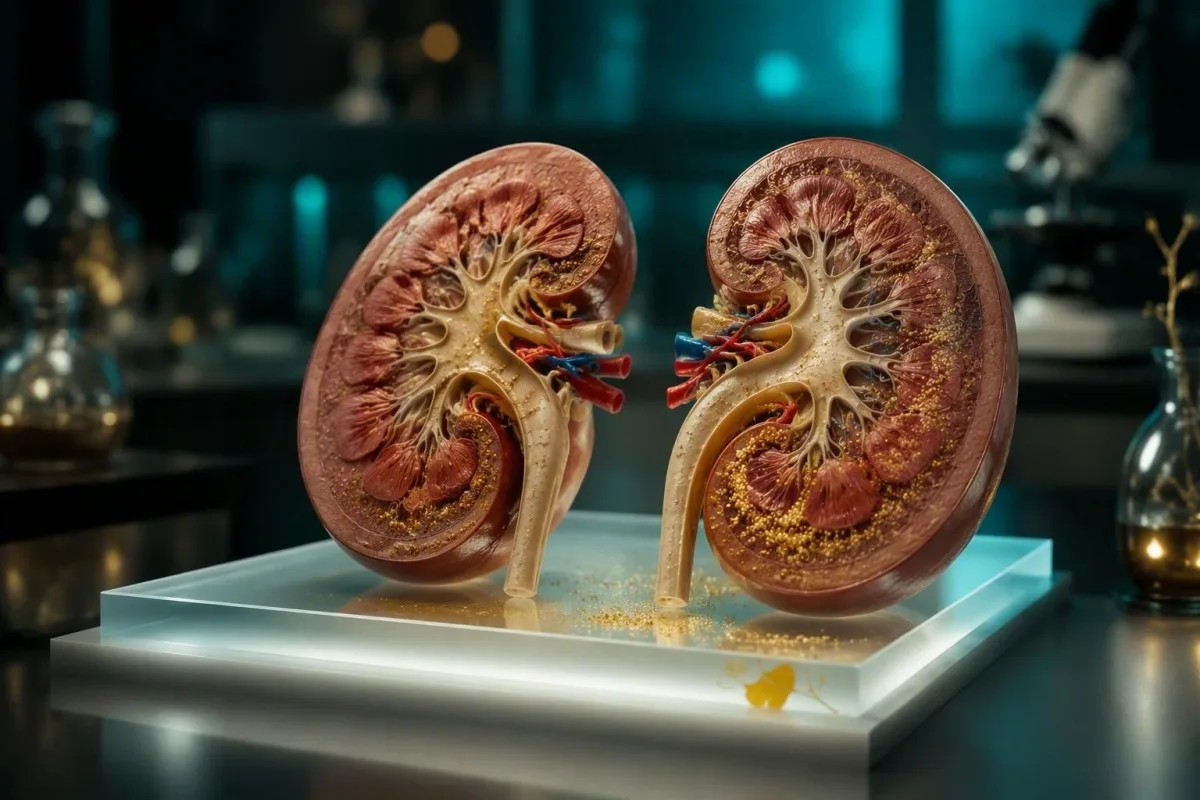
አብዛኛዎቹ ቀላል የፎስፌት ውጤቶች ምልክቶች አያስከትሉም ፣ ነገር ግን ዝቅተኛ ካልሲየም ፣ የኩላሊት ውድቀት ፣ ከባድ ድክመት ፣ ግራ መጋባት ፣ ወይም የጡንቻ መወጠር ያለበት ከፍተኛ ፎስፌት ፈጣን ክሊኒካዊ ግምገማ ይፈልጋል። ቁጥሩ ከእሱ ጋር ባለው ንድፍ ያነሰ ጠቀሜታ አለው።.


- ብዙውን ጊዜ ዝምተኛ፡ የአዋቂዎች ፎስፌት በብዛት ከ 0.80-1.50 mmol/L (2.5-4.5 mg/dL) አካባቢ ነው ፣ እና ትንሽ ለይቶ የመጣ ጭማሪ ብዙ ጊዜ ምልክቶችን አያስከትልም።.
- አስቸኳይ ንድፍ፡ ከ 2.0 mmol/L (6.2 mg/dL) በላይ የፎስፌት ውጤት ከዝቅተኛ ካልሲየም ፣ ድክመት ፣ መንቀጥቀጥ ፣ ግራ መጋባት ፣ ወይም የሽንት ውፅዓት መቀነስ ጋር በተመሳሳይ ቀን ክሊኒካዊ ምክር ይፈልጋል።.
- የኩላሊት ፍንጭ፡ የማያቋርጥ የደም ማነስ ችግር ብዙውን ጊዜ የኩላሊት መወገድ በመቀነሱ ምክንያት ነው ፣ በተለይም eGFR ከ 30 mL/min/1.73 m² በታች ሲሆን።.
- ራስዎ አይታከሙ፡ የካልሲየም ተጨማሪዎች ፎስፌት በሚበዛበት ጊዜ ደህንነቱ የተጠበቀ ላይሆን ይችላል ምክንያቱም የካልሲየም-ፎስፌት ክምችት አደጋ በሁለቱም ውጤቶች ላይ የተመሠረተ ነው።.
- የተደበቀ ምክንያት፡ አስቸጋሪ የናሙና ስዕል የሕዋስ አካላትን ሊያፈርስ እና የውሸት የከፍተኛ ፎስፌት ውጤትን ሊፈጥር ይችላል ፤ ተደጋጋሚ ምርመራ ጥያቄውን ሊፈታ ይችላል።.
- የካንሰር ሕክምና ድንገተኛ አደጋ፡ የደም ማነስ ፣ ፖታሲየም ፣ ዩሬት እና የ creatinine መዛባት ጋር ፈጣን የፎስፌት ጭማሪ የ ዕጢ lysis syndrome ምልክት ሊሆን ይችላል እና የአደጋ ጊዜ እንክብካቤ ይፈልጋል።.
- ምርጥ የሚቀጥሉ ምርመራዎች፡ ፎስፌትን ከካልሲየም ፣ አልቡሚን ፣ creatinine/eGFR ፣ ማግኒዥየም ፣ ቢካርቦኔት ፣ parathyroid hormone ፣ እና የመድኃኒት ግምገማ ጋር ይድገሙት።.
የትኞቹ የከፍተኛ ፎስፌት ምልክቶች አሁን እንክብካቤ ይፈልጋሉ?
የከፍተኛ ፎስፌት ምልክቶች አለመመቸት ሲሆን የፖታስየም መጠን ሲጨምር, ነገር ግን ከፍተኛ ድክመት፣ የጡንቻ ህመም መኮማተር፣ በአፍ ዙሪያ መደንዘዝ፣ መናወጥ፣ ግራ መጋባት፣ የደረት ብልጭታ፣ ወይም የሽንት መጠን በከፍተኛ ሁኔታ መቀነስ አስቸኳይ ግምገማ ያስፈልጋቸዋል— በተለይም ከታወቀ የኩላሊት በሽታ ጋር። ውጤቱ ብቻውን የአደጋ ምርመራ አይደለም፤ ተያያዥ ካልሲየም፣ ፖታስየም፣ የኩላሊት ተግባር እና የመለዋወጥ ፍጥነት አደጋውን ይወስናሉ።.
ለመናወጥ እንቅስቃሴ፣ ለመሳት፣ ለአዲስ ግራ መጋባት፣ ለከፍተኛ የትንፋሽ እጥረት፣ ለዘላቂ የልብ ምት መዛባት፣ ወይም ፎስፌት ከተገኘ በኋላ ለከፍተኛ ድክመት አሁን ወደ ድንገተኛ እንክብካቤ ይሂዱ። እነዚህ ምልክቶች ሊያንጸባርቁ ይችላሉ ዝቅተኛ ionised calcium, ፣ ከፍተኛ ፖታስየም፣ አሲዶሲስ፣ ወይም የኩላሊት ውድቀት ከፎስፌት ብቻ ይልቅ፤ እያንዳንዳቸው በሰዓታት ውስጥ የልብ እና የነርቭ ጡንቻ ተግባር ላይ ተጽዕኖ ሊያሳድሩ ይችላሉ።.
ፎስፌት ከ 2.0 mmol/L (6.2 mg/dL) በላይ ከሆነ፣ ወይም የ eGFR ከ 30 mL/min/1.73 m² በታች ከሆነ በፍጥነት እየጨመረ ከሆነ ሐኪሙን ወይም የኩላሊት ቡድኑን በተመሳሳይ ቀን ይደውሉ። ኪሞቴራፒ፣ የደም ካንሰር ሕክምና፣ ወይም የዳያሊስስ ሕክምና የሚወስዱ ሰዎች የፍጥነት ሴል መበስበስ ፎስፌት እና ፖታስየም አንድ ላይ ሊለቁ ስለሚችሉ መደበኛ ግምገማ መጠበቅ የለባቸውም፤ የእኛን ይመልከቱ የዕጢ lysis የላብ መመሪያ.
በእኔ ክሊኒካዊ ልምድ፣ 1.55 mmol/L ፎስፌት ያለው እና ምንም የኩላሊት በሽታ የሌለበት ሰው የድንገተኛ ክፍል ጉብኝት ብዙ ጊዜ አያስፈልገውም። የማሳስበው ሰው 2.35 mmol/L፣ ማቅለሽለሽ፣ የሽንት መጠን መቀነስ፣ 1.82 mmol/L ካልሲየም፣ እና በድንገት እየባሰ የሚሄድ ክሬቲኒን ያለው ሰው ነው፤ ይህም ሙሉ በሙሉ የተለየ ሁኔታ ነው።.
ምልክቶቹ ከባድ ከሆኑ እራስዎን አያሽከርክሩ
መናወጥ፣ መውደቅ፣ ከፍተኛ ግራ መጋባት፣ ወይም ሽባ የሚያደርጉ መኮማተር ከመዘግየት የውጪ የደም ምርመራ ይልቅ የአካባቢውን ድንገተኛ አገልግሎቶች ያረጋግጣሉ። የላቦራቶሪ ሪፖርት እና አሁን ያሉ የመድኃኒቶች ዝርዝር፣ የሆድ መድኃኒቶችን፣ enemas፣ ተጨማሪ ምግቦችን እና የካንሰር መድኃኒቶችን ጨምሮ ያምጡ።.
የከፍተኛ ፎስፌት መጠን በእውነቱ ምን ማለት ነው
ሃይፐርፎስፌትሚያ ማለት የፎስፌት መጠን ከላቦራቶሪ ማጣቀሻ ክልል በላይ ነው።, ፣ በአዋቂዎች ውስጥ በተለምዶ ከ 1.50 mmol/L ወይም 4.5 mg/dL በላይ። የማጣቀሻ ክልሎች እንደ ምርመራው፣ ዕድሜው እና ጊዜው ይለያያሉ፣ ስለዚህ ባንዲራ ምርመራ እንጂ ምርመራ ለመጀመርያ ነጥብ ነው።.
ፎስፌት በATP የኃይል ማስተላለፊያ፣ የሕዋስ ሽፋን፣ የአጥንት ማዕድን እና የአሲድ-መሠረት መከላከያ ውስጥ ጥቅም ላይ የሚውል ክፍያ ያለው ማዕድን ነው። አዋቂዎች በተለምዶ 0.80-1.50 mmol/L (2.5-4.5 mg/dL) ሲኖራቸው፣ ልጆችና ታዳጊዎች የሚያድግ አጥንት ፎስፌትን በፍጥነት ስለሚቀይር በተለምዶ ከፍ ያለ እሴት አላቸው።.
የድንበር ውጤት ለማግኘት የጾም የጠዋት ናሙና ብዙ ጊዜ ይመረጣል ምክንያቱም ፎስፌት ዕለታዊ ምት ስለሚከተል እና በሌሊት የመጨመር አዝማሚያ ስላለው። ከምሽት ምግብ በኋላ ወይም ከባድ የአካል ብቃት እንቅስቃሴ በኋላ 1.55 mmol/L ያለው እሴት ከጾም ምርመራዎች ጋር እየቀነሰ በሄደ eGFR 1.55 mmol/L ከተለያየ ሁኔታ ጋር ይተረጎማል፤ የእኛ የፎስፌት ማጣቀሻ ክልል መመሪያ ለምን የላቦራቶሪ ማስጠንቀቂያ ምልክቶች አውድ (context) እንደሚፈልጉ ያብራራል።.
ካንቴስቲ እ.ኤ.አ. AI የደም ምርመራ ተንታኝ ያም የካልሲየም፣ ክሬቲኒን፣ eGFR፣ አልቡሚን፣ እና ቀዳሚ ውጤቶች ጎን ለጎን ፎስፌትን የሚያነብ እንጂ አንድ ጎልቶ የወጣ መስመርን ፍርድ አድርጎ የማይቆጥር ነው። ያ የፓተርን-ఆధారిత አቀራረብ፣ አሃዶች በmmol/L እና mg/dL መካከል ሲለያዩ በተለይ ጠቃሚ ነው።.
ለምን ቀላል የደም ማነስ ችግር ዝምተኛ የሆነው
Mild hyperphosphatemia symptoms are usually absent, because phosphate itself does not reliably produce a sensation at modest elevations. Symptoms emerge mainly when phosphate lowers ionised calcium, accompanies kidney failure, or rises abruptly.
Phosphate can bind calcium, but total calcium may still appear normal while biologically active ionised calcium shifts downward. That is why tingling, cramps, or carpopedal spasm are more informative than vague fatigue when deciding whether a mineral imbalance is clinically active.
A useful practical distinction: tiredness, headache, and poor concentration are nonspecific and should not automatically be blamed on a phosphate of 1.58 mmol/L. In a patient with chronic kidney disease, those same symptoms deserve broader review for anaemia, acidosis, uraemia, medicines, and fluid status; our low eGFR symptom guide maps the warning signs better than phosphate alone.
Dr. Thomas Klein's approach is to ask what changed: an acute illness, phosphate-containing bowel preparation, dehydration, new vitamin D treatment, dialysis timing, or a laboratory artifact. A stable isolated result over years is not managed like a sharp rise over 48 hours.
Symptoms may be caused by the underlying condition
Reduced appetite, itch, nausea, and sleep disruption in advanced kidney disease are often attributed to high phosphate online, but they are not specific phosphate symptoms. Treating only the number can miss the underlying kidney and mineral-bone disorder.
የካልሲየም ወይም የኤሌክትሮላይት ችግሮችን የሚያመለክቱ ምልክቶች
Muscle cramps, twitching, tingling, spasms, seizures, and rhythm symptoms are the high phosphate symptoms that deserve fastest attention, because they can signal falling ionised calcium or another simultaneous electrolyte disorder. New neurological symptoms should never be monitored casually at home.
Low calcium symptoms often begin with pins-and-needles around the lips or fingers, hand stiffness, calf cramping, and facial twitching. Severe hypocalcaemia can prolong the QT interval on ECG and provoke seizures, which is why a clinician may request ionised calcium rather than relying only on corrected total calcium; compare our low calcium urgent signs.
Chest discomfort, racing heart, or a skipped-beat sensation is not a recognised reliable symptom of phosphate alone, but it needs urgent assessment when potassium is also raised. A potassium above 6.0 mmol/L is commonly treated as an urgent threshold, although ECG changes and the speed of rise matter more than a single cutoff.
Kantesti AI ከዚህ በመመልከት ይተረጉማል elevated phosphate level by checking linked electrolyte patterns, including potassium, bicarbonate, calcium, and magnesium. It cannot replace an ECG, examination, or emergency evaluation when symptoms suggest electrical instability.
The calcium-phosphate product is not a home calculation
Clinicians sometimes consider calcium-phosphate burden in advanced CKD, but modern treatment decisions do not rely on one product threshold in isolation. KDIGO recommends serial assessment of phosphate, calcium, and parathyroid hormone together rather than chasing one biochemical target (KDIGO CKD-MBD Update Work Group, 2017).
የኩላሊት በሽታ የመሆን ዕድሉ መቼ ነው
Chronic kidney disease is the most common cause of persistent high phosphate in adults, because functioning nephrons excrete less phosphate as GFR falls. Levels often remain normal until later CKD because FGF23 and parathyroid hormone compensate for a time.
Phosphate commonly rises more clearly when eGFR falls below about 30 mL/min/1.73 m², although individual timing varies with diet, residual kidney function, parathyroid hormone, and dialysis. KDIGO's 2017 CKD-MBD guideline advises lowering elevated phosphate toward the normal range in CKD, based on repeated measurements rather than preventive treatment of a normal result (KDIGO CKD-MBD Update Work Group, 2017).
In dialysis care, the pre-dialysis phosphate number also depends on the gap since the last session and whether the sample was drawn before or after treatment. Missing a session, constipation, poor binder timing, or a recent dietary change can move phosphate substantially without representing a permanent new baseline; see the CKD stages overview.
ካንቴስቲ እ.ኤ.አ. የAI ባዮማርከር ትርጓሜ መድረክ that can display phosphate and eGFR trends across reports, helping patients notice a persistent pattern before a clinic visit. Trend review is useful, but dialysis prescriptions and phosphate binders must remain with the renal team.
High phosphate is not proof of CKD
A normal creatinine does not exclude every kidney issue, yet a single high phosphate result does not diagnose CKD either. CKD requires evidence of kidney abnormality lasting at least 3 months, usually interpreted with eGFR and urine albumin results.
ፈጣን የፎስፌት ጭማሪዎች ፤ የሕዋስ መበላሸት እና ድንገተኛ አደጋዎች
A rapid phosphate rise can be an emergency when many cells break down at once, releasing intracellular phosphate, potassium, urate, and nucleic acids into circulation. This pattern is most concerning after chemotherapy, during severe crush injury, or with major tissue injury.
Tumour lysis syndrome is classically assessed around cancer treatment, particularly in high-burden blood cancers, but it can occasionally occur spontaneously. The Cairo-Bishop definition uses phosphate at or above 1.45 mmol/L (4.5 mg/dL) in adults or a 25% rise from baseline as one laboratory criterion, interpreted with urate, potassium, calcium, timing, and clinical complications (Cairo & Bishop, 2004).
An adult with chemotherapy exposure plus vomiting, weakness, reduced urine, palpitations, or confusion should seek emergency assessment rather than trying to lower phosphate through diet. The dangerous issue is the combined metabolic cascade—hyperkalaemia, acute kidney injury, and hypocalcaemia—not simply the phosphate value.
Other acute causes include rhabdomyolysis, severe haemolysis, major burns, and phosphate-containing bowel preparations or enemas. New severe muscle pain and dark urine after extreme exertion warrants urgent testing for creatine kinase, creatinine, potassium, and phosphate; our high CK danger guide covers this separate but related emergency.
Why symptoms can arrive late
The laboratory change may precede symptoms by several hours, especially after cancer therapy. This is one reason oncology teams schedule repeat chemistry panels rather than waiting for a patient to feel unwell.
የሆርሞን ፣ የቫይታሚን ዲ እና የአጥንት ምክንያቶች ሊታሰብባቸው ይገባል
Low parathyroid hormone and excessive active vitamin D activity can raise phosphate, because both normally influence kidney phosphate handling and intestinal absorption. These causes are less common than CKD but matter when kidney function is preserved.
Hypoparathyroidism commonly produces the combination of low calcium, high phosphate, tingling, cramps, and sometimes a history of neck surgery or autoimmune disease. PTH should be interpreted beside calcium, magnesium, 25-hydroxyvitamin D, and kidney function; an isolated low PTH can be misleading if calcium was not measured simultaneously.
Vitamin D toxicity usually causes high calcium more prominently than high phosphate, but calcitriol and related active vitamin D medicines can raise both calcium and phosphate, especially in CKD. Do not stop prescribed calcitriol abruptly without advice, but report supplements and dose changes accurately; our high vitamin D symptom guide የማስጠንቀቂያ ንድፍ ያብራራል።.
Rare inherited renal phosphate-handling disorders exist, yet they are not the first explanation for a newly abnormal adult result. In my experience, medication reconciliation and a repeat specimen answer more cases than exotic testing.
Bone turnover is not the same as serum phosphate
A fracture or high alkaline phosphatase does not automatically explain high phosphate. Bone metabolism is dynamic, but serum phosphate is governed predominantly by renal excretion and hormonal signals; see our ALP after fracture explanation.
አመጋገብ ፣ ተጨማሪዎች ፣ ተጨማሪዎች እና መድኃኒቶች
Diet alone rarely causes marked hyperphosphatemia in people with normal kidneys, because healthy kidneys increase phosphate excretion after higher intake. Processed-food phosphate additives, cola-type drinks, and supplements matter more when kidney clearance is reduced.
Organic phosphate in beans, nuts, whole grains, dairy, fish, and meat is absorbed variably; plant phytate phosphate is generally less absorbable than additive phosphate. In advanced CKD, clinicians often focus first on highly absorbable additives rather than indiscriminately removing all nutritious protein foods.
Phosphate-containing laxatives, enemas, and bowel preparations can cause dangerous acute hyperphosphatemia in older adults, people with CKD, and those taking medicines that affect kidney perfusion. Tell the prescriber about ACE inhibitors, ARBs, diuretics, NSAIDs, vitamin D products, and all over-the-counter preparations before a colonoscopy preparation is chosen.
Do not start calcium-based phosphate binders after reading a single result online. KDIGO advises restricting calcium-based binder dose in adults receiving phosphate-lowering treatment, partly because positive calcium balance and vascular calcification are genuine concerns in CKD (KDIGO CKD-MBD Update Work Group, 2017); our kidney supplement safety guide adds practical cautions.
Read ingredients, not marketing claims
Ingredient terms such as phosphoric acid, phosphate, polyphosphate, and pyrophosphate can identify additives, although labels vary internationally. A renal dietitian can preserve adequate protein, calories, and cultural food preferences while reducing the highest-yield sources.
የፎስፌት ውጤቱ በሐሰት ከፍ ሊል ይችላል?
A falsely high phosphate result can occur when the sample is haemolysed, because cellular elements contain phosphate that can leak into serum or plasma after collection. A surprising result should be repeated before major treatment unless symptoms or related abnormalities demand immediate action.
Laboratories often flag haemolysis, but not every degree of sample interference produces a clear patient-facing note. Delayed separation, difficult collection, prolonged tourniquet time, or rough transport can alter chemistry results, and a repeat clean sample may be more useful than a broad scan of rare diseases.
Pseudohyperphosphatemia is also described with marked hyperlipidaemia, hyperglobulinaemia, or some assay interferences, though it is uncommon. If phosphate is high while calcium, kidney function, clinical state, and repeat result are all reassuring, ask whether the lab can review analytical interference; our repeat test after haemolysis guide explains what a redraw can clarify.
Kantesti AI flags discordant results for follow-up but does not label a high phosphate result as harmless merely because one value looks out of place. The safest rule is simple: repeat an unexpected mild result soon, while escalating immediately if symptoms or kidney markers are concerning.
Do not deliberately dehydrate before retesting
Arrive normally hydrated unless your clinician gives different instructions, and avoid unusually heavy exercise immediately beforehand. Bring the first report so the laboratory and clinician can compare method, sample quality, and timing.
ምክንያቱን ለማወቅ ሐኪሞች የሚጠቀሟቸው ምርመራዎች
The first follow-up panel for high phosphate usually includes repeat phosphate, calcium, albumin, creatinine/eGFR, potassium, bicarbonate, magnesium, and sometimes ionised calcium. The correct additional tests depend on whether the pattern suggests CKD, low PTH, acute cell breakdown, or a laboratory artifact.
PTH, 25-hydroxyvitamin D, alkaline phosphatase, and urinary phosphate can help distinguish hormonal from renal causes. In selected cases, clinicians assess tubular phosphate reabsorption, but this is not useful during an unstable acute illness or before confirming the serum result.
If kidney dysfunction is present, urine albumin-creatinine ratio, urinalysis, blood pressure, medication review, and renal imaging may be more revealing than repeated phosphate alone. Creatinine needs interpretation in context of muscle mass, diet, hydration, and recent exercise; review our BUN እና ክሬአቲኒን መመሪያ ይመልከቱ.
Kantesti's neural network groups mineral, renal, and acid-base results into a structured question list for the appointment. Kantesti is an በ AI የተጎላበተ የደም ምርመራ ትንተና መሳሪያ designed to support understanding of laboratory patterns, not to diagnose hypoparathyroidism or prescribe treatment.
Bring a complete list
Include the exact name and dose of prescribed drugs, supplements, antacids, protein powders, laxatives, and bowel preparations. In renal medicine, a product's formulation can matter as much as its brand category.
ሐኪሞች ፎስፌትን በደህና ሁኔታ እንዴት እንደሚቀንሱ
Treatment targets the cause of high phosphate rather than simply suppressing the number. Acute severe hyperphosphatemia may require intravenous fluids, treatment of low calcium or high potassium, phosphate binders, and sometimes dialysis under hospital supervision.
For CKD, treatment commonly includes dietary counselling, improved dialysis adequacy where relevant, and phosphate binders taken with meals. Binder choice is individual: calcium-containing agents, sevelamer, lanthanum, iron-based agents, and others have different side-effect profiles, pill burdens, costs, and calcium implications.
Never take a phosphate binder away from meals and expect the same benefit; it works by binding dietary phosphate in the gut. Conversely, taking extra doses can cause constipation, diarrhoea, calcium disturbances, or interactions with medicines such as levothyroxine and some antibiotics.
A 52-year-old patient I reviewed had phosphate of 1.92 mmol/L despite a reasonable diet; the issue was taking every binder dose two hours after meals to avoid nausea. Adjusting timing with the renal pharmacist—not radically restricting food—produced a safer and more sustainable plan; our kidney diet AI safety article explains why diet advice must stay clinically supervised.
Rapid correction can be risky
Treating symptoms of hypocalcaemia may require calcium, but indiscriminate calcium administration can increase calcium-phosphate precipitation risk when phosphate is very high. Emergency clinicians balance immediate neuromuscular safety against this risk using repeat results and ECG monitoring.
ሕፃናት ፣ እርግዝና እና የደም ንፅህና ፤ የተለያዩ ህጎች
Children normally have higher phosphate levels than adults, and dialysis patients require individualized targets and timing-aware interpretation. A number that is high for a 45-year-old may be normal during adolescence, while the same number in a dialysis patient can require a treatment review.
Paediatric reference intervals vary substantially by age and laboratory, often exceeding adult upper limits during infancy and growth spurts. Parents should use the range printed for the child's age, not an adult internet cutoff; high phosphate with poor feeding, lethargy, vomiting, or twitching still warrants prompt paediatric advice.
Pregnancy can change kidney filtration and mineral metabolism, but phosphate is not a stand-alone screening test for pregnancy complications. Persistent vomiting, reduced urine output, severe headache, visual change, or swelling should be assessed based on the full clinical picture, not attributed to phosphate.
For people on dialysis, a pre-dialysis result should be interpreted with the day of the dialysis cycle, missed sessions, residual urine output, and binder adherence. Kantesti supports secure longitudinal review, while our የሕክምና አማካሪ ቦርድ emphasizes that renal teams set individualized treatment goals.
Older adults need medication scrutiny
Older adults are disproportionately affected by phosphate-containing bowel products and acute kidney injury from dehydration or interacting medicines. A medication review after an abnormal result is often more productive than an extreme diet change.
ለሚቀጥለው ክሊኒካዊ ግምገማዎ በፊት ምን ማድረግ አለበት
For a mild, symptom-free elevated phosphate level, arrange repeat testing and bring context rather than making abrupt dietary or supplement changes. The most useful details are timing, fasting status, hydration, illness, exercise, medicines, kidney history, and previous results.
Write down whether you had a bowel preparation, enema, laxative, chemotherapy, major exercise session, vomiting illness, or missed dialysis within the preceding 7 days. Ask for the actual phosphate unit and reference range, because 1.8 mmol/L and 1.8 mg/dL are radically different clinical values.
Request copies of calcium, albumin, creatinine, eGFR, potassium, bicarbonate, and magnesium from the same draw. A physician can usually triage the result more accurately from this cluster than from phosphate alone; our የኤሌክትሮላይት ፓነል መመሪያ ይረዳል የተለመዱ ጥምሮችን ያሳያል።.
As of September 12, 2026, I would advise most asymptomatic adults with a small isolated elevation to contact their usual clinician within days, not months. If a report is difficult to read, Kantesti's clinical validation approach explains how our AI organizes source values and uncertainty for discussion with a qualified professional.
የመጠየቅ ዋጋ ያላቸው ጥያቄዎች
Ask whether the result was repeated, whether the sample was haemolysed, whether calcium was corrected or ionised, and whether kidney function has changed from baseline. Those four questions often prevent both false reassurance and unnecessary alarm.
ለከፍተኛ ፎስፌት ውጤቶች ምልክት-መጀመሪያ እቅድ
Most people with slightly high phosphate feel nothing and need confirmation plus a cause-focused review; people with neurological, muscle, cardiac, or kidney-failure symptoms need urgent care. The safest response is proportional to the number, the trend, the companion tests, and how unwell the person is.
Use emergency services for seizures, collapse, severe confusion, disabling spasms, severe weakness, or palpitations with a known major electrolyte abnormality. Seek same-day medical advice for phosphate above 2.0 mmol/L (6.2 mg/dL), new oliguria, active cancer treatment, advanced CKD, or a rapidly changing result.
For a borderline isolated result, repeat testing under ordinary conditions and avoid self-prescribed binders, calcium, or drastic protein restriction. The goal is not a perfect-looking report; it is finding whether renal clearance, hormonal regulation, an acute event, intake, or sample quality explains the change.
Dr. Thomas Klein recommends treating a laboratory result as a clinical clue, never as a diagnosis delivered by an app or search result. Our የAI ቴክኖሎጂ መመሪያ describes how Kantesti AI preserves the original laboratory values and highlights questions for clinician review rather than replacing medical care.
መደምደሚያ
High phosphate is usually a sign to look wider—particularly at kidney function and calcium—not a symptom diagnosis by itself. Prompt review is most valuable when the number is marked, rising, paired with abnormal electrolytes, or accompanied by real symptoms.
በተደጋጋሚ የሚጠየቁ ጥያቄዎች
ከፍተኛ ፎስፌት ምን ይሰማዋል?
High phosphate usually causes no specific symptoms when the elevation is mild, such as 1.51-1.80 mmol/L (4.6-5.6 mg/dL). When phosphate rises substantially or lowers ionised calcium, people may develop tingling around the mouth, muscle cramps, twitching, hand spasms, weakness, or seizures. Nausea, itch, and fatigue are more often related to advanced kidney disease or another accompanying problem than to phosphate alone. New neurological, cardiac, or severe muscle symptoms need urgent medical assessment.
የ1.6 mmol/L የፎስፌት መጠን አደገኛ ነው?
A phosphate level of 1.6 mmol/L, approximately 5.0 mg/dL, is a mild elevation and is often not immediately dangerous in a well adult with normal calcium and kidney function. It should still be confirmed and interpreted with creatinine/eGFR, calcium, albumin, potassium, bicarbonate, medicines, and sample quality. Persistent results above the laboratory range can be an early clue to kidney or hormone-related phosphate imbalance. Urgency increases if symptoms occur, kidney function is declining, or phosphate rises above 2.0 mmol/L (6.2 mg/dL).
ከፍተኛ ፎስፈረስ መቼ ወደ ድንገተኛ ክፍል መሄድ አለቦት?
High phosphorus warrants emergency care when it occurs with seizure, fainting, confusion, severe muscle spasms, sustained palpitations, severe weakness, or markedly reduced urine output. Emergency assessment is also appropriate after chemotherapy or a major muscle injury when phosphate rises alongside potassium, urate, or creatinine. A phosphate concentration above 2.0 mmol/L (6.2 mg/dL) merits same-day clinical advice even without symptoms, especially in chronic kidney disease. The emergency risk comes from the whole electrolyte and kidney-function pattern, not phosphate alone.
ድርቀት ከፍተኛ ፎስፌት ሊያስከትል ይችላል?
Dehydration can concentrate laboratory values and can contribute to a mildly elevated phosphate result, but it should not be assumed to explain a persistent or marked elevation. Dehydration may also reduce kidney perfusion and temporarily worsen creatinine, which makes the result more clinically relevant rather than less. Normal hydration and a repeat chemistry panel can help clarify a borderline value. Severe vomiting, diarrhoea, dizziness, low urine output, or an eGFR decline needs medical review rather than self-management alone.
ምግቦች በጤናማ ኩላሊት ውስጥ ከፍተኛ ፎስፌት ሊያስከትሉ ይችላሉ?
Diet alone rarely causes major hyperphosphatemia when kidney function is normal because healthy kidneys increase phosphate excretion. Highly absorbable inorganic phosphate additives in processed foods, cola-type beverages, supplements, and bowel preparations become more consequential when eGFR is reduced. A persistent phosphate above 1.50 mmol/L (4.5 mg/dL) should prompt review for kidney disease, medicines, hormonal causes, and laboratory interference before imposing severe dietary restrictions. A renal dietitian can tailor changes without compromising protein and calorie intake.
ከፍ ያለ ፎስፌት ምን ምርመራዎች ጋር ይጣራል?
Clinicians commonly repeat phosphate with calcium, albumin, creatinine, eGFR, potassium, bicarbonate, and magnesium to assess high phosphate safely. Parathyroid hormone, 25-hydroxyvitamin D, alkaline phosphatase, urinalysis, and urinary phosphate may be added when the initial pattern suggests a hormonal or renal cause. Ionised calcium is especially useful when tingling, cramps, spasms, or seizures suggest biologically active hypocalcaemia. In suspected tumour lysis syndrome, urate, potassium, creatinine, calcium, phosphate, and urine output are assessed urgently.
ዛሬ የAI-የኃይል የደም ምርመራ ትንተና ያግኙ
በፍጥነት እና ትክክለኛ የላቦራቶሪ ምርመራ ትንተና ለማግኘት Kantestiን የሚያምኑ ከ2 ሚሊዮን በላይ ተጠቃሚዎችን ይቀላቀሉ። የደም ምርመራ ውጤትዎን ይስቀሉ እና በ15,000+ ባዮማርከሮች ላይ የተሟላ ትርጓሜን በሰከንዶች ውስጥ ይቀበሉ።.
📚 የተጠቀሱ የምርምር ህትመቶች
📖 ውጫዊ የሕክምና ማጣቀሻዎች
⚕️ የሕክምና ማስተባበያ
ይህ ጽሑፍ ለትምህርታዊ ዓላማ ብቻ ነው እና የሕክምና ምክር አይደለም። ለምርመራ እና ለሕክምና ውሳኔዎች ሁልጊዜ ብቁ የጤና ባለሙያን ያማክሩ።.
የE-E-A-T እምነት ምልክቶች
ልምድ
በሐኪም መሪነት የላቦራቶሪ ትርጓሜ የስራ ፍሰቶች ክሊኒካዊ ግምገማ።.
ባለሙያነት
በክሊኒካዊ አውድ ውስጥ ባዮማርከሮች እንዴት እንደሚሰሩ ላይ የላቦራቶሪ ሕክምና ትኩረት።.
ስልጣን ያለው
በዶክተር ቶማስ ክላይን የተፃፈ ከዶክተር ሳራ ሚቸል እና ፕሮፌሰር ዶክተር ሃንስ ዌበር ግምገማ ጋር።.
አስተማማኝነት
ለማስጠንቀቂያ ምላሽ መቀነስ ግልጽ የቀጣይ መንገዶች ያለው በማስረጃ የተደገፈ ትርጓሜ።.