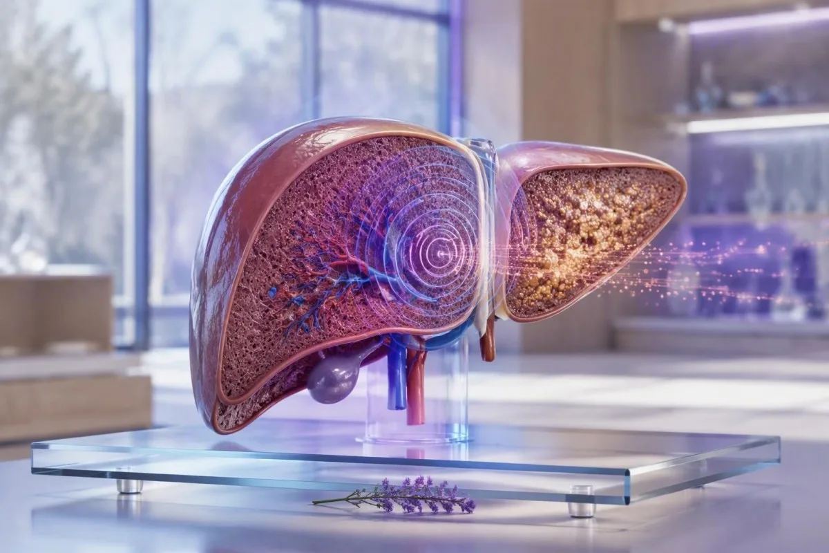
FibroScan results estimate liver stiffness in kPa and liver fat with a CAP score; they do not diagnose fibrosis or cirrhosis alone. As of September 28, 2026, the most useful report combines the number, scan quality, fasting status, liver blood tests, and the reason you were scanned.


- Liver stiffness measurement below 8 kPa usually makes advanced fibrosis unlikely in metabolic liver disease when the scan is reliable.
- FibroScan kPa score of 8-12 kPa is an indeterminate zone that commonly needs blood-test context or repeat assessment.
- Higher stiffness above 12-15 kPa raises concern for advanced fibrosis or cirrhosis, but congestion, hepatitis, cholestasis, and meals can also elevate it.
- CAP score meaning relates to liver fat, not scar tissue; roughly 238, 260, and 290 dB/m are commonly used research thresholds for increasing steatosis.
- உண்ணாவிரதம் for at least 3 hours before transient elastography reduces meal-related false elevation of liver stiffness.
- Reliable scans usually include at least 10 valid readings and an IQR/median ratio of 30% or less.
- அவசரமாக மதிப்பாய்வு செய்யவும் is warranted for jaundice, confusion, vomiting blood, black stools, or abdominal swelling, regardless of the FibroScan number.
- Specialist follow-up is more appropriate than self-diagnosis when stiffness is 8 kPa or higher, platelets are low, or liver enzymes remain abnormal.
What the Two Main Numbers on a FibroScan Report Measure
FibroScan results usually show two different measurements: liver stiffness in kilopascals (kPa) and fat attenuation in decibels per metre (dB/m). A higher kPa value can reflect fibrosis, but it can also rise temporarily from a recent meal, active hepatitis, bile blockage, or fluid pressure around the liver.
Liver stiffness measurement uses vibration-controlled transient elastography: a small mechanical pulse travels through the liver, and the device estimates how quickly that wave moves. Stiffer tissue transmits the wave faster. In my clinic, the common mistake is treating a 9.5 kPa result as a diagnosis rather than a probability signal that must be checked against the person's cause of liver disease.
CAP, or controlled attenuation parameter, estimates how much the ultrasound signal weakens as it passes through the liver. CAP is reported in dB/m, typically on a scale of about 100-400 dB/m, and primarily reflects steatosis. A high CAP score does not prove steatohepatitis, and a low CAP score does not rule out fibrosis from alcohol, viral hepatitis, autoimmune disease, or iron overload.
கான்டெஸ்டி என்பது ஒரு AI இரத்த பரிசோதனை பகுப்பாய்வி that places liver enzymes, platelets, glucose, lipids, and iron markers beside imaging results rather than reading either in isolation. That distinction matters: a 7.2 kPa result with normal platelets and normal bilirubin means something quite different from 7.2 kPa with falling platelets and an elevated INR. Start with the FIB-4 கணக்கீடு when you have the required laboratory values.
Why one number cannot name the disease
FibroScan does not identify the cause of stiffness. A person with heart failure, acute alcohol-related hepatitis, or an obstructed bile duct can have a kPa value in a range otherwise associated with substantial fibrosis. The clinical question is therefore not “What stage am I?” but “Is this result credible, persistent, and consistent with my wider liver assessment?”
How to Read a FibroScan kPa Score Without Overcalling Fibrosis
A FibroScan kPa score measures resistance to a shear wave, not scar tissue directly. In many adults with metabolic dysfunction-associated steatotic liver disease, a reliable value below 8 kPa has a good negative predictive value for advanced fibrosis.
The kPa scale is not linear in clinical meaning. A change from 5 to 7 kPa may reflect ordinary biological or technical variation, whereas a persistent shift from 9 to 15 kPa is more concerning—provided the same probe, preparation, and disease context apply. EASL guidance treats liver stiffness below 8 kPa as useful for ruling out compensated advanced chronic liver disease in several common settings (EASL, 2021).
Different conditions need different cutoffs. In untreated chronic hepatitis C, values around 7-8 kPa have often been used as possible significant fibrosis thresholds, while cirrhosis thresholds commonly fall around 12-14 kPa. In metabolic liver disease, obesity and the XL probe alter performance, so transplanting a cutoff from a hepatitis study can mislead.
I have reviewed scans in lean patients with a 6.5 kPa reading and substantial metabolic risk, then in larger patients with 10 kPa readings obtained using poor-quality M-probe attempts. Neither should be casually labelled. For the broader clinical context, review our MASLD பரிசோதனை வழிகாட்டி.
Why the unit is kPa
A kilopascal is a pressure unit. FibroScan converts wave-propagation data into an estimate of tissue stiffness, which is why results are reported in kPa rather than a percentage of fibrosis. A liver stiffness measurement of 15 kPa is not “twice as scarred” as 7.5 kPa.
Practical Fibrosis-Risk Bands for Liver Stiffness Results
For many adults with suspected metabolic liver disease, less than 8 kPa is a low-risk band, 8-12 kPa is indeterminate, and above 12-15 kPa is a high-risk band for advanced fibrosis. These are triage bands, not universal fibrosis stages or a substitute for specialist assessment.
EASL's 2021 non-invasive test guidance uses 8 kPa க்கும் குறைவாக to help exclude compensated advanced chronic liver disease and 12-15 kPa or higher to help rule it in, depending on the underlying disease and test quality (EASL, 2021). Values around 8-12 kPa are where I most often avoid reassuring or alarming language until I see platelets, AST, ALT, bilirubin, albumin, and the scan reliability metrics.
A reliable reading of 20-25 kPa or more can raise concern for clinically significant portal hypertension, especially when platelets are below 150 × 10^9/L. It still does not diagnose varices or replace clinical examination, ultrasound, or endoscopy. The combination of a high kPa score and thrombocytopenia is more meaningful than either finding alone.
A 52-year-old endurance runner I saw had a stiffness result of 11.8 kPa after a viral illness, AST 89 IU/L, and ALT 104 IU/L. A repeat scan after recovery and appropriate fasting was 7.1 kPa. If liver enzymes remain high, use our guide to எல்லைக்கோடு ALT பின்தொடர்வு (follow-up) rather than assuming that one scan settled the question.
CAP Score Meaning: What the dB/m Result Says About Liver Fat
A CAP score estimates liver fat, with higher dB/m values generally indicating more steatosis. It does not measure fibrosis, liver-cell injury, or whether fat is causing progressive disease.
Research thresholds often place mild steatosis near 238 dB/m, moderate steatosis near 260 dB/m, and more marked steatosis near 290 dB/m. Karlas and colleagues' individual-patient meta-analysis found useful CAP cutoffs, but also showed substantial overlap between categories (Karlas et al., 2017). A score of 275 dB/m is therefore an estimate, not a precise liver-fat percentage.
CAP can run higher with greater body mass, diabetes, and use of the XL probe; probe-specific reference ranges are not perfectly interchangeable. The reading also has less clarity at the individual level than many reports imply. I tell patients that CAP is best used to monitor direction over time under comparable conditions, not to chase a single “ideal” dB/m target.
Fatty liver often travels with triglyceride elevation, glucose dysregulation, and high fasting insulin. Our review of fasting insulin signals explains why insulin resistance can matter even when an HbA1c is not yet in the diabetes range.
CAP is not a severity score for liver damage
A CAP score of 320 dB/m may reflect substantial fat while stiffness remains 5.5 kPa, which is reassuring about current advanced fibrosis risk but not a free pass. Conversely, a CAP score below 238 dB/m can coexist with advanced fibrosis from hepatitis C, alcohol exposure, autoimmune hepatitis, or previous steatotic injury.
Why Fasting Changes FibroScan Results and How Long to Fast
Fasting for at least 3 hours before FibroScan is the usual practical standard because eating can temporarily increase liver stiffness. Water is normally acceptable, but follow the testing centre's instructions if medication timing or diabetes management is involved.
After a meal, blood flow through the portal circulation increases and can push stiffness readings upward for roughly 1.5-3 hours. The effect may be clinically relevant in people near an 8 or 12 kPa decision boundary. This is one reason a non-fasting 9.0 kPa result should not automatically trigger a fibrosis label.
Avoiding a heavy meal is not the same as fasting. Most centres ask for no food or caloric drinks for 3 மணி நேரம், and some use longer intervals. I also prefer patients avoid vigorous exercise on the day of the scan when possible; the evidence is less settled than for food, but it removes one avoidable source of physiological noise.
If your scan and routine blood draw are planned together, preparation differs between tests. Our இரத்த பரிசோதனை உண்ணாவிரத வழிகாட்டி covers supplements, coffee, and medication questions that can affect a same-day laboratory panel.
Do not stop prescribed medicines without advice
Diabetes medicines, blood-pressure medicines, and essential therapies should not be skipped simply to prepare for elastography. A person using insulin or a sulfonylurea may need an individual fasting plan to avoid hypoglycaemia. Call the scanning unit beforehand rather than making a last-minute medication change.
How to Tell Whether a FibroScan Reading Is Reliable
A credible FibroScan report usually records at least 10 valid measurements and an interquartile-range-to-median ratio, often called IQR/Med. An IQR/Med of 30% or less is commonly considered acceptable when the median stiffness is in the clinically important range.
The median is the central stiffness value from repeated acquisitions; the IQR shows how widely those individual readings scattered. A median of 10.0 kPa with an IQR of 1.5 kPa is generally more persuasive than the same median with an IQR of 5 kPa. In practical terms, a noisy scan is not rescued by a dramatic-looking final number.
The M probe is commonly used in standard body habitus, while the XL probe improves feasibility when the skin-to-liver distance is greater. Do not directly compare an old M-probe value with a new XL-probe value as though they were identical instruments. The report should state the probe type, fasting status, and whether the operator obtained an adequate number of valid shots.
கான்டெஸ்டி என்பது ஒரு AI ஆய்வகப் பரிசோதனை விளக்கம் சேவை that can organise the relevant liver-panel and platelet context, but it does not replace the technical quality statement from the scan provider. Our மருத்துவ சரிபார்ப்பு அணுகுமுறை treats low-quality source data as a reason for caution, not a reason to manufacture certainty.
The often-missed reliability clue
A report can look normal at first glance while being technically limited. Ask for the median kPa, CAP, IQR/Med, number of valid measurements, success rate if supplied, and probe type. Those five details make a repeat result far easier for a hepatology team to interpret.
Conditions That Raise kPa Without Permanent Liver Scarring
Acute liver injury, bile obstruction, venous congestion, recent meals, and heavy alcohol exposure can raise a FibroScan kPa score without representing fixed fibrosis. These reversible causes are particularly relevant when the stiffness result and the rest of the clinical picture disagree.
ALT or AST values more than five times the laboratory upper limit of normal make stiffness harder to interpret because active hepatocyte injury can increase tissue rigidity. For example, an ALT of 450 IU/L during acute viral hepatitis is a poor moment to stage chronic fibrosis by elastography. Repeat assessment after the acute process settles is usually more informative.
Cholestasis can elevate stiffness through increased biliary pressure. A person with jaundice, dark urine, pale stools, bilirubin above 50 µmol/L, or a rising alkaline phosphatase needs clinical assessment for obstruction rather than a casual fibrosis discussion. Our guide to cholestatic liver patterns explains the laboratory clues.
Right-sided heart failure and severe fluid overload can also elevate liver stiffness through congestion. I have seen a high kPa value fall substantially after cardiac treatment, which is why breathlessness, ankle swelling, raised jugular venous pressure, and medication changes belong in the history. A FibroScan is measuring a physical property of the liver at that moment.
Alcohol changes the interpretation
Recent sustained alcohol use can elevate GGT, AST, and liver stiffness through steatohepatitis and inflammation. A period of abstinence may improve values, but a lower repeat score does not erase prior risk. See our explanation of biomarker trends after alcohol cessation for realistic timing.
Why Fibrosis Cutoffs Change With the Cause of Liver Disease
FibroScan cutoffs vary because hepatitis B, hepatitis C, alcohol-related liver disease, MASLD, cholestatic disease, and congestive hepatopathy alter stiffness in different ways. A number that is concerning in one setting can be less specific in another.
In chronic hepatitis B, viral activity can raise ALT and stiffness, so timing relative to a flare matters. In chronic hepatitis C, published cirrhosis cutoffs often cluster around 12-14 kPa, but cure with antiviral therapy can lower stiffness through reduced inflammation before established scar tissue has fully regressed. The ஹெபடைடிஸ் பி அறிகுறி வழிகாட்டி explains why normal appearance does not exclude infection.
In MASLD, stiffness is interpreted alongside obesity, type 2 diabetes, hypertension, triglycerides, and FIB-4. The 2023 AASLD guidance recommends an initial FIB-4-based risk assessment in appropriate adults and secondary assessment with elastography or ELF when risk is not low (Rinella et al., 2023). This staged approach reduces unnecessary referral while missing fewer people with advanced fibrosis.
Cholestatic conditions and infiltrative disorders can produce stiffness values that overestimate fibrosis. If alkaline phosphatase is disproportionately high, autoimmune markers or imaging may matter more than a generic kPa cutoff. Patients with relevant exposure or symptoms should also understand hepatitis C testing rather than relying on elastography alone.
Pregnancy and paediatric scans need separate expertise
Pregnancy changes blood volume, liver physiology, and the differential diagnosis of abnormal liver tests; adult fibrosis bands are not a self-management tool in this situation. Children also require age-appropriate probes and specialist interpretation. A high result in either group deserves clinician-led evaluation.
Blood Tests That Make FibroScan Results More Useful
Platelets, AST, ALT, bilirubin, albumin, INR, glucose, lipids, ferritin, and viral hepatitis tests can materially change the meaning of a FibroScan result. A kPa value becomes safer to interpret when it agrees with these independent markers.
platelet count 150 × 10^9/L-க்கு கீழே, albumin below the local reference interval, bilirubin elevation, and a prolonged INR can suggest more advanced liver dysfunction or portal hypertension when paired with high stiffness. Normal ALT does not rule out advanced fibrosis; I have seen quiet enzyme panels in patients whose platelets and imaging told the more important story.
Ferritin is an acute-phase protein and can rise with metabolic inflammation, but a ferritin above 1,000 µg/L or persistently high transferrin saturation deserves a structured iron-overload assessment. This is particularly useful when CAP is low but stiffness is unexpectedly elevated. Review iron studies in context before assuming raised ferritin means iron overload.
கான்டெஸ்டி என்பது ஒரு AI பயோமார்க்கர் விளக்க தளம் that reads liver results as patterns across time, including whether platelet count, GGT, ALT, and triglycerides are moving together. The liver panel reference guide can help you bring the correct values to a GP or hepatology appointment.
A useful discrepancy pattern
High stiffness with normal CAP should prompt a search beyond fatty liver, including viral hepatitis, alcohol-related injury, congestion, cholestasis, medication injury, autoimmune disease, and iron overload. High CAP with low stiffness usually shifts the immediate focus toward cardiometabolic risk reduction and periodic fibrosis-risk reassessment.
When an Abnormal FibroScan Needs Repeat Testing or Specialist Referral
A reliable FibroScan result of 8 kPa or more commonly warrants clinician review when liver tests are abnormal, metabolic risk is present, or there is known liver disease. A value above 12-15 kPa generally merits hepatology assessment rather than watchful waiting alone.
Repeat testing is reasonable when fasting was inadequate, IQR/Med was high, ALT was acutely elevated, alcohol intake changed recently, or the result conflicts with the clinical picture. For low-risk MASLD, repeat non-invasive fibrosis assessment is often considered every 1-3 ஆண்டுகள், with shorter intervals for type 2 diabetes or several metabolic risk factors. The exact schedule is individual.
Refer sooner if kPa is above 12-15, FIB-4 is elevated, platelets fall, bilirubin rises, or ultrasound suggests nodularity, splenomegaly, or ascites. A specialist may choose ELF testing, MR elastography, targeted imaging, or occasionally tissue examination if the answer will change management. Our ELF score guide compares one common second-line option.
Kantesti AI can flag a mismatch—such as 14 kPa with normal enzymes but platelets of 118 × 10^9/L—as a follow-up trigger rather than a diagnosis. In my experience, that is the useful role for automated interpretation: preparing better questions for the clinician, not replacing the consultation.
சந்திப்புக்கு எடுத்துச் செல்ல வேண்டியது
Bring the complete FibroScan report, not only the kPa and CAP figures. Include the scan date, fasting duration, probe type, IQR/Med, recent illness, alcohol history, medication and supplement list, and liver tests from the previous 12 months. This often avoids an unnecessary repeat.
What Specialists Check After a High Liver Stiffness Reading
After a high liver stiffness measurement, specialists usually confirm the result, identify the cause, and assess for complications rather than immediately assigning a fibrosis stage. The work-up often includes repeat laboratory testing, abdominal ultrasound, viral serology, medication review, and fibrosis-risk calculations.
A targeted work-up may include hepatitis B surface antigen, hepatitis C antibody with RNA testing if positive, iron saturation, ferritin, autoimmune markers, immunoglobulins, alpha-1 antitrypsin testing, and coeliac testing when indicated. Not everyone needs every test. The history—family disease, alcohol, metabolic risk, medicines, and ethnicity—guides sensible selection.
If cirrhosis is suspected, clinicians may assess platelet trends, spleen size, portal-vein features, kidney function, and whether liver-cancer surveillance is indicated. Ultrasound surveillance is commonly performed at 6-month intervals for people with established cirrhosis who are appropriate candidates, but a high FibroScan alone does not automatically establish that programme.
Unexpectedly high stiffness with emphysema, family liver history, or unexplained abnormal liver tests can make alpha-1 antitrypsin testing relevant. Kantesti's AI தொழில்நுட்ப வழிகாட்டி explains how our system presents possible follow-up themes without claiming to determine a diagnosis.
Why biopsy is now less common, not obsolete
Tissue examination is no longer required for every patient because combined blood and imaging tests classify many people well enough for care decisions. It remains useful when non-invasive tests disagree, an alternative diagnosis is plausible, or treatment eligibility depends on histological detail.
What Can Improve CAP, Liver Enzymes, and Fibrosis Risk Over Time
Weight loss, regular physical activity, diabetes control, and avoiding alcohol when liver disease is present can reduce liver fat and improve liver risk markers. A falling CAP or ALT is encouraging, but it does not by itself prove that fibrosis has reversed.
For many people with MASLD, sustained loss of உடல் எடையின் 5% கல்லீரல் கொழுப்பை மேம்படுத்துகிறது, அதே நேரத்தில் 7-10% is more often associated with improvement in steatohepatitis and fibrosis-related outcomes. Response varies: a person starting at 100 kg may see metabolic benefit from 5 kg lost, but their individual treatment plan should account for nutrition, medicines, and muscle mass.
குறைந்தது குறிக்கோள் வாரத்திற்கு 150 நிமிடங்கள் of moderate aerobic activity plus resistance work where safe. Exercise can reduce liver fat even without major weight loss. Avoid “liver detox” supplements; concentrated green-tea extract, anabolic products, and multi-ingredient weight-loss remedies are recurrent causes of drug-induced liver injury.
Food choices work best when they are specific and sustainable: fibre-rich plants, minimally processed protein, unsweetened drinks, and fewer refined carbohydrates are reasonable starting points. Our evidence-based liver-supportive food guide separates practical dietary measures from detox marketing.
Do not use a scan to punish yourself
CAP and kPa are clinical measurements, not report cards. I have had patients dramatically restrict food before a scan in an effort to improve the number; that only makes serial comparison less useful. Use the same preparation each time and focus on changes that remain workable after the appointment.
Symptoms That Need Urgent Care Regardless of Your FibroScan Number
Jaundice, confusion, vomiting blood, black tarry stools, new abdominal swelling, severe drowsiness, or fever with upper abdominal pain require urgent medical assessment regardless of FibroScan results. Elastography does not assess acute liver failure, gastrointestinal bleeding, infection, or bile-duct obstruction safely enough to delay care.
New confusion, sleep-wake reversal, or marked drowsiness in someone with known liver disease can signal hepatic encephalopathy or another serious condition. Vomiting blood or passing black stools can indicate upper gastrointestinal bleeding. Call emergency services or seek emergency care rather than waiting for a repeat scan or online interpretation.
Yellow eyes with dark urine, pale stools, fever, and right-upper abdominal pain may indicate cholestasis, gallbladder disease, or acute hepatitis. Bilirubin can rise quickly in these settings, while kPa may be temporarily inflated by biliary pressure. Our guide to பிலிரூபின் எப்போது அவசர சிகிச்சை தேவைப்படுகிறது எச்சரிக்கை முறையை உள்ளடக்கியது.
As Chief Medical Officer, Dr. Thomas Klein advises against using any result app as emergency triage. Kantesti can preserve longitudinal records and help list relevant abnormalities, but symptoms and deterioration always outrank a reassuring older number.
Pregnancy is a special case
Itching of the palms or soles, jaundice, headache with visual changes, severe nausea, or upper abdominal pain during pregnancy needs same-day maternity assessment. Pregnancy-related liver disorders can evolve quickly, and standard FibroScan thresholds are not designed to exclude them.
How to Track FibroScan Results Safely Over Time
The best comparison is a repeat FibroScan done with the same fasting conditions, similar probe selection, and the same supporting blood tests. One isolated kPa result is less informative than a trend that remains consistent after technical and clinical confounders are checked.
Record the date, kPa, CAP, probe, IQR/Med, fasting interval, weight, alcohol exposure, recent illness, and ALT value each time. A shift from 6.4 to 7.0 kPa may be ordinary variation, while a confirmed move from 7.0 to 13.5 kPa alongside falling platelets needs prompt review. Trends only mean something when the conditions are comparable.
Kantesti AI supports longitudinal review of uploaded laboratory reports and can identify changes in enzyme, platelet, glucose, and lipid patterns across visits. Kantesti is used by people in more than 127 countries, but local laboratory ranges, access to elastography, and referral pathways still matter. Dr. Thomas Klein's practical rule is simple: preserve the original report and ask what changed physiologically before assuming fibrosis progressed.
Our doctors and clinical standards are described through the மருத்துவ ஆலோசனைக் குழு. For readers who want supplementary lab-interpretation publications, the two DOI records below concern iron and coagulation testing; they do இல்லை validate FibroScan or replace liver-specific guidelines. They are included for transparent research indexing only.
A final practical question for your clinician
Ask: “Does my kPa result agree with my platelet count, liver function, metabolic risk, ultrasound, and scan quality?” That question usually produces a more useful plan than asking for a fibrosis stage from a single number. Persistently abnormal results deserve follow-up, while a technically flawed scan deserves repetition.
அடிக்கடி கேட்கப்படும் கேள்விகள்
சாதாரண FibroScan kPa மதிப்பெண் என்ன?
வளர்சிதை மாற்ற கல்லீரல் நோய் சந்தேகிக்கப்படும் பெரியவர்களில், ஸ்கேன் தொழில்நுட்ப ரீதியாக நம்பகமானதாக இருக்கும்போது, 8 kPa க்கும் குறைவான ஒரு FibroScan கல்லீரல் விறைப்பு அளவீடு, மேம்பட்ட ஃபைப்ரோஸிஸ் அபாயத்தைக் குறைக்கும் ஒரு முடிவாகக் கருதப்படுகிறது. பல ஆரோக்கியமான பெரியவர்கள் 2-7 kPa அளவில் வாசிப்புகளைக் கொண்டுள்ளனர், இருப்பினும் வயது, உடல் அளவு, ஆய்வு வகை மற்றும் கல்லீரல் நிலை ஆகியவை விளக்கத்தை பாதிப்பதால் ஒரு தனிப்பட்ட உலகளாவிய இயல்பான வரம்பு இல்லை. 8 kPa க்கும் குறைவான ஒரு மதிப்பு, குறிப்பாக கடுமையான ஹெபடைடிஸ் அல்லது ஃபைப்ரோசிஸ் அல்லாத நோய் போன்ற ஒவ்வொரு கல்லீரல் நிலையையும் நிராகரிக்காது. அறிக்கையின் IQR/Med, உண்ணாவிரத நிலை மற்றும் இரத்த பரிசோதனைகள் எண்ணுடன் சேர்ந்து மதிப்பாய்வு செய்யப்பட வேண்டும்.
FibroScan இல் 7 kPa மோசமானதா?
பல MASLD பாதைகளில் மேம்பட்ட ஃபைப்ரோஸிஸ் ஆபத்துக்கான பொதுவாகப் பயன்படுத்தப்படும் 8 kPa வரம்பிற்குக் கீழே இருப்பதால், 7 kPa இன் நம்பகமான FibroScan முடிவு பெரும்பாலும் திருப்திகரமாக இருக்கும். இது ஒவ்வொரு சூழலிலும் தானாகவே இயல்பானது அல்ல: சிகிச்சையளிக்கப்படாத வைரஸ் ஹெபடைடிஸ், அசாதாரண கல்லீரல் சோதனைகள் அல்லது குறைந்து வரும் பிளேட்லெட் எண்ணிக்கை அதன் முக்கியத்துவத்தை மாற்றும். சாப்பிட்ட பிறகு அல்லது கடுமையான நோய்வாய்ப்பட்டிருக்கும் போது பெறப்பட்ட 7 kPa முடிவு குறைவாக விளக்கப்படலாம். பெரும்பாலான நோயாளிகள் ஸ்கேன் ஏன் ஆர்டர் செய்யப்பட்டது என்பதை அறிந்த மருத்துவருடன் மதிப்பெண்ணைப் பற்றி விவாதிக்க வேண்டும்.
கொழுப்பு கல்லீரலை CAP மதிப்பெண் என்றால் என்ன?
சுமார் 238 dB/m அல்லது அதற்கு மேற்பட்ட CAP மதிப்பெண் லேசான கல்லீரல் கொழுப்பைக் குறிக்கலாம், அதேசமயம் மிதமான மற்றும் குறிப்பிடத்தக்க ஸ்டீட்டோசிஸ்க்கு பொதுவாக 260 dB/m மற்றும் 290 dB/m போன்ற ஆராய்ச்சி வரம்புகள் பயன்படுத்தப்படுகின்றன. CAP என்பது கல்லீரல் கொழுப்பு சதவீதத்தின் நேரடி அளவீடு அல்ல, மாறாக அல்ட்ராசவுண்ட் குறைப்பின் மதிப்பீடு ஆகும். உடல் அளவு, நீரிழிவு, ப்ரோப் வகை மற்றும் தொழில்நுட்ப காரணிகள் முடிவைப் பாதிக்கலாம். அதிக CAP மதிப்பெண் ஃபைப்ரோஸிஸ் உள்ளதா என்பதைக் கூறாது, எனவே kPa மதிப்பெண் மற்றும் கல்லீரல் இரத்த பரிசோதனைகள் இன்னும் முக்கியமானவை.
FibroScan-க்கு முன் சாப்பிடுவது kPa மதிப்பை அதிகமாக காட்ட முடியுமா?
ஆம், சாப்பிடுவது கல்லீரல் அழற்சியை தற்காலிகமாக உயர்த்தக்கூடும், ஏனெனில் உணவு கல்லீரல் இரத்த ஓட்டத்தை அதிகரிக்கும் மற்றும் கல்லீரல் இரத்த ஓட்டத்தை மாற்றுகிறது. பெரும்பாலான மையங்கள் பரிசோதனைக்கு குறைந்தது 3 மணி நேரத்திற்கு முன் உணவு மற்றும் கலோரி பானங்களைத் தவிர்க்குமாறு நோயாளிகளைக் கேட்கின்றன. முடிவு 8 kPa அல்லது 12 kPa முடிவுப் பட்டைகளுக்கு அருகில் இருக்கும்போது இந்த விளைவு போதுமானதாக இருக்கும். தண்ணீர் பொதுவாக அனுமதிக்கப்படுகிறது, ஆனால் குளுக்கோஸ் குறைக்கும் மருந்துகளை உட்கொள்பவர்கள் பரிசோதனை மையத்துடன் தங்கள் தயாரிப்பு திட்டத்தை உறுதிப்படுத்த வேண்டும்.
12 kPa என்ற FibroScan மதிப்பெண் கல்லீரல் இழைநார் வளர்ச்சியைக் குறிக்குமா?
12 kPa என்ற FibroScan மதிப்பெண் கல்லீரல் இழைநார் வளர்ச்சியை நிரூபிக்காது, ஆனால் பல கல்லீரல் நோய் சூழல்களில் முறையான மருத்துவப் பின்தொடர்தலுக்கு போதுமானதாக உள்ளது. வளர்சிதை மாற்ற கல்லீரல் நோயில், 12-15 kPa என்பது மேம்பட்ட கல்லீரல் நார்ப்பிற்கான அதிக ஆபத்து வரம்பாகக் கருதப்படுகிறது; வைரஸ் ஹெபடைடிஸில், வெளியிடப்பட்ட கல்லீரல் இழைநார் வளர்ச்சி அளவுகள் ஒத்ததாக இருக்கலாம் ஆனால் ஆய்வுக்கேற்ப மாறுபடும். சமீபத்திய உணவுகள், கடுமையான கல்லீரல் அழற்சி, பித்த நாள அடைப்பு மற்றும் இதயத் தொடர்பான நெரிசல் ஆகியவை நிலையான கல்லீரல் இழைநார் வளர்ச்சி இல்லாமலே தசைநார் உறுதியை அதிகரிக்கலாம். ஒரு நிபுணர் பிளேட்லெட்டுகள், FIB-4, ELF, இமேஜிங் அல்லது MR எலாஸ்டோகிராஃபி ஆகியவற்றைப் பயன்படுத்தி ஆபத்தை உறுதிப்படுத்தலாம்.
How often should FibroScan be repeated?
Repeat FibroScan timing depends on the cause of liver disease and the first result, but low-risk metabolic liver disease is often reassessed every 1-3 years. People with type 2 diabetes, several metabolic risk factors, abnormal liver enzymes, or indeterminate stiffness may need earlier reassessment. A technically limited scan, non-fasting scan, or scan performed during acute illness is often repeated sooner once the confounder has resolved. Repeating under the same fasting and probe conditions makes the trend more meaningful.
இன்று AI இயக்கப்படும் இரத்த பரிசோதனை பகுப்பாய்வைப் பெறுங்கள்
உடனடி, துல்லியமான ஆய்வக பரிசோதனை பகுப்பாய்வுக்காக Kantesti-ஐ நம்பும் உலகம் முழுவதும் 2 மில்லியனுக்கும் மேற்பட்ட பயனர்களுடன் சேருங்கள். உங்கள் இரத்த பரிசோதனை முடிவுகளை பதிவேற்றி, சில வினாடிகளில் 15,000+ பயோமார்க்கர்களின் முழுமையான விளக்கத்தை பெறுங்கள்.
📚 மேற்கோள் காட்டப்பட்ட ஆராய்ச்சி வெளியீடுகள்
Klein, T., Mitchell, S., & Weber, H. (2026). Kantesti LTD. (2026). Iron Studies Guide: TIBC, Iron Saturation & Binding Capacity. Zenodo. https://doi.org/10.5281/zenodo.18248745. Kantesti AI மருத்துவ ஆராய்ச்சி.
Klein, T., Mitchell, S., & Weber, H. (2026). Kantesti LTD. (2026). aPTT Normal Range: D-Dimer, Protein C Blood Clotting Guide. Zenodo. https://doi.org/10.5281/zenodo.18262555. Kantesti AI மருத்துவ ஆராய்ச்சி.
📖 வெளிப்புற மருத்துவ குறிப்புகள்
⚕️ மருத்துவ மறுப்பு
இந்த கட்டுரை கல்வி நோக்கங்களுக்காக மட்டுமே; இது மருத்துவ ஆலோசனையாகாது. நோயறிதல் மற்றும் சிகிச்சை முடிவுகளுக்காக எப்போதும் தகுதியான சுகாதார வழங்குநரை அணுகுங்கள்.
E-E-A-T நம்பிக்கை சிக்னல்கள்
அனுபவம்
ஆய்வக விளக்க (lab interpretation) பணிச்சூழல்களின் மீது மருத்துவர் வழிநடத்தும் மருத்துவ மதிப்பாய்வு.
நிபுணத்துவம்
மருத்துவ சூழலில் உயிர்க்குறிகள் (biomarkers) எவ்வாறு நடக்கின்றன என்பதில் ஆய்வக மருத்துவத்தின் கவனம்.
அதிகாரம்
டாக்டர் தாமஸ் க்ளைன் எழுதியது; டாக்டர் சாரா மிட்செல் மற்றும் பேராசிரியர் டாக்டர் ஹான்ஸ் வெபர் மதிப்பாய்வு செய்தது.
நம்பகத்தன்மை
எச்சரிக்கையை குறைக்க தெளிவான பின்தொடர்பு பாதைகளுடன் ஆதார அடிப்படையிலான விளக்கம்.