Nowe powiększynie piersi u chłopa może uzasadnić badania testosteronu, estradiolu, LH, FSH i hCG; prolaktyna, SHBG i badania tarczyce zależą ôd wynikōw badania klinicznego. Na piyrszym placu je badanie i przeglōnd lekōw, bo wyniki hormonōw niy mogōm wykluczyć niepokojōncej zmiany we piersi abo jōdrach.

Najważniejsze informacje
- Podstawowe badania hormonōw mogōm ôbejmować testosterōn, estradiol, LH, FSH i ilościowe hCG, kej badanie potwierdzi niewyjaśnione powiększynie tkanki gruczołowyj.
- Testosteron rano nojlepi ôdebrać kole godziny 7–10 rano; do zdiagnozowanio niedoboru testosterōnu potrzebne sōm pasujōnce ôbjawy i stale niskie wyniki z 2 ôsobnych porōnkōw.
- Estradiol zwykle mo u dorosłych chłopōw zakres referencyjny kole 10–40 pg/mL, ale metoda laboratoryjno mo znaczenie i żodyn pojedynczy próg niy diagnozuje ginekomastii.
- Dodatkowe badanie mogōm ôbejmować SHBG, prolaktynã, TSH, AFP i badania wōntroby abo nerek; szeroki panel niy je automatycznie potrzebny u kożdego nastolatka.
- Badanie klatki piersiowyj ôdrōżnio twardy abo gumowaty krążek pod brodawkōm ôd rozlanego tłōszczu, chocioż tkanka gruczołowo i tłōszcz mogōm wystympować społym.
- Przeglōnd lekōw powinno ôbejmować przepisane leki, testosterōn, sterydy anaboliczne i suplementy; niy przestŏwej brać ważnego leczenio bez porady.
- Powiększynie w czasie dojrzewanio czynsto ustympuje w czasie ôkoło 1–2 rokōw, ale poczōntek przed dojrzewaniym, wartki postymp abo utrzymywanie sie dłużyj, niż sie ôczekuje, uzasadnio ponowne badanie.
- Pilna ocena je potrzaebne przi twardym guzie niy w środku cyca, wciągniyntym sutku abo wycieku z niego, powiynkszōnych wōłach pod pachōm abo nowym guzie jōdra; nagły, srogi bōl jōdra to nagły przipadek.
Kere hormōny powinny być we krwi badane przy ginekomastii?
A badanie krwi przi ginekomastii może ôbejmować całkowity testosterōn, estradiol, LH, FSH i ilościowe hCG, kej niy idzie wyjaśnić powiynkszynio gruczołu. SHBG, prolaktyna, TSH i inksze badania zależōm ôd badania, ôbjawōw i historii zażywanych lekōw — niy kożdy potrzebuje tego samego zestawu badań.
Wytyczne European Academy of Andrology ôpisujōm potencjalnie szeroki zestaw badań, w tym testosterōn, estradiol, SHBG, LH, FSH, TSH, prolaktyna, hCG, AFP, funkcjo wōntroby i nerek (Kanakis et al., 2019). To je ramowy plan do ôceny prziczyny, a niy zalecynie, coby bez badania kupować kożdy test; typowe powiynkszynie w czasie dojrzewanio i nowe, niewyjaśniōne powiynkszynie u dorosłego majōm inksze prawdopodobiyństwo wyjściowe.
Badania ôdpowiadajōm na 3 roztōmajte pytania: czy aktywność androgenōw je ôgraniczōno, czy stymulacjo estrogenowo je zwiykszōno, i czy trzeba zbadać choroba ogōlnoustrojowo abo narośl produkujōnco hormōny. Prawidłowy wynik estradiolu niy wykluczo piyrszej możliwości, a prawidłowy testosterōn niy wykluczo wpływōw lekōw na receptor w tkance piersi.
Kantesti je Analizatōr podszukowań krwi sztucznyj inteligyncyje co może pōmōc wyjaśnić, jak te wyniki hormonōw majōm sie do siebie, w tym czamu mogōm być potrzebne 2 pomiary testosterōnu. Nasz przewodnik po biomarkerach hormonalnych podowo dodatkowy kōntekst laboratoryjny, ale ani naszo interpretacjo, ani prawidłowy zestaw badań niy zastōmpiōm badania nowego guza.
Jak badanie ôdrōżnio ginekomastiōm ôd tłōszczu na klatce piersiowyj?
Ginekomastia je powiynkszyniem gruczołowyj tkanki piersi, zwykle wyczuwanym jako gumowaty abo twardy krōżek pod sutkym. Pseudoginekomastio to przede wszystkim rozlana tkanka tłuszczowo; oba stany mogōm wystympować razym, tōż sam wyglōnd abo waga niy wystarczōm, coby ustalić rozpoznanie.
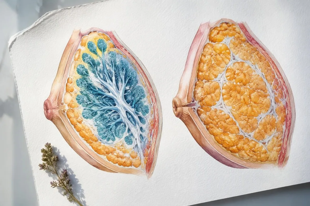
Lekorz ôcenio umiejscowiynie, konsystyncjo, ruchomość i symetryjo tkanki, a potym sprawdzo sutki i wōły pod pachōm. Na przikłod hipotetyczny, ruchōmy i bolesny krōżek pod sutkym ô wielkości 2 cm mo inkszy obraz w badaniu niż twardy guzek ô wielkości 2 cm dalij ôd sutka; sama wielkość niy ôdrōżnio łagodnego powiynkszynio ôd raka.
Świeże rozrośniyńcie gruczołu może być bolesne, a długo utrzymujōnco sie tkanka może z czasym stować sie twardszo, kej przez miesiōnce rozwijo sie włóknienie. Bolesność je tōż wskazōwkom co do czasu trwanio, a niy dowodym nieszkodliwyj prziczyny; bolesny guzek ze zmianami skōry abo szybkim postympym dalij wymago ôceny, nawet kej ôbjawy trwajōm ino 2 tydnie.
Utrata wagi może zmniejszyć tłuszczowo składowo, ale niy usunie ukształtowanego już gruczołowego krōżka, a człowiek może mieć prawdziwo ginekomastijo przy normalnym BMI. Naszo ômowa ô decyzjach ô badaniach zwiōnzanych z wagōm ôbjśnio, czamu badania endokrynologiczne majōm wynikać ze klinicznych wskazōwek, a niy z założynio, że kożdo utrzymujōnco sie zmiana klatki piersiowyj je spowodowano nadwagōm.
Kedy nowe powiększynie piersi u chłopa uzasadnio badania hormonōw?
Nowo, niewyjaśniōno ginekomastijo u dorosłego, powiynkszynie przed dojrzewaniym, szybki postymp abo towarzyszōnce ôbjawy endokrynologiczne wymagajōm ôceny klinicznyj i czynsto celowanych badań. Typowo ginekomastijo w czasie dojrzewanio może być ino ôbserwowano, bo czynsto sie poprawio w czasie ôkoło 1–2 rokōw bez utrzymujōncego sie zaburzenia hormonalnego.
A reassuring adolescent pattern consists of subareolar tissue arising during otherwise normal puberty, without a suspicious lump, systemic illness or abnormal genital examination. Persistence for more than about 2 lata, substantial distress or continued growth merits reassessment; that interval is a practical review point, not a rule that earlier investigation is unnecessary.
Prepubertal glandular enlargement is unusual and should not be dismissed as ordinary puberty. Clinicians look for hormone exposure, unusual pubertal development and other endocrine signs; our guide to early puberty hormone testing explains why age, growth velocity and developmental stage change the meaning of an LH or testosterone result.
In an adult, reduced libido, erectile difficulties, infertility, small testes, weight loss, tremor or features of liver disease make laboratory testing more informative. The 2019 EAA guideline also advises looking for underlying disease even when a medication seems plausible, because a drug explanation and a second medical cause can occur together (Kanakis et al., 2019).
Czamu przeglōnd lekōw je ważny przed badaniami hormonōw?
Medication-related gynecomastia may arise from reduced androgen action, altered hormone production or increased estrogen exposure. A useful review includes prescription drugs, nonprescription products and anabolic steroids, with doses and start dates; even a plausible drug cause does not cancel the need to examine a new lump.
Spironolactone, antiandrogens, finasteride, some antipsychotics and certain HIV treatments are recognised possibilities, although the strength of evidence differs between drugs. Finasteride is used at 1 mg or 5 mg daily for different indications; those doses belong in the history, but neither dose establishes causation or predicts whether an individual will develop enlargement.
Testosterone can be converted to estradiol, while stopping anabolic steroids may leave endogenous testosterone production suppressed. A hypothetical person developing tenderness 6 tygodni after changing a testosterone regimen needs review of formulation, dose and sampling time—not an automatic aromatase inhibitor; our estradiol review during TRT explains why symptoms and numbers can disagree.
Supplements marketed as testosterone boosters may contain undeclared hormonal ingredients, so bring photographs of the packaging and a complete ingredient list. A practical history covers at least the preceding 3–6 miesiōncōw, including stopped products; our skōrzonych efektōw u starzych ludzi also addresses why prescribed treatment should be adjusted with the prescriber rather than stopped independently.
Jak trza badać i interpretować testosterōn?
Całkowity testosteron should usually be measured fasting in the morning, often around 7–10 am, when investigating possible deficiency. A low result requires confirmation on a separate morning, and diagnosis depends on compatible symptoms rather than breast enlargement or a single laboratory flag alone.
The Endocrine Society guideline gives a harmonised total testosterone reference interval of 264–916 ng/dL, w przybliżeniu 9.2–31.8 nmol/L, for healthy, nonobese men aged 19–39 when appropriately standardised assays are used (Bhasin et al., 2018). The lower boundary is population- and method-specific; it is not a universal diagnosis threshold for every age group, laboratory or clinical situation.
Niskie abo wysokie SHBG can make total testosterone misleading, so clinicians may request calculated free testosterone using total testosterone, SHBG and albumin, or equilibrium dialysis where available. Direct analogue free-testosterone immunoassays are less reliable; our low free testosterone explanation shows why a value such as 280 ng/dL needs its binding-protein context.
Kantesti AI can help organise 2 separate testosterone results, their collection times and the accompanying LH values, but it cannot diagnose hypogonadism from a screenshot alone. Shift workers may need sampling aligned with their main sleep period rather than a rigid clock time; our przewodnik ô porannym pobieraniu prōbek hormonōw explains how sleep and recent illness affect preparation.
Co tak naprowdy pokazujōm poziōmy estradiolu przy ginekomastii?
Gynecomastia estradiol levels may be normal or elevated, because glandular growth reflects estrogen action relative to androgen action at the tissue. Many laboratories use an adult male estradiol interval around 10–40 pg/mL, but the assay-specific range—not an internet cutoff—should guide interpretation.
An estradiol interval of 10–40 pg/mL ôdpowiado w przybliżyniu 37–147 pmol/L; converting units prevents the same result from appearing nearly 4 times higher on a different report. Some laboratories have wider intervals, and immunoassays can be less accurate at the low concentrations typical in men, so unexpected results may need confirmation with a suitably validated LC–MS/MS method.
A hypothetical estradiol result of 48 pg/mL with a laboratory upper limit of 40 is a mild elevation, not evidence by itself of a hormone-producing growth. The next questions are whether testosterone treatment, increased aromatisation, liver dysfunction or assay interference could explain it; rapid enlargement, substantially abnormal results or a testicular finding changes the urgency.
Nie ma no validated universal estradiol-to-testosterone ratio that diagnoses gynecomastia, particularly when the hormones are reported in different units. SHBG also changes free hormone availability, which is why our przewodnik po SHBG i testosteronie is relevant; lowering estradiol solely to place a number below 40 pg/mL can create problems for bone and sexual health.
| Poziōm | Zakres | Co to znaczy |
|---|---|---|
| Illustrative adult male interval | 10–40 pg/mL; approximately 37–147 pmol/L | Common laboratory interval, not universal; normal estradiol does not exclude gynecomastia. |
| Above the laboratory upper limit | For example, 48 pg/mL when the upper limit is 40 | Review medications, testosterone exposure and assay accuracy before drawing conclusions. |
| Clearly elevated and unexplained | No universal numerical threshold | Clinical findings determine confirmation, endocrine referral and investigation for a hormone-producing source. |
Co ôdkrywajōm LH i FSH ô podłożu prziczyny?
Niski testosteron przy wysokim LH suggests primary testicular dysfunction, while low testosterone with low or inappropriately normal LH suggests a hypothalamic–pituitary or functional cause. FSH adds information about sperm-producing function; interpreting all 3 hormones together is more useful than reading isolated reference-range flags.
Illustrative adult male intervals are LH 1.7–8.6 IU/L i FSH 1.5–12.4 IU/L, but age, assay and laboratory affect the limits. An LH of 4 IU/L may be normal when testosterone is normal yet inappropriately normal when testosterone is repeatedly very low, because the expected compensatory response is an increase in LH.
Small, firm testes, infertility and a high-gonadotropin pattern may prompt consideration of zespōł Klinefeltera, usually associated with a 47,XXY karyotype. Chromosome testing is not required for every person with gynecomastia; our Klinefelter chromosome testing guide explains why physical findings and reproductive history determine whether it belongs in the workup.
Low testosterone with suppressed LH and FSH may also follow anabolic steroid exposure, opioid use, severe illness or significant energy deficit. A hypothetical athlete with testosterone 190 ng/dL, LH 0.8 IU/L and recent steroid withdrawal has a different starting explanation from someone with the same testosterone and LH 18 IU/L; neither pattern justifies treatment without confirmation and assessment.
Kedy trza zbadać prolaktynã i makroprolaktynã?
Prolactin testing is particularly useful when breast enlargement accompanies low testosterone with low or normal LH, reduced libido, nipple milk secretion or pituitary symptoms. Mild elevations should usually be reassessed in context; many adult male laboratory intervals have an upper limit around 15–20 ng/mL, but ranges vary.
Raised prolactin generally contributes indirectly by suppressing reproductive hormone signalling rather than acting as the usual direct driver of glandular enlargement. A hypothetical result of 28 ng/mL against an upper limit of 18 may reflect stress, medication, hypothyroidism or a persistent endocrine problem; our mild prolactin elevation guide explains why one modest result rarely settles the question.
For a repeat sample, follow the laboratory's instructions and consider a quiet rest of 20–30 minutes beforehand, particularly if the first collection was stressful. A fall from 28 do 14 ng/mL would change the interpretation, whereas a persistent elevation requires medication and thyroid review; headaches with visual changes should be assessed promptly rather than waiting for routine retesting.
Kantesti je platforma do interpretacji wyników badań krwi AI that can help distinguish a total prolactin flag from a report of monomeric prolactin after laboratory processing. Persistent elevation with few compatible symptoms may lead to makroprolaktyna testing; our macroprolactin result explanation describes why this larger, often less biologically active complex can prevent an unnecessary leap to pituitary imaging.
Czamu hCG i AFP mogōm być włączōne do diagnostyki?
Quantitative hCG can identify a hormone signal associated with certain testicular or other growths that may cause gynecomastia; AFP provides additional information when a germ-cell process is suspected. Neither marker is a stand-alone cancer diagnosis, and normal results do not exclude a testicular mass.
Some laboratories use an adult male hCG reference limit below 2 IU/L, while others report different cutoffs, including below 5; the exact method and reference interval must be checked. A clearly unexpected result, such as 38 IU/L in a hypothetical man without prescribed hCG, warrants prompt clinician review, confirmation and investigation rather than an assumption that the assay is only relevant to pregnancy.
A small unexpected hCG elevation can occasionally reflect assay interference, including heterophile antibodies, and clinicians may use a different analytical platform or other laboratory checks. IU/L and mIU/mL are numerically equivalent for hCG, so changing between those units does not explain a discrepancy; prescribed fertility treatment or bodybuilding-related hCG use must also be disclosed.
AFP reference limits are often around 8–10 ng/mL, but liver disease can also elevate AFP and some testicular cancers do not produce it. Our AFP interpretation and limits explains this overlap; a new testicular lump requires examination and usually ultrasound even if both hCG and AFP fall within the laboratory range.
Kere badania tarczyce, wōntroby i nerek mogōm być przidatne?
TSH, liver function and kidney function may help identify systemic causes of gynecomastia, particularly when symptoms or examination suggest illness. Hyperthyroidism can alter SHBG and hormone balance, while liver disease or advanced kidney disease can disrupt hormone handling; a mildly abnormal result alone rarely explains new enlargement.
A common adult TSH interval is approximately 0.4–4.0 mIU/L, although laboratories differ. A suppressed TSH with elevated free T4 or T3 supports hyperthyroidism, which can raise SHBG and shift effective androgen–estrogen balance; our TSH and thyroxine explanation helps distinguish that pattern from a single borderline TSH result.
Liver assessment may include ALT, AST, bilirubin, albumin and other tests selected for the clinical situation; normal transaminases do not exclude advanced chronic liver disease. Albumin is commonly around 35–50 g/L in adults, and our przewodnik interpretacji białek surowicy explains why binding proteins and synthetic function can matter more than a small isolated enzyme rise.
Kidney tests usually begin with creatinine and estimated GFR, interpreted alongside history rather than as a direct gynecomastia test. An eGFR of 58 mL/min/1,73 m² on one occasion does not establish chronic kidney disease or explain breast enlargement; our cirrhosis symptom and laboratory guide also shows why systemic causes need a recognisable clinical pattern, not merely one abnormal number.
Jak trza sie przigotować do badań hormonōw przy ginekomastii?
Preparation for gynecomastia hormone testing depends on the assays and any hormone treatment: morning fasting collection is usually appropriate for diagnostic testosterone, while treatment monitoring requires formulation-specific timing. Record the collection time, last dose and recent illness so 2 results can be compared meaningfully.
Poor sleep, acute illness and major calorie restriction can temporarily lower testosterone, so routine diagnostic testing is often deferred until recovery when clinically safe. If a hypothetical result of 220 ng/dl was collected after an overnight shift and fever, a properly timed repeat may be more useful than immediately labelling permanent deficiency; our piyrszy przewodnik przigotowanio do pobranio prōbki covers what to document.
Biotin can interfere with certain immunoassays, with the direction and magnitude depending on the assay design. Supplements containing 5–10 mg, co odpowiada 5,000–10,000 micrograms, deserve particular attention, but there is no universal safe washout period; ask the laboratory or clinician because dose, renal function and platform determine how long withholding may be required.
When reviewing results with Kantesti AI, retain the laboratory's units, reference intervals and any sample-quality comments, together with the time since the last testosterone dose. A sample collected 24 hours after an injection is not automatically comparable with one collected immediately before the next dose; unexplained discordance may require laboratory confirmation rather than a medication change.
Kere zmiany we piersi abo jōdrach potrzebujōm szybkigo badania?
A hard off-centre breast lump, nipple retraction or discharge, skin distortion, enlarged armpit nodes, or a new testicular lump needs prompt clinical evaluation. Do not wait for a gynecomastia blood test to reassure you; sudden severe testicular pain requires emergency care because torsion is time-sensitive.

Gynecomastia usually produces tissue centred beneath the nipple, but unilateral enlargement can still be benign and bilateral enlargement is not a guarantee of safety. A hypothetical 1 cm hard, fixed lump away from the nipple deserves a different pathway from a soft, mobile subareolar disc; blood tests cannot reliably distinguish these conditions without examination and, when needed, imaging.
When the examination is clearly typical of gynecomastia, routine breast imaging may not be necessary; an indeterminate or suspicious finding may require ultrasound, mammography and tissue sampling according to the clinical assessment. The 2019 EAA guideline supports imaging when examination is equivocal and direct tissue assessment for suspicious findings (Kanakis et al., 2019), rather than using normal estradiol as a reason to dismiss a lump.
A painless testicular lump, new firmness, size change or heaviness also deserves prompt review, while sudden severe pain should bypass routine outpatient hormone testing. Our family cancer risk testing guide explains why symptom-directed assessment differs from screening; a person with a new mass should not wait 3–6 miesiōncōw for the next planned gynecomastia follow-up.
Jak wzory hormonōw zmieniajōm dalsze postympowanie?
Hormone patterns guide the search for a cause; they do not choose treatment on their own. Low testosterone needs confirmation and clinical correlation, unexpected hCG needs prompt investigation, and an isolated mild estradiol rise may need review rather than suppression; the examination determines which pathway takes priority.
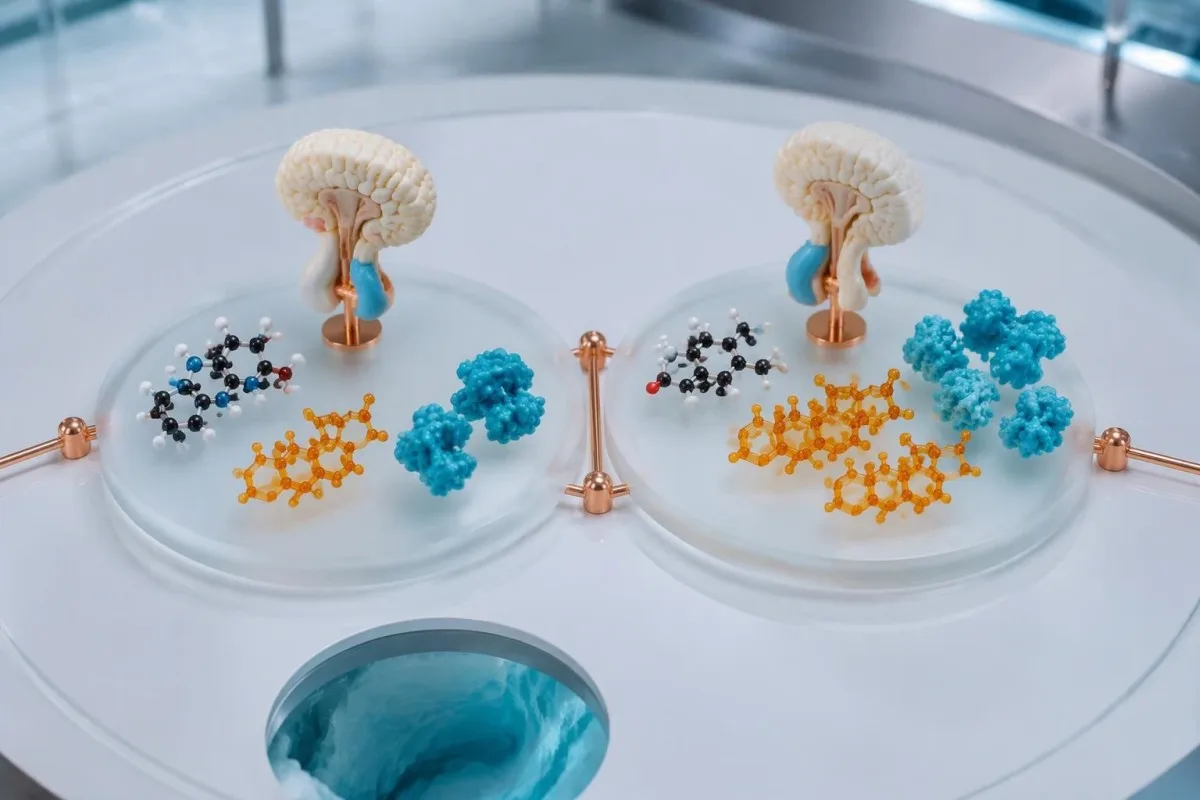
Consider a clearly hypothetical 52-year-old with tender subareolar tissue, testosterone 210 ng/dL, LH 15 IU/L, estradiol 26 pg/mL and no suspicious mass. Repeating testosterone and assessing primary testicular dysfunction is more logical than treating normal estradiol as the problem; a low androgen signal can permit glandular enlargement even when estrogen lies inside its reference interval.
A second hypothetical adult has testosterone 520 ng/dL, estradiol 48 pg/mL and enlargement beginning after a medication change, while examination is reassuring and hCG is normal. Medication review and possible estradiol confirmation may come before treatment; if the same person instead has hCG 38 IU/L or a testicular lump, the priority changes to prompt investigation regardless of testosterone.
The EAA guideline does not recommend routine use of aromatase inhibitors, selective estrogen receptor modulators or non-aromatisable androgens for gynecomastia in general (Kanakis et al., 2019), although specialists may consider selected off-label treatment in particular circumstances. Kantesti can support discussion of 2 or more related results, podczas gdy nasz validation and clinical limits explain why an AI interpretation should not initiate testosterone, tamoxifen or estrogen-lowering medication.
Dalsze kontrole, ograniczynia AI i publikacyje naukowe
Follow-up should track tissue changes and the underlying cause, not repeatedly order every hormone without a reason. A clinician may use a review interval around 3–6 miesiōncōw for a reassuring, stable case, with earlier reassessment for progression or new breast, testicular or systemic symptoms.
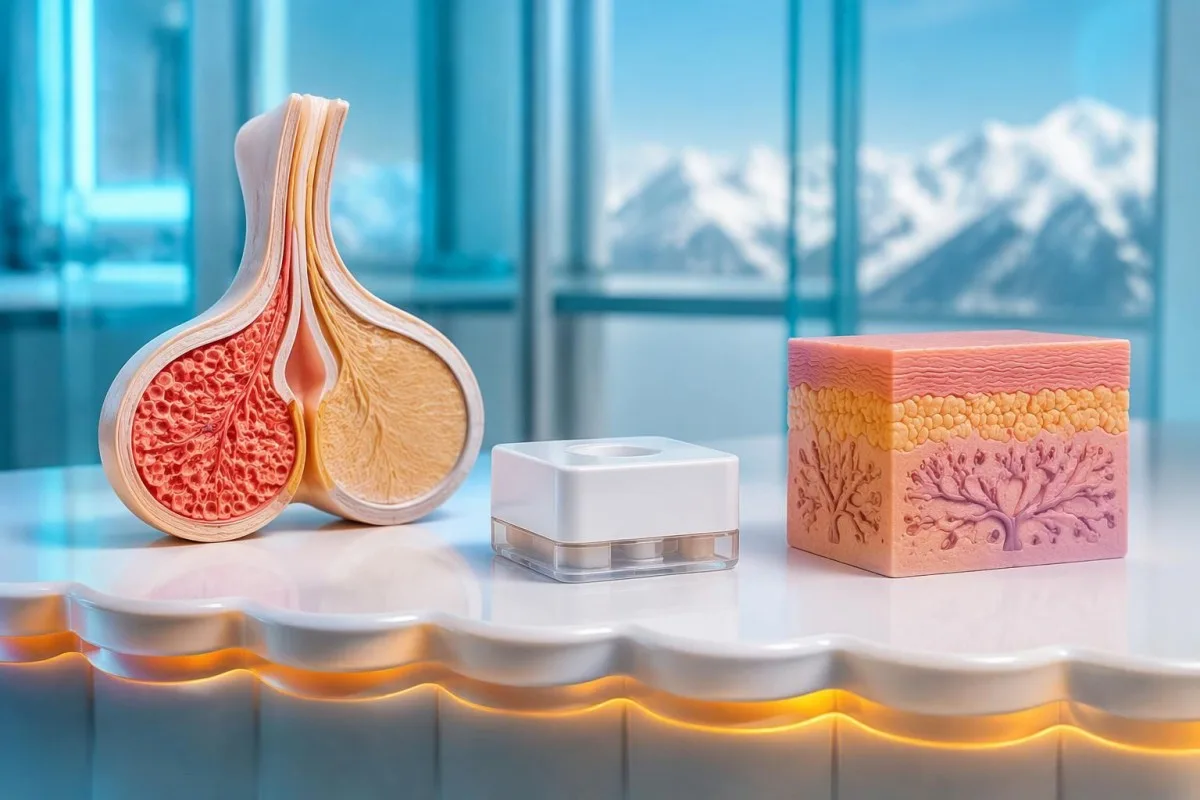
A useful follow-up record includes symptom onset, medication changes, examination findings and whether a glandular disc is increasing or becoming less tender. Longstanding tissue may persist after the trigger is corrected, particularly after approximately 12 miesiōncach when fibrosis can limit regression; persistent discomfort or distress may justify specialist discussion even after the laboratory cause has been addressed.
Kantesti je Narzędzie do analizy badań krwi oparte na AI that helps organise uploaded hormone reports and trends, including comparisons across 2 separate collection dates, without examining breast or testicular tissue. Kantesti LTD is the organisation behind the service, described on our company background page; nasze zasady ôdpowiedzialnego używanio AI explain why new physical findings and urgent symptoms require human clinical assessment.
Te 2 publikacyje Zenodo listed below are additional laboratory education resources covering iron studies and coagulation, not evidence sources for gynecomastia diagnosis or hormone treatment. The topic-specific recommendations in this article are supported by the 2019 EAA gynecomastia guideline i Wytyczne Towarzystwa Endokrynologicznego z 2018 roku wsōm testosteron; a DOI identifies a deposited publication but does not, by itself, establish independent peer review or clinical validation.
Często zadawane pytania
Jakie badania krwi mo chłop mieć przi ginekomastii?
Գինեկոմաստիայի գնահատումը կրնա ներառել ընդհանուր տեստոստերոն, էստրադիոլ, LH, FSH ու քանակական hCG՝ SHBG, պրոլակտին, TSH, AFP ու լյարդի կամ երիկամներու հետազոտություններն ընտրելով՝ կախված կլինիկական գտածոներեն։ Եվրոպական անդրոլոգիայի ակադեմիայի 2019 թվի ուղեցույցը կնկարագրե հնարավոր հետազոտություններու լայն շրջանակ մը, բայց ամեն դեռահասի ամեն հետազոտությունը պետք չէ։ Ախտորոշիչ տեստոստերոնը սովորաբար կը չափվի առավոտը՝ քաղցած փորին, ու եթե ցածր է, կը կրկնվի երկրորդ առավոտ մը։ Զննումն ու դեղերու վերանայումը անհրաժեշտ են, որովհետեւ արյան հետազոտությունները չեն կրնա պարզել կրծքի նոր գունդի ամեն պատճառը։.
Czy możesz mieć ginekomastyjo ze normalnym estradiolem?
Gynekomastijo może wystąpić przō normalnym estradiolu, bo rozwōj grōnzołōw zależy ôd aktywności estrogenōw wzgłyndem aktywności androgenōw we tkance. Wiela laboratorijōw stosuje przedział estradiolu dlō dorosłych chłopōw ôkoło 10–40 pg/mL, ale wynik w tym przedziale niy wykluczo niskigo testosterōnu, efektōw lekōw ani wrażliwości tkanki. Normalny wynik estradiolu tyż niy może wykluczyć niepokojōcego guza w piersi. Leczynie powinno być skierowane na klinicznō prziczyna, a niy ôpiyrać sie na założyniu, że kożdy przipadek wymago tłumienia estrogenōw.
Ôznaczo wysoki wynik estradiolu, że potrzebuja inhibitora aromatazy?
Wysoki wynik estradiolu niy uzasadnio automatycznie użycio inhibitora aromatazy. Na przikład 48 pg/mL przi górnyj granicy laboratoryjnyj 40 pg/mL to lekki wzrost, co może wymagać przejrzenio leków, oceny ekspozycyje na testosteron abo potwierdzenio wyniku odpowiednim testym. Wytyczne EAA z 2019 roku niy zalecajōm inhibitorōw aromatazy jako rutynowego, ogōlnego leczenio ginekomastii. Nadmierne obniżanie estrogenōw może szkodzić zdrowiu kości i funkcyji seksualnyj, tōż decyzje ô leczeniu należōm do klinicysty.
Jak mōm poznać, eli powiynkszynie piersiōw je ôd fetu abo ginekomastyjo?
Gynekomastijo ôbyczajowo ôdczuwo sie jak gumowaty abo twardy krążek pod sutkym, kej zaś powiększynie głōwnie ôd fetu je bardzij rozlane. Ôbydwa mogōm wystympować naroz, a guzek wielkości 2 cm niy idzie bezpiecznie zakwalifikować ino po jego wielkości abo zdjyncu. Lekorz sprawdzo umiejscowiynie tkanki, jej ruchliwość, zmiany sutka i węzły chłonne pod pachōm, coby ôsōndzić, eli trza robić bildowanie. Twardy guzek poza środkiym, wciągniyncie sutka abo wydzielina wymagajōm rychłego ôcyny, a niy samodiagnozy, że to tłuszcz na klacie.
Czy nastolatek z ginekomastiōm mo mieć zrobione badania hormonalne krwi?
Nastolatka ze typowym pubertalnym ginekomastiom i inkszym normalnym rozwojym może być ôbserwowany bez rozszerzōnego panelu hormonalnego. Pubertalnie powiynkszynie zwykle sie cofŏ w czasie ôkoło 1–2 rokōw, ale dalszy postymp, utrzymywanie sie poza ôczekiwanym przebiegym abo znaczny dyskomfort wymogajō ponownego ôcyniania. Předpubertalnie powiynkszynie, niyprawidłowy rozwōj, podejrzano gruda abo objawy układowe powinny być sprawdzōne pryndzyj. Wyniki hormonalne trza interpretować z uwzglydniyniym wieku i stadium dojrzewanio, a niy ino dorosłych zakresōw referencyjnych.
0 Czy normalne wyniki badań hormonalnych mogōm wykluczyć rak piersi abo rak jōder?
Normalne wyniki badań hormonalnych niy mogōm wykluczyć raka piersi abo jōder. I hCG, i AFP mogōm być normalne u ôsoby ze guzem jōdra, a rak piersi może sie pokozać bez niynormalnego testosterōnu abo estradiolu. Nowo twardo gruda w piersi, zmiana sutka abo gruda w jōdrze wymago bezto badania i ôdpowiednich ôbrazań bez wzglyndu na wyniki panelu laboratoryjnego. Nagły, silny bōl jōdra to nagły przipadek i niy powinien czekać na rutynowo kontrola abo wizyta laboratoryjno za 3–6 miesiyncy.
Dowiydz się, jak czytać wyniki badańo krwi
Wgraj raport laboratoryjny, a AI Kantesti wyjaśni kożdy biomarker prostym jynzykiym w pora sekund. Używane bezma 2 miliony ludzi na cołkim świecie.
Ôdniesiynia
- Klein, T., Mitchell, S., & Weber, H. (2026). Przewodnik po badaniach żelaza: TIBC, wysycenie żelazym i zdolność wiōnzania. Badania medyczne AI Kantesti. DOI
- Klein, T., Mitchell, S., & Weber, H. (2026). Norma aPTT: D-dimer, białko C — przewodnik po krzepniyniu krwi. Badania medyczne AI Kantesti. DOI
- Kanakis GA et al. (2019). EAA clinical practice guidelines—gynecomastia evaluation and management. Andrology. DOI
- Bhasin S et al. (2018). Testosterone Therapy in Men With Hypogonadism: An Endocrine Society Clinical Practice Guideline. The Journal of Clinical Endocrinology & Metabolism. DOI PubMed
- 2M+Ônalyzowane badania
- 127+Kroje
- 100+Jynzyki
Ô zespole medycznym


Sygnały zaufanio E-E-A-T
Doświadczynie
Kliniczny przeglōnd prowadzōny przez lekarza w ramach procydur interpretacyje wynikōw laboratorijnych.
Ekspertyza
Skupiyńce na medycynie laboratorijnej: jak biomarkery zachowujōm sie w klinicznym kontekście.
Autorytetność
Zweryfikowane medycznie przez dr. Thomasa Kleina, przy współudziale dr Sarah Mitchell i prof. dr. Hansa Webera.
Godność
Interpretacyja na bazie dowodōw z jasnymi ścieżkami dalszego postępowania, coby zredukujōć alarm.

Zastrzeżynie medyczne
Ten artykuł je wyłōncznie do celōw edukacyjnych i nie stanowi porady medyczno. Zawsze skonsultuj się z wykwalifikowanym pracownikiem ochrony zdrowio w sprawie decyzji o diagnozie i leczeniu.
Opublikowane · Skontaktuj sie z redakcyjōm
Opublikowane przez Kantesti Ltd, zarejestrowane w Anglii i Walii, numer firmy. 17090423, Londyn, Zjednoczone Królestwo.
