Um alerta de cálcio urinário elevado precisa de confirmação antes do tratamento. A questão útil é se a perda de cálcio é persistente, por que está ocorrendo e se o restante do perfil de risco de cálculo altera o plano.


- Hipercalciúria geralmente significa mais de 250 mg/dia de cálcio na urina em mulheres ou 300 mg/dia em homens, mas os laboratórios e o tamanho corporal importam.
- Cálcio na urina spot pode ser distorcido pela hidratação e pelo momento; não pode diagnosticar de forma confiável a perda persistente de cálcio por si só.
- cálcio urinário de 24 horas é o teste de confirmação preferido para formadores recorrentes de cálculo de cálcio e deve ser interpretado com sódio, citrato, oxalato, volume e pH da urina.
- Ingestão de sal aumenta o cálcio urinário porque o sódio e o cálcio são parcialmente reabsorvidos juntos no rim; um alvo prático é abaixo de 2.300 mg de sódio por dia.
- Cálcio alimentar normal de aproximadamente 1.000 a 1.200 mg/dia é geralmente mais seguro para a prevenção de cálculo de oxalato de cálcio do que restringir a ingestão de alimentos com cálcio.
- Testes paratireoides são apropriados quando o cálcio urinário elevado acompanha o cálcio sanguíneo elevado ou normal-alto, baixo teor de fósforo, perda óssea ou cálculos recorrentes.
- Medicamentos tiazídicos reduzem o cálcio urinário, mas requerem monitoramento de potássio, sódio, glicose e ácido úrico.
- Avaliação urgente é necessária para febre com dor nos flancos, vômitos com baixo débito urinário, um rim único funcionante ou dor intensa com possível obstrução.
O que o cálcio urinário elevado realmente significa
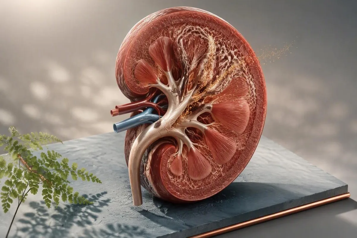
O cálcio urinário elevado significa que os rins estão liberando mais cálcio na urina do que o esperado, não necessariamente que o cálcio no sangue está elevado. Um resultado pontual é uma pista de triagem; a hipercalciúria persistente geralmente é confirmada com uma coleta completa de 24 horas antes que alguém a rotule como um distúrbio de cálculo ou inicie o tratamento.
Em adultos, muitas clínicas de cálculo utilizam mais de 250 mg por 24 horas para mulheres e mais de 300 mg por 24 horas para homens como limites práticos de hipercalciúria. Um limite baseado no peso acima de 4 mg/kg/dia é útil para um adulto menor, enquanto alguns laboratórios usam 200 mg/dia como limite superior. O número só é significativo se a coleta estiver completa; consulte nosso guia de coleta de 24 horas.
Uma concentração de cálcio urinário pontual, como 9 mg/dL, diz quase nada sem o contexto de creatinina urinária e diluição. Uma relação cálcio-creatinina pode ser útil em crianças e em exames endócrinos selecionados, mas seu papel na prevenção de cálculos em adultos é limitado. Uma amostra matinal muito concentrada pode parecer alarmante, mesmo quando a excreção diária de cálcio é normal.
Kantesti é um Analisador de teste de sangue de IA que coloca cálcio sérico, creatinina, bicarbonato, fósforo, vitamina D e hormônio paratireoidiano no mesmo quadro clínico. Na minha prática, o erro mais evitável é tratar um sinal urinário isolado como um diagnóstico em vez de perguntar se a pessoa tem cálculos, risco de fratura, alta ingestão de sódio ou uma explicação medicamentosa.
Um resultado pode ser elevado sem um nível elevado de cálcio no sangue
Cerca de 99% do cálcio corporal é armazenado nos ossos, e os rins filtram aproximadamente 10.000 mg de cálcio diariamente antes de reabsorver quase tudo. Portanto, alguém pode ter cálcio sérico normal e excesso de cálcio urinário ao mesmo tempo; este padrão é comum na hipercalciúria idiopática. Para informações de fundo sobre um valor sanguíneo elevado, leia cálcio ligeiramente elevado.
Alertas de urina spot versus um teste de cálcio urinário de 24 horas
Um teste de cálcio urinário de 24 horas é mais confiável do que um resultado de urina pontual porque mede a perda total diária de cálcio em condições de dieta e fluidos da vida real. É o próximo teste usual após cálculos de cálcio recorrentes, cálcio pontual inexplicavelmente elevado ou um distúrbio metabólico suspeito de cálculo.
Para uma coleta válida, descarte a primeira urina no horário de início, colete todas as micções pelas próximas 24 horas e inclua a micção final exatamente no horário de término. Perder até mesmo uma micção diurna grande pode dar uma falsa sensação de segurança; coletar por mais de 24 horas pode elevar falsamente o total. A creatinina urinária é o controle de qualidade silencioso do laboratório: um débito muito baixo geralmente sinaliza uma coleta incompleta.
O estudo ideal mede volume, cálcio, oxalato, citrato, sódio, ácido úrico, pH e creatinina do mesmo recipiente. Volume urinário abaixo de 2,0 L/dia é independentemente relevante porque os sais de cálcio se tornam mais concentrados, enquanto um débito urinário alvo de pelo menos 2,0 a 2,5 L/dia reduz a supersaturação. Razões de creatinina urinária explicam por que a diluição altera a interpretação de amostras únicas.
Do not deliberately eat a calcium-free diet before testing unless the ordering clinician gives that instruction. A 52-year-old patient I saw had reduced dairy and calcium-fortified foods for three days before collection; her calcium appeared normal, but oxalate rose substantially. The clinically useful test reflects habitual intake, not a one-day attempt to pass the exam.
Como o cálcio urinário elevado aumenta o risco de cálculo renal
High urine calcium kidney stones form when calcium combines with oxalate or phosphate in a urine environment that is too concentrated or chemically favorable for crystals. Calcium output alone predicts risk imperfectly; low urine volume and low citrate often matter just as much.
Most stones are calcium based, commonly calcium oxalate with or without calcium phosphate. The risk rises as urinary supersaturation rises, which is why a calcium result of 320 mg/day has a different meaning at 1.1 L urine volume than at 3.0 L/day. In the American Urological Association guideline, higher fluid intake sufficient to produce at least 2.5 L urine daily is a core prevention recommendation (Pearle et al., 2014).
Urine citrate below 320 mg/day is a useful warning sign because citrate binds free calcium and slows crystal growth. Low citrate plus high calcium is a much more actionable combination than either result alone; our guide to low urine citrate covers this pairing in detail.
Stone composition changes the workup. Calcium phosphate stones are more associated with urine pH persistently above 6.5, distal renal tubular acidosis, and sometimes primary hyperparathyroidism, while calcium oxalate stones often track with oxalate exposure, low calcium intake at meals, or intestinal malabsorption. A retrieved stone should be analysed at least once rather than guessed from an ultrasound report.
Crystals on a urinalysis do not prove a stone
Calcium oxalate crystals can appear after dehydration, a spinach-heavy meal, or delayed processing of urine and do not diagnose a stone by themselves. Repeated crystals plus pain, haematuria, or a stone history deserve more attention. Our discussion of cristais de oxalato de cálcio explains the common false alarms.
Causas alimentares de hipercalciúria que você pode alterar
High sodium intake is one of the most modifiable hypercalciuria causes because sodium loss in urine pulls calcium loss upward. Reducing salt is usually more useful than removing calcium-rich foods from the diet.
A practical sodium target for many stone formers is under 2,300 mg/day, and some people with hypertension benefit from closer to 1,500 mg/day if their clinician agrees. Packaged soups, sauces, breads, restaurant meals, and sports electrolyte products are more common sources than the salt shaker. A fall in urine sodium on repeat collection is often the best evidence that this change actually happened.
Dietary calcium should generally remain around 1,000 to 1,200 mg/day, preferably divided between meals. Calcium in food binds oxalate in the gut, reducing oxalate absorption; in Borghi's randomized trial, a normal-calcium, low-salt, lower-animal-protein diet prevented recurrent stones better than a low-calcium diet (Borghi et al., 2002). Kidney-focused nutrition guidance can help translate that into meals.
Very high animal-protein intake can increase calcium excretion, acid load, and uric acid while lowering citrate. I do not tell active patients to avoid protein; I ask them to avoid the bodybuilding-style extreme of 2.5 to 3.0 g/kg/day without a clear reason. Vitamin C supplements above 1,000 mg/day may also increase urinary oxalate in susceptible people.
Medicamentos e suplementos que podem aumentar o cálcio urinário
Loop diuretics, excessive vitamin D, calcium supplements taken away from meals, and some carbonic anhydrase inhibitors can increase urinary calcium or alter stone chemistry. Never stop a prescribed medicine solely because of a collection result; the reason for the medicine often outweighs stone risk.
Furosemide and related loop diuretics increase calcium excretion by reducing calcium reabsorption in the thick ascending limb. Topiramate and acetazolamide can raise urine pH and reduce citrate, making calcium phosphate stones more likely even if calcium is only modestly elevated. Review every prescribed medicine, over-the-counter product, protein powder, and antacid with the clinician ordering the test.
Vitamin D toxicity is uncommon at standard doses, yet high-dose regimens can uncover or worsen hypercalciuria in sensitive people. A 25-hydroxyvitamin D above 150 ng/mL (375 nmol/L) raises concern for toxicity, but urine calcium can rise well below that level in sarcoidosis or primary hyperparathyroidism. See vitamin D safety cutoffs before escalating a supplement.
Calcium supplements are not automatically forbidden. When needed for osteoporosis prevention, taking a modest dose with a meal is usually preferable to taking it at bedtime, because meal calcium binds dietary oxalate. Kantesti is an Ferramenta de análise de exames de sangue com IA that can identify a supplement-related pattern across calcium, 25-OH vitamin D, creatinine, and previous results, but a clinician must decide whether dose changes are safe.
Contribuições da paratireóide, vitamina D e exames de sangue
Primary hyperparathyroidism should be considered when high urine calcium occurs with repeatedly high or high-normal serum calcium and a parathyroid hormone level that is not suppressed. This matters because surgery can prevent recurrent stones and protect bone in selected patients.
In true hypercalcaemia, parathyroid hormone should normally be low. A PTH that remains normal-range or elevated alongside adjusted calcium above the laboratory limit is “inappropriately normal” and warrants endocrine assessment. Useful companion tests include albumin, ionized calcium when appropriate, phosphate, creatinine/eGFR, magnesium, 25-OH vitamin D, and often a 24-hour urine calcium.
The calcium-to-creatinine clearance ratio can help distinguish primary hyperparathyroidism from familial hypocalciuric hypercalcaemia, a rare inherited condition in which urine calcium is usually low rather than high. A ratio below 0.01 supports FHH, whereas a ratio above 0.02 supports primary hyperparathyroidism; values between them overlap, especially with vitamin D deficiency or thiazide use.
Dr. Thomas Klein's clinical rule is simple: do not call a calcium result “normal” until albumin and the assay range have been checked. Patients can review the distinction between a blood calcium issue and urine loss in our calcium result explainer, but symptoms and trend data decide urgency.
Other systemic explanations
Sarcoidosis and some lymphomas can increase active vitamin D production, while prolonged immobilisation, hyperthyroidism, and severe bone turnover can increase filtered calcium. These are uncommon explanations for an isolated mild urine result. Weight loss, thirst, constipation, bone pain, or a changing serum calcium pattern raises the priority for direct medical review.
Condições renais e intestinais por trás da perda de cálcio
Idiopathic hypercalciuria is the most common explanation when blood calcium is normal, but kidney tubular disorders and intestinal diseases can create distinctive patterns. The accompanying bicarbonate, potassium, urine pH, citrate, and stone composition narrow the possibilities quickly.
Distal renal tubular acidosis often presents with a non-anion-gap metabolic acidosis, low potassium, urine pH above 5.5, low citrate, and calcium phosphate stones. Serum bicarbonate below 22 mmol/L is a meaningful clue, although diarrhoea and chronic kidney disease can also lower it. Renal tubular acidosis patterns are worth reviewing when several of these clues cluster.
Inflammatory bowel disease, coeliac disease, bowel resection, and chronic fatty diarrhoea more often produce high urine oxalate than high urine calcium. Yet dehydration and low citrate can coexist, so the net stone risk remains substantial. A stool history is not trivial here: loose, oily stools after meals point the investigation toward intestinal absorption rather than simply blaming diet.
Chronic kidney disease usually lowers urinary calcium as filtration falls, so marked hypercalciuria with reduced eGFR deserves a careful review rather than a casual assumption. Conversely, a normal creatinine does not exclude stone-related obstruction. Estágios da doença renal crônica provide the context for interpreting eGFR alongside urine tests.
Como se preparar para uma coleta precisa
An accurate 24-hour urine calcium test requires an ordinary diet, ordinary fluid intake, and every urine sample during the timed interval. Acute diarrhoea, a marathon, major dietary change, or a newly started supplement can make the result less representative.
Ask the laboratory whether preservative is already in the container and whether it must be refrigerated. Do not urinate directly onto a preservative powder, and keep the container away from children. Start on a day without travel when possible; many missed samples happen during commuting, not because a patient is careless.
Maintain typical calcium, sodium, and protein intake for 3 a 7 dias beforehand unless your clinician gives different instructions. Avoid collecting immediately after a 10-kilometre race or during a gastrointestinal illness because sweat loss and temporary low urine volume can exaggerate saturation. The wider context of fitas reagentes para urina may help distinguish hydration clues from disease.
A collection should not be judged by calcium alone. Kantesti, an serviço de interpretação de testes do laboratório de IA, can organise laboratory trends and highlight missing companion markers for discussion, but it cannot verify that every void entered the container. Write down supplements, unusually salty meals, pain episodes, and fluid intake; these details often explain a borderline result better than a repeat test does.
Leia cálcio com sódio, citrato, oxalato, pH e volume
The highest-risk profile is not simply high calcium; it is high calcium combined with low volume, low citrate, high oxalate, or an unfavorable urine pH. A complete profile tells clinicians which change is most likely to reduce supersaturation for that individual.
Urine sodium above 100 mmol/day often signals a salt intake that can sustain calcium loss, although athletes and people in hot climates need individualized advice. If calcium and sodium both fall on a repeat collection, a salt-focused intervention is probably working. This is more persuasive than assuming an herbal product or alkaline water has fixed the problem.
Urine oxalate above 40 mg/dia deserves a separate dietary and intestinal review. The practical step is usually pairing calcium-containing food with oxalate-containing meals—not trying to eliminate all plant foods. Interpretação do pH da urina adds value because calcium phosphate risk rises in more alkaline urine, while uric acid stones favour persistently acidic urine.
A 24-hour collection is a snapshot, and day-to-day calcium output varies. In my experience, repeating the collection after 8 to 12 weeks of one targeted change is more instructive than changing salt, calcium, fluids, supplements, and medication all at once. You need to know which lever moved the chemistry.
Prevenção de primeira linha para cálcio urinário persistentemente elevado
First-line treatment for persistent hypercalciuria is higher fluid intake, lower sodium intake, normal food calcium, and individualized protein moderation. These measures reduce stone supersaturation without compromising bone health in most adults.
Aim for enough fluid to make 2.0 to 2.5 L of urine daily, not merely to drink a fixed number of glasses. For many adults this means 2.5 to 3.0 L of fluid intake, with more needed in heat or exercise. Pale urine is a rough clue, but a measured 24-hour volume is more dependable.
Keep calcium-containing foods at meals and reduce sodium gradually over two to four weeks. The calcium target differs for adolescents, pregnancy, osteoporosis, and chronic kidney disease, so broad internet advice can be counterproductive. Kidney-safe supplement choices are particularly relevant if eGFR is reduced.
Citrate from fruits and vegetables can help some calcium stone formers, but lemon water is not a substitute for measuring low citrate or correcting severe metabolic acidosis. Sweetened lemonade can worsen metabolic risk, and potassium-containing products require caution with ACE inhibitors, ARBs, spironolactone, or impaired kidney function. Small, sustained changes beat heroic restriction.
Quando o tratamento com tiazidas é considerado
Thiazide or thiazide-like diuretics may be used for recurrent calcium stones with persistent hypercalciuria after diet and fluid measures, especially when hypertension is also present. These medicines lower urine calcium by increasing calcium reabsorption in the kidney.
Hydrochlorothiazide, chlorthalidone, and indapamide are commonly considered, but dose choice depends on blood pressure, kidney function, glucose, gout history, and adverse effects. The AUA guideline supports offering thiazides to recurrent calcium stone formers with high or relatively high urine calcium (Pearle et al., 2014). Benefit is not immediate proof that every stone was calcium-driven.
Thiazides can lower potassium and sodium, raise uric acid, and modestly worsen glucose tolerance. A basic metabolic panel is often checked about 1 a 2 semanas after initiation or a meaningful dose change, then again based on clinical stability. Potassium citrate is sometimes added, but only after checking kidney function and serum potassium.
Esta é uma das razões pelas quais a nossa medication trend tracker focuses on trends rather than a single “normal” flag. A potassium of 3.4 mmol/L after a new thiazide may be more clinically relevant than a stable value of 3.4 mmol/L that has been discussed and monitored for years.
Saúde óssea e cálcio urinário elevado
Persistent idiopathic hypercalciuria can coexist with lower bone mineral density, particularly when dietary calcium is restricted or menopause, low body weight, or steroid exposure adds risk. Losing calcium in urine does not automatically mean bone is being lost, but the association deserves attention.
A history of low-trauma fracture, height loss, early menopause, chronic steroid use, or recurrent stones may justify bone-density testing. Hypercalciuria can be a marker of altered bone turnover, but osteoporosis has many causes; a DEXA result should not be attributed to urine calcium without checking thyroid status, vitamin D, PTH, nutrition, and medication history.
Avoiding calcium food sources can paradoxically harm both bone and stone prevention. Adults generally need 1.000 mg/dia, increasing to 1,200 mg/day for many women over 50 and men over 70, though national guidance varies. The dietary approach should be adapted if there is advanced CKD or a documented calcium balance disorder.
Kantesti é um plataforma de interpretação de biomarcadores por IA that can bring vitamin D, phosphate, alkaline phosphatase, calcium, and kidney markers into a longitudinal view. For patients with an unexpectedly high alkaline phosphatase, our fracture-related ALP guide explains why bone and liver sources need to be separated.
Quando o cálcio urinário elevado requer um especialista
Specialist evaluation is appropriate for recurrent stones, urine calcium above 400 mg/day, high blood calcium, reduced kidney function, osteoporosis, or a suspected tubular disorder. A urologist, nephrologist, or endocrinologist may each be the right clinician depending on the pattern.
Refer sooner when there are two or more stone events, a stone before age 25, bilateral stones, a family history of inherited stone disease, or calcium phosphate stones. A high urine calcium result with serum calcium above the laboratory limit makes endocrine evaluation particularly important. Imaging can identify stone burden, but it does not replace metabolic testing.
Pregnancy, childhood, a solitary kidney, bowel disease, or eGFR below 60 mL/min/1,73 m² changes the safety margin for diet and medication advice. Children use age-specific calcium-to-creatinine ratios rather than adult 24-hour thresholds in many settings. A one-size-fits-all stone diet is frankly unsafe in these groups.
De Kantesti conselho consultivo médico supports physician-led clinical review standards for results that may need escalation. Bring the original laboratory report, collection timing, stone analysis, current medicines, and any imaging report; that package prevents repeat appointments devoted to reconstructing basic facts.
Sintomas que necessitam de atendimento de urgência em vez de um novo teste
Fever or chills with flank pain, persistent vomiting, very low urine output, confusion, or pain in a person with one kidney can signal an obstructed infected urinary system and needs urgent assessment. High urine calcium itself is rarely an emergency, but a stone complication can be.
Seek same-day urgent care for a temperature of 38,0°C ou mais with suspected renal colic, especially with shaking chills, weakness, or rapid heart rate. Infection behind an obstruction can deteriorate quickly and needs imaging, urine testing, and prompt treatment. Do not wait for a 24-hour collection result when red flags are present.
Visible blood in urine deserves medical evaluation even when a stone is likely, particularly after age 40, with smoking history, painless bleeding, or persistent symptoms. Stones are common, but they should not become an automatic explanation for every episode. Our sangue na urina covers the usual evaluation pathway.
Severe pain that is not controlled with prescribed treatment, inability to keep fluids down for 12 to 24 hours, or markedly reduced urine output also warrants rapid review. Home urine strips cannot rule out obstruction, sepsis, or kidney injury. A normal spot calcium would not make those symptoms safe.
Um plano prático de acompanhamento após um resultado elevado
After confirmed high urine calcium, most patients need one targeted change, repeat urine testing in 8 to 12 weeks, and blood monitoring tailored to the suspected cause. The plan should be driven by stone history and the full chemistry profile, not a generic “avoid calcium” instruction.
Step one is to confirm the collection and review urine volume, sodium, citrate, oxalate, pH, and creatinine. Step two is usually increasing fluids and reducing sodium while keeping ordinary food calcium. Step three is repeat testing after enough time for the new routine to become real life rather than a short-lived experiment.
If serum calcium is high, arrange PTH-based evaluation rather than repeating urine studies indefinitely. If serum bicarbonate is low, potassium is low, or urine pH stays high, investigate renal tubular acidosis. If stones recur despite good volume and sodium control, medication and specialist input become more reasonable.
As of September 21, 2026, the most defensible approach remains individualized metabolic assessment followed by measurable prevention targets. Kantesti's abordagem de validação clínica emphasizes that AI interpretation supports, rather than replaces, diagnosis by a qualified clinician. Dr. Thomas Klein recommends saving each collection report beside the corresponding blood results and medication list; pattern recognition is where the answer usually emerges.
Publicações de pesquisa e evidências por trás desta abordagem
The strongest evidence for high urine calcium management supports full metabolic urine testing in recurrent stone formers, higher urine volume, sodium reduction, normal dietary calcium, and selective thiazide use. Evidence for individual supplements and alkaline drinks is far less certain, so claims should be checked against the measured urine pattern.
Pearle and colleagues' American Urological Association guideline ties recurrent calcium stone prevention to urine volume, dietary sodium reduction, normal calcium intake, and pharmacologic options when indicated (Pearle et al., 2014). Borghi and colleagues showed that normal dietary calcium paired with lower salt and animal protein outperformed a low-calcium diet for recurrent calcium oxalate stones (Borghi et al., 2002).
For related laboratory literacy, our kidney function research guide explains why serum markers cannot be substituted for a metabolic urine profile. Kantesti's clinical team uses research evidence as a starting point, then checks whether the person's actual stone type, collection quality, comorbidities, and medicines fit the evidence population.
Formal supplementary publications are listed below for transparency and broader laboratory-health context. They are not primary evidence for diagnosing hypercalciuria, and neither should be used to replace a stone clinic, endocrine assessment, or emergency evaluation when symptoms suggest obstruction.
Perguntas frequentes
Qual nível de cálcio na urina é considerado alto?
High urine calcium is commonly defined as more than 250 mg per 24 hours in adult women or more than 300 mg per 24 hours in adult men, although laboratory limits and body size vary. Some clinicians also use more than 4 mg/kg/day as a weight-based threshold. A value above 400 mg/day is more clearly abnormal and usually warrants review for diet, supplements, parathyroid disease, recurrent stones, and collection accuracy. A spot urine value cannot reliably establish the diagnosis because urine concentration changes through the day.
O alto nível de cálcio na urina pode causar pedras nos rins?
High urine calcium increases the chance of calcium oxalate and calcium phosphate stones by raising calcium salt supersaturation in urine. Risk is greatest when urine volume is below 2.0 L/day, citrate is below about 320 mg/day, oxalate is high, or urine pH favors calcium phosphate crystallization. Many people with hypercalciuria never form a stone, so clinicians interpret calcium alongside the whole 24-hour profile. Producing at least 2.0 to 2.5 L of urine daily is a common prevention target.
Devo parar de comer cálcio se o meu cálcio na urina estiver alto?
Most adults with high urine calcium should not stop eating calcium-rich foods. A dietary calcium intake of roughly 1,000 to 1,200 mg/day, taken with meals, can reduce gut oxalate absorption and may prevent calcium oxalate stones better than a low-calcium diet. Calcium supplements may need timing or dose review, especially if taken away from meals. People with high blood calcium, advanced kidney disease, or a diagnosed parathyroid disorder need individualized instructions.
Quais exames de sangue devem ser verificados com cálcio urinário elevado?
The usual blood tests for high urine calcium include serum calcium with albumin, creatinine and eGFR, bicarbonate, phosphate, potassium, magnesium, 25-hydroxyvitamin D, and parathyroid hormone when serum calcium is high or high-normal. A bicarbonate level below 22 mmol/L or low potassium can suggest a renal tubular disorder in the right setting. PTH should usually be suppressed when blood calcium is genuinely high; a normal or raised PTH then needs endocrine review. Test selection changes with symptoms, medicines, kidney function, and stone type.
How do you prepare for a 24-hour urine calcium test?
For a 24-hour urine calcium test, keep your usual diet and fluid intake for several days, discard the first urine at the starting time, then collect every urine sample for exactly 24 hours. Do not intentionally avoid calcium foods or load up on water unless the clinician specifically instructs you to do so. Ask whether the collection container needs refrigeration and whether it contains preservative. A missed sample can make a high calcium result look falsely normal, which is why urine creatinine is often checked for collection completeness.
Quando o cálcio elevado na urina é uma emergência?
High urine calcium alone is not usually an emergency, but a stone complication can be. Fever of 38.0°C or higher with flank pain, chills, persistent vomiting, minimal urine output, confusion, or pain in a person with a solitary kidney needs urgent assessment because infection behind an obstructing stone can damage the kidney quickly. Severe uncontrolled pain also needs prompt care. Do not wait for repeat urine testing when these symptoms are present.
Faça hoje a análise de exame de sangue com IA
Junte-se a mais de 2 milhões de usuários no mundo todo que confiam na Kantesti para análise instantânea e precisa de exames laboratoriais. Envie seus resultados de exame de sangue e receba uma interpretação abrangente de biomarcadores 15,000+ em segundos.
📚 Publicações de pesquisa referenciadas
Klein, T., Mitchell, S., & Weber, H. (2026). Guia de tipo sanguíneo B negativo, teste de sangue de LDH e contagem de reticulócitos. Pesquisa Médica por IA da Kantesti.
Klein, T., Mitchell, S., & Weber, H. (2026). Diarreia após jejum, pontos pretos nas fezes e guia GI 2026. Pesquisa Médica por IA da Kantesti.
📖 Referências Médicas Externas
⚕️ Aviso Médico
Este artigo é apenas para fins educacionais e não constitui aconselhamento médico. Consulte sempre um profissional de saúde qualificado para decisões de diagnóstico e tratamento.
Sinais de confiança E-E-A-T
Experiência
Revisão clínica orientada por médicos dos fluxos de interpretação de exames laboratoriais.
Especialização
Foco em medicina laboratorial sobre como os biomarcadores se comportam no contexto clínico.
Autoridade
Escrito pelo Dr. Thomas Klein, com revisão da Dra. Sarah Mitchell e do Prof. Dr. Hans Weber.
Confiabilidade
Interpretação baseada em evidências, com caminhos de acompanhamento claros para reduzir alarmes.