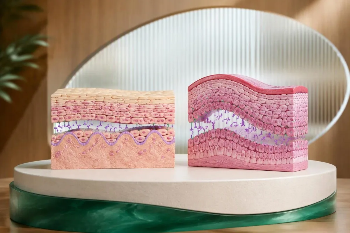
Позитивниот тест за антитела на десмоглеин ја поддржува дијагнозата на пемфигус само кога е во согласност со симптомите и наодот од прегледот. Антителата на десмоглеин 1 обично укажуваат на зафатеност на кожата; антителата на десмоглеин 3 обично укажуваат на зафатеност на устата. За потврда на дијагнозата често е потребна биопсија со директна имунофлуоресценција, особено кога наодите не се совпаѓаат.


- Десмоглеин 1 антителата обично поддржуваат образец на пемфигус со претежна зафатеност на кожата, особено кога антителата на десмоглеин 3 се негативни.
- Десмоглеин 3 антителата обично поддржуваат мукозен пемфигус вулгарис; ерозиите во устата може да се појават пред какви било кожни меури.
- Позитивни антитела на двата типа често укажуваат на мукокутан пемфигус вулгарис, но наодот од прегледот може да се разликува од очекуваниот образец.
- Гранични вредности за позитивен резултат зависат од анализата: некои ELISA-тестови користат вредност поголема од 20 U/mL, додека други лаборатории користат различни прагови или индексни вредности.
- Само позитивен резултат не потврдува пемфигус и не оправдува започнување кортикостероиди или ритуксимаб без процена од специјалист.
- Два примерока ткиво вообичаено се земаат: еден од зафатениот раб за рутинска хистологија и еден од блиското незасегнато ткиво за директна имунофлуоресценција.
- Директната имунофлуоресценција бара антитела наталожени меѓу епителните клетки; примерокот не смее да се стави во формалин за рутинска обработка.
- Негативни антитела не исклучуваат пемфигус кога убедливите ерозии или кревките меури перзистираат, особено по терапија.
- Потребна е итна проценка е потребна при тешкотии со голтање течности, болка во очите, отежнато дишење, треска со ширење на зафатените области или брзо лупење на кожата.
Што мери тестот за антитела на десмоглеин?
На тест за антитела на десмоглеин ги мери циркулирачките IgG антитела против десмоглеин 1 и десмоглеин 3, протеини што им помагаат на епителните клетки да се држат една за друга. Позитивниот резултат укажува на автоимуно создавање меури, но за дијагностицирање пемфигус е потребно совпаѓање на образецот на антителата, клиничките наоди и, вообичаено, испитувањето на ткивото.
Десмоглеин 1 и 3 се адхезивни протеини, а не општи маркери за воспаление или алергија. Тие се наоѓаат во десмозомите — мали структури за прицврстување меѓу соседните клетки — а антителата што ја попречуваат нивната функција може да предизвикаат акантолиза, односно губење на врската меѓу клетките.
Повеќето лаборатории ги мерат овие 2 антитела одделно со ензимски имуносорбентен тест, односно ELISA. Може да се нарача и тест за индиректна имунофлуоресценција, но тој поставува друго прашање: дали антителата во серумот препознаваат карактеристичен образец на лабораториски ткивен супстрат.
Јас сум Thomas Klein, MD, и пишувам во својство на главен медицински директор. Мојот пристап кон овој резултат е намерно претпазлив: лице со перзистентни ерозии во устата треба да се процени поинаку од лице со едвај позитивен скрининг резултат и без симптоми, дури и кога лабораторијата ги означува двата извештаја.
Кантести е анализатор на крвна слика со вештачка интелигенција што помага да се објаснат резултатите за десмоглеин 1 и 3 заедно со нивните лабораториски референтни вредности и пријавените симптоми. Нашите организација и клинички фокус даваат контекст за таа улога; нашето објаснување на позитивни резултати од антитела исто така прави разлика меѓу откривање антитела и потврдена дијагноза.
Што значи позитивен резултат за антитела на десмоглеин 1?
A позитивен резултат за антитела на десмоглеин 1 резултатот најчесто укажува на зафатеност на кожата кај пемфигус. Кога антителата на десмоглеин 3 се негативни, а прегледот покажува површни, кревки меури или ерозии со красти без зафатеност на устата, образецот особено упатува на пемфигус фолијацеус.
Пемфигус фолијацеус обично не ја зафаќа устата, и покрај понекогаш широко распространетата зафатеност на кожата. Неговите меури се толку површни што пациентите можеби никогаш нема да забележат неоштетен меур; наместо тоа, може да забележат перзистентно лупење, красти или дамки што изгледаат како да се бришат премногу лесно.
Скалпот, лицето, горниот дел од градниот кош и грбот се корисни места за преглед кога постои сомневање за ваков образец. Хипотетички пациент на 58 години со неколкунеделни промени со красти на скалпот и градниот кош, десмоглеин 1 од 76 U/mL и негативен десмоглеин 3 треба да се процени за површен пемфигус — не да му се постави дијагноза само врз основа на вредноста 76.
Позитивноста на десмоглеин 1 може да се јави и кај пемфигус вулгарис, especially when skin disease accompanies mucosal disease. The distinction between foliaceus and vulgaris comes from the clinical picture and the level of epithelial separation on histology, rather than from the antibody name alone.
Itching is not a reliable sorting tool: pemphigus may itch, burn, or simply feel tender, and several more common conditions produce similar complaints. Our guide to тестирање за чешање на кожата explains why broad laboratory screening cannot replace examination of an actual affected area.
Што значи позитивен резултат за антитела на десмоглеин 3?
A desmoglein 3 antibody positive result most often supports mucosal pemphigus vulgaris, particularly with persistent mouth erosions and negative desmoglein 1 antibodies. Mouth involvement can precede skin disease, so an otherwise normal skin examination does not dismiss this antibody pattern.
Oral pemphigus often presents as erosions rather than visible blisters, because the blister roof breaks quickly during eating or brushing. Persistent areas on the inner cheeks, palate, or gums may be mistaken for dental irritation, thrush, or ordinary canker sores before the pattern becomes clear.
Consider a hypothetical 46-year-old with 8 weeks of painful gum peeling, desmoglein 3 of 112 U/mL, and desmoglein 1 below the laboratory cutoff. That combination raises suspicion for mucosal pemphigus vulgaris, but a tissue sample remains valuable because erosive lichen planus and mucous membrane pemphigoid can look surprisingly similar.
Desmoglein 3 positivity does not measure swallowing safety or nutritional status. A patient who is drinking very little needs assessment of hydration, weight change, and possibly electrolytes even if the antibody value is modest; a higher value in someone eating normally does not automatically imply an emergency.
Recurrent discrete mouth ulcers with genital ulcers, eye symptoms, or characteristic skin findings suggest a different diagnostic route. Our discussion of Behçet mouth sore patterns explains those clues; desmoglein testing is not a screening test for Behçet disease.
Како помагаат комбинираните обрасци на десмоглеин 1 и 3?
Both desmoglein 1 and 3 antibodies positive often supports pemphigus vulgaris affecting both skin and mucosa. The desmoglein compensation model explains this tendency, but it is a useful clinical framework—not a rule that predicts every patient’s examination.
Desmoglein distribution differs between skin and oral mucosa. In skin, desmoglein 1 is abundant toward the surface while desmoglein 3 is concentrated more deeply; oral mucosa relies heavily on desmoglein 3, which helps explain why losing its function can expose the mouth before the skin.
The compensation idea is that one functioning desmoglein can partly preserve adhesion when antibodies interfere with the other. That model fits many cases, but antibody binding site, antibody pathogenicity, treatment, and other adhesion mechanisms can all make the clinical pattern less tidy than a 2-marker diagram suggests.
Murrell et al. (2020), in the international pemphigus recommendations, describe desmoglein 1 and 3 ELISA as useful for diagnosis and follow-up while emphasizing clinical and immunopathological correlation. The practical lesson is simple: antibodies help classify a convincing phenotype; they should not manufacture one in an asymptomatic person.
Total IgG is not a substitute for anti-desmoglein IgG, and a normal total IgG concentration does not exclude pathogenic antibodies against either protein. Our immunoglobulin result guide explains the difference between measuring the size of an antibody class and identifying its particular target.
Кои се граничните вредности за позитивен резултат и нормалните опсези?
Desmoglein antibody cutoffs are laboratory-specific, and there is no universal normal range that applies to every assay. Some ELISA systems classify both antibodies as positive above 20 U/mL while using different negative and borderline bands for desmoglein 1 and desmoglein 3.
The table below illustrates one commonly used ELISA reporting convention, not a universal standard. A report using a 20 U/mL threshold, a 30 U/mL threshold, or an index value must be read using its own instructions; apparently identical numbers can have different meanings across assay systems.
A result of 21 U/mL is close to a cutoff of greater than 20 U/mL and deserves more caution than the laboratory’s positive flag may suggest. It does not mean there is a 21% probability of pemphigus, and 100 U/mL does not mean five times as much tissue damage as 20 U/mL.
No desmoglein concentration is a universal emergency threshold. Urgency comes from rapidly spreading skin involvement, inability to drink, eye symptoms, breathing symptoms, or systemic illness; a laboratory report marked high without those findings calls for clinical interpretation, not automatic emergency treatment.
Before comparing 2 reports, check the antibody target, units, laboratory, assay method, and whether either value exceeded the instrument’s measuring range. Our explanation of квалитативни и квантитативни тестови shows why a positive label and a measured concentration carry different kinds of information.
Зошто позитивниот резултат не ја потврдува дијагнозата на пемфигус?
A positive desmoglein result does not establish pemphigus because detectable antibodies may be low-level, nonpathogenic, or inconsistent with the patient’s symptoms. The probability that a positive result represents disease also depends on why the test was ordered in the first place.
Pretest probability strongly affects the meaning of antibody positivity. A positive result in someone with months of characteristic mouth erosions is more persuasive than the same result found during broad screening for fatigue, where the clinical picture provides little support for pemphigus.
Here is an illustrative calculation—not an estimate of desmoglein assay performance: if a test has 95% sensitivity and 99% specificity, testing 10,000 people in a population with 1% disease prevalence produces about 95 true positives and 99 false positives. Only about 49% of positive results would represent disease in that hypothetical setting.
Nonpathogenic anti-desmoglein antibodies can recognize parts of the protein that do not meaningfully disrupt adhesion. This helps explain why antibody concentration is not a direct measurement of blister formation, and why specialists sometimes investigate an unexpected positive with another laboratory method rather than simply repeating the same assay.
A similar principle applies to other disease-specific autoantibodies, although the confirmation pathway is different for each condition. Our guide to interpreting MOG antibody positivity illustrates why a recognizable syndrome and an appropriate assay matter; its neurological criteria should not be transferred to pemphigus.
Може ли да се јави пемфигус со негативни антитела на десмоглеин?
Negative desmoglein 1 and 3 results do not completely exclude pemphigus, especially after treatment or when the clinical findings are strongly suggestive. Persistent fragile blisters or mucosal erosions may still require histology and direct immunofluorescence despite negative serum testing.
Serum testing measures circulating antibodies at one time point, not every antibody present within tissue. Low concentrations, assay limitations, or antibodies against additional epithelial targets can help explain a mismatch between the laboratory result and the examination.
Treatment history changes the interpretation of a negative result. If a person began systemic corticosteroids or received B-cell-directed treatment before the sample was collected, a low antibody concentration may reflect treatment effect rather than establish that the original erosions were unrelated to pemphigus.
Repeating serum testing alone is not the best next step when the clinical suspicion is high. A dermatologist may arrange 2 appropriately sited tissue specimens, add indirect immunofluorescence, or request a broader blistering-disease panel while reviewing medication exposures and the original onset of symptoms.
A negative test is most reassuring when the symptoms, examination, and other investigations also point away from the disease. Our discussion of negative autoimmune neuromuscular tests explains the same general reasoning, although the relevant antibodies and confirmatory procedures are entirely different.
Кога е потребна биопсија со директна имунофлуоресценција?
Biopsy with direct immunofluorescence is usually recommended during the initial assessment of suspected pemphigus, especially with borderline antibodies, atypical symptoms, or disagreement between serum testing and examination. Routine histology and direct immunofluorescence answer different questions, so clinicians commonly obtain 2 separate specimens.
Routine histology identifies where epithelial cells separate, while direct immunofluorescence identifies immunoreactants deposited within the patient’s tissue. International recommendations from Murrell et al. (2020) support combining clinical findings, histology, and immunological evidence rather than relying on serum positivity alone.
The routine histology specimen is usually taken from the edge of a fresh affected area so it includes useful epithelium. The direct immunofluorescence specimen comes from nearby intact tissue; many services use a site a few millimeters from the affected area, with the exact choice adapted for skin, oral mucosa, and local laboratory instructions.
A direct immunofluorescence specimen must not go into routine formalin, because that can compromise the examination. It generally needs the receiving laboratory’s specified transport medium, such as Michel’s medium, or another validated handling pathway; arranging this before sampling prevents an avoidable repeat procedure.
Occasionally, a specialist can establish the diagnosis using a strongly compatible presentation, characteristic histology, and convincing serology when direct immunofluorescence is unavailable. Kantesti’s клиничка рамка за валидација describes our interpretation standards, not a replacement for specialist tissue diagnosis; antibodies and kidney biopsy illustrate how biopsy exceptions differ by disease.
Што покажуваат хистологијата и имунофлуоресценцијата кај пемфигус?
Pemphigus direct immunofluorescence typically shows intercellular IgG, often with C3, in a net-like epithelial pattern. Routine histology usually shows suprabasal separation in pemphigus vulgaris and more superficial separation in pemphigus foliaceus, helping distinguish conditions with similar-looking erosions.
The classic pemphigus vulgaris split occurs above the basal epithelial layer. Basal cells may remain attached to the underlying basement membrane, creating the histological appearance often described as a row of tombstones; this is a tissue-level clue, not something a patient can recognize from a photograph.
A linear band along the basement membrane suggests a different family of autoimmune blistering disorders, including pemphigoid conditions. The distinction between an intercellular net and a basement-membrane line can redirect antibody testing toward targets such as BP180 rather than desmoglein 1 or 3.
A negative direct immunofluorescence result is not always definitive if sampling or handling was suboptimal. Sampling an eroded center, obtaining too little intact epithelium, using the wrong medium, or testing after substantial treatment can reduce the diagnostic yield; the specialist may request another specimen if the discrepancy remains convincing.
C3 detected in epithelial tissue is not interchangeable with a serum C3 concentration. Our complement and autoimmune testing guide explains circulating complement measurements; a normal serum C3 result does not cancel an intercellular C3 finding on direct immunofluorescence.
Што друго може да предизвика ерозии во устата и кожни меури?
Pemphigoid disorders, erosive lichen planus, infections, medication reactions, and mechanical injury can resemble pemphigus. A desmoglein-positive result should not stop the differential diagnosis, especially when the distribution, blister firmness, scarring, or onset after a new medicine points elsewhere.
Bullous pemphigoid often produces firmer, tense blisters and prominent itching, whereas pemphigus blisters are generally more fragile. Mucous membrane pemphigoid deserves particular attention with eye symptoms or scarring because protecting vision and other vulnerable mucosal sites may be more urgent than resolving the serum antibody label.
Painful grouped changes in a one-sided distribution suggest a viral cause rather than a typical pemphigus pattern. Our guide to early shingles symptom clues explains that distribution; unusual, widespread, or oral presentations still need examination rather than a photo-based conclusion.
Blisters on sun-exposed hands with skin fragility can suggest porphyria cutanea tarda, which follows a different testing pathway. Our porphyria testing during symptoms article distinguishes biochemical testing from autoimmune antibody testing; urinary porphyrin assessment may be relevant to a blistering porphyria phenotype.
Severe, persistent stomatitis with an unusual widespread eruption can raise concern for paraneoplastic pemphigus, also called paraneoplastic autoimmune multiorgan syndrome. Specialist testing may include antibodies to envoplakin or periplakin and selected indirect immunofluorescence methods; a routine 2-antibody desmoglein panel does not fully assess that possibility.
Како треба да се подготвите и дали лековите влијаат врз тестирањето?
Desmoglein antibody testing usually does not require fasting, although other tests collected at the same visit may have separate instructions. Tell the clinician about immunosuppressive treatment, recent infusions, and new medicines; do not stop prescribed treatment just to make an antibody test easier to interpret.
The date of treatment initiation matters as much as the medication name. Recording whether the sample preceded corticosteroids, rituximab, or other immunosuppression helps the dermatologist interpret a later negative or falling result; keep the original report even when a newer report appears more reassuring.
Some medicines, including penicillamine and captopril, have been associated with drug-induced pemphigus. A new medication does not automatically explain a blistering eruption, but its start date, dose changes, and the interval before symptoms are useful; never discontinue an essential medicine without a prescribing clinician’s plan.
Infection-risk assessment is separate from confirming pemphigus, but becomes important when immunosuppression is being considered. For people with relevant residence or travel exposure, our guide to Strongyloides пред имуносупресија explains why this parasite deserves attention before substantial corticosteroid treatment.
Azathioprine safety assessment commonly includes TPMT and, where appropriate, NUDT15 testing, alongside blood counts and liver monitoring. Our explanation of тестирање на TPMT пред примена на азатиоприн covers that preparation; a positive desmoglein result alone is not a reason to start this medication or any other systemic treatment.
Може ли нивото на антитела да следи ремисија или да предвиди релапс?
Desmoglein antibody trends can support pemphigus monitoring, but they cannot define remission or predict every relapse. New affected areas, healing, oral symptoms, and treatment exposure remain central; a persistently positive result in a clinically well patient does not automatically require more immunosuppression.
Desmoglein 1 levels often track skin activity more closely than desmoglein 3 levels track oral activity. The relationship is imperfect, and some patients remain desmoglein 3 positive during clinical remission; Joly et al. (2020), in the EADV management guideline, place serological monitoring within a broader clinical assessment.
Murrell et al. (2020) suggest checking antibodies at treatment initiation, after approximately 3 months, and then every 3–6 months or with relapse, adjusted to the clinical course. That is a follow-up framework for diagnosed pemphigus—not a recommendation to repeatedly screen asymptomatic people with an incidental low-positive result.
A change from 80 to 40 U/mL is a numerical decline, not proof that disease activity has halved. Compare results from the same assay when possible, note whether the original result exceeded the measuring range, and record the timing of treatment; our водич за трендови на безбедност на лекови explains why timelines matter.
Кантести е AI платформа за толкување крвна слика that can organize desmoglein results with dates, reference intervals, and other uploaded laboratory findings. Our водич за технологијата за AI толкување explains the workflow, but the platform cannot inspect mucosal surfaces, measure a validated clinical activity score, or authorize a medication taper.
Што треба да направите по позитивен резултат за десмоглеин?
Arrange clinical review after a positive desmoglein result, with dermatology or oral medicine assessment when erosions or blisters are present. Seek urgent care for inability to swallow fluids, eye pain or vision change, breathing difficulty, fever with spreading skin changes, or rapidly peeling skin; do not wait for repeat antibodies.
An asymptomatic low-positive result and widespread active blistering need different timelines. For an unexpected result without symptoms, contact the ordering clinician to review the indication and assay; for ongoing oral or skin changes, ask specifically whether histology and direct immunofluorescence should be arranged rather than simply repeating the panel.
Bring 2 kinds of evidence: the complete laboratory report and a dated symptom timeline. Photographs of changing affected areas, a current medication list, approximate weight change, and whether eating or drinking has become difficult can help the specialist choose a useful sampling site and assess immediate support needs.
My practical recommendation as Thomas Klein, MD, is to ask: “What finding would confirm or refute pemphigus in my case?” Our медицински советодавен одбор describes the physicians involved in clinical oversight; an individual diagnosis still belongs with the clinician who examines you and evaluates the relevant tissue evidence.
Кантести е услугата за интерпретација на тестови на AI that can help translate a desmoglein report into questions for that appointment, not certify pemphigus from a PDF. Our guide to checking AI medical explanations recommends verifying units, source text, and limitations; never let an automated reassurance override difficulty drinking, eye symptoms, or rapid deterioration.
Научни публикации и ограничувањата на доказите за толкување
Pemphigus diagnostic recommendations come from disease-specific clinical literature, not from general AI interpretation benchmarks. As of October 17, 2026, the international recommendations by Murrell et al. (2020) and the EADV guideline by Joly et al. (2020) remain relevant references for the diagnostic and monitoring principles discussed here.
The 2 publications below describe Kantesti materials and do not establish desmoglein test accuracy or prove pemphigus diagnostic capability. A Zenodo DOI identifies a deposited record; it does not, by itself, show peer review, prospective clinical validation, or reliable performance in autoimmune blistering disease.
Kantesti. (n.d.). Clinical validation framework v2.0 (Medical validation page). Zenodo. https://doi.org/10.5281/zenodo.17993721. The DOI record is the primary reference; the accompanying ResearchGate and Academia.edu links are discovery searches, not claims that this record is hosted or independently reviewed there.
Kantesti. (2026). AI blood test analyzer: 2.5M tests analyzed | Global health report 2026. Zenodo. https://doi.org/10.5281/zenodo.18175532. The test count in the publication title should not be interpreted as the number of confirmed pemphigus cases, desmoglein assays, or diagnostic comparisons.
General laboratory abnormalities can identify complications without confirming pemphigus. Albumin and other proteins may be relevant when extensive skin involvement or restricted intake affects nutrition, but our водич за толкување на серумски протеини addresses those supportive measurements; histology, immunofluorescence, and appropriate antibody testing remain a separate diagnostic pathway.
Често поставувани прашања
Дали позитивниот тест за антитела на десмоглеин значи дека имам пемфигус?
Позитивен тест за антитела против десмоглеин не значи, сам по себе, дека имате пемфигус. Резултатот мора да соодветствува на наодите од прегледот, симптомите и, обично, на наодите од ткивото при рутинска хистологија и директна имунофлуоресценција. Кај некои системи ELISA, како позитивен праг се користи вредност поголема од 20 U/mL, но лабораториските прагови се разликуваат, а гранично позитивен резултат без симптоми е помалку убедлив. Не започнувајте имуносупресивна терапија само врз основа на резултатот за антитела.
Која е разликата помеѓу антителата на дезмоглеин 1 и дезмоглеин 3?
Антителата против дезмоглеин 1 обично укажуваат на зафатеност на кожата, додека антителата против дезмоглеин 3 обично укажуваат на зафатеност на слузницата, особено на упорни ерозии во устата. Позитивноста на дезмоглеин 1 со негативен дезмоглеин 3 често одговара на пемфигус фолијацеус, додека позитивноста само на дезмоглеин 3 често одговара на мукозен пемфигус вулгарис. Позитивноста на овие 2 антитела често одговара на мукокутан пемфигус вулгарис. Овие обрасци го насочуваат проценувањето, но не ги предвидуваат сигурно симптомите кај секој пациент.
Кое ниво на антитела против десмоглеин се смета за позитивно?
Позитивното ниво на антитела против дезмоглеин зависи од лабораторијата и тестот. Според една често користена конвенција за ELISA, вредностите над 20 U/mL се сметаат за позитивни, при што граничните вредности за дезмоглеин 1 се 14–20 U/mL, а за дезмоглеин 3 се 9–20 U/mL. Другите тестови користат различни гранични вредности или индексни вредности, па затоа предност има опсегот наведен во вашиот извештај. Ниту една концентрација на антитела не претставува универзален праг за итна состојба или тежина на болеста.
Дали ми е потребна биопсија ако имам позитивни антитела на десмоглеин?
Биопсија обично се препорачува кога за првпат постои сомнеж за пемфигус, особено ако симптомите се атипични, антителата се гранични или наодите од прегледот и серолошкото тестирање не се совпаѓаат. Клиничарите најчесто земаат 2 примероци: од засегнатиот раб за рутинска хистологија и од блиско неоштетено ткиво за директна имунофлуоресценција. Примерокот за директна имунофлуоресценција бара ракување според упатствата на лабораторијата и не смее да се стави во вообичаен формалин. Специјалист повремено може да ја постави дијагнозата врз основа на карактеристични клинички наоди, хистологија и серологија кога директната имунофлуоресценција не е достапна.
0. Може ли да имам пемфигус со негативни тестови за десмоглеин 1 и 3?
Пемфигус може да се јави и покрај негативните серумски тестови за дезмоглеин 1 и 3, особено по лекување или кога концентрациите на антителата се под прагот на детекција на тестот. Негативните резултати за овие 2 антитела се посигурни кога и прегледот упатува на нешто друго, а не на пемфигус. Постојаните кревки меури или ерозиите во устата сепак може да бараат хистологија, директна имунофлуоресценција или дополнително специјалистичко тестирање на антитела. Не ја одложувајте процената на влошените симптоми додека чекате друг серумски тест.
Дали антителата против десмоглеин можат да останат позитивни кога пемфигусот е во ремисија?
Антителата на десмоглеин може да останат позитивни за време на клиничка ремисија, особено антителата на десмоглеин 3. Меѓународните препораки наведуваат тестирање при започнување на терапијата, по приближно 3 месеци, а потоа на секои 3–6 месеци или при релапс, во зависност од текот на болеста кај пациентот. Трендовите на антителата помагаат во следењето, но не ја заменуваат процената на новите зафатени подрачја, заздравувањето, јадењето и пиењето. Само перзистентно позитивен резултат не оправдува зголемување на терапијата.
Дали треба да бидам гладен пред тест за антитела на десмоглеин?
Гладувањето обично не е потребно за тестирање на антитела на десмоглеин, иако за други тестови земени при истиот термин може да биде потребно. Пријавете лекување со кортикостероиди, ритуксимаб или други имуносупресивни лекови, бидејќи тие можат да влијаат врз толкувањето на двете мерења на антитела. Забележете дали примерокот бил земен пред или по започнувањето на лекувањето. Не ги прекинувајте препишаните лекови освен ако лекарот што ви ги препишал не ви даде конкретен план.
Добијте AI анализа на крв со моќ на вештачка интелигенција денес
Придружете се на над 2 милиони корисници ширум светот кои му веруваат на Kantesti за инстантна, точна анализа на лабораториски тестови. Поставете ги вашите резултати од крвна слика и добијте сеопфатно толкување на 15,000+ биомаркери за секунди.
📚 Реферирани научни публикации
Клајн, Т., Мичел, С., и Вебер, Х. (2026). Клиничка рамка за валидација v2.0 (страница за медицинска валидација). AI Medical Research.
Клајн, Т., Мичел, С., и Вебер, Х. (2026). AI анализатор на крвни тестови: 2.5M анализирани тестови | Глобален здравствен извештај 2026. AI Medical Research.
📖 Надворешни медицински референци
Murrell DF et al. (2020). Diagnosis and management of pemphigus: Recommendations of an international panel of experts. Весник на Американската академија за дерматологија.
Joly P et al. (2020). Updated S2K guidelines on the management of pemphigus vulgaris and foliaceus initiated by the European Academy of Dermatology and Venereology (EADV). Зборник на Европската Академија за Дерматологија и Венерологија.
⚕️ Медицинско одрекување од одговорност
Овој напис е само за едукативни цели и не претставува медицински совет. Секогаш консултирајте се со квалификуван здравствен работник за одлуки за дијагноза и третман.
Сигнали за доверба E-E-A-T
Искуство
Клиничка ревизија водена од лекар за работните текови на толкување на лабораториски резултати.
Експертиза
Фокус на лабораториска медицина за тоа како биомаркерите се однесуваат во клинички контекст.
Авторитивност
Напишано од д-р Thomas Klein, со ревизија од д-р Sarah Mitchell и проф. д-р Hans Weber.
Доверливост
Толкување засновано на докази со јасни патеки за следење за да се намали алармирањето.