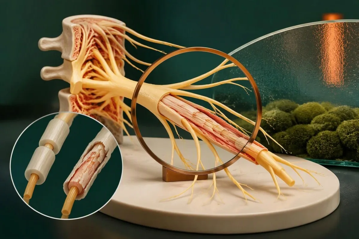
Weakness that spreads or changes your ability to walk needs assessment—not reassurance from a normal blood panel. Trouble breathing, swallowing, or holding your head up makes the situation an emergency.


- Progressive weakness that worsens over hours or days, particularly in both legs, needs urgent in-person assessment; rapidly spreading weakness warrants emergency care.
- Neatliekamie simptomi include breathlessness, a weak cough, choking, difficulty swallowing saliva, or inability to stand or walk safely. Call your local emergency number.
- Nesenā infekcija precedes approximately two-thirds of Guillain-Barre syndrome cases, usually within the previous 6 weeks; an infection is not required for diagnosis.
- Ascending weakness often begins in the legs and spreads upward, but facial, arm-predominant, and other variants can occur.
- Normal bloodwork cannot exclude Guillain-Barre syndrome, even when the CBC, CRP, electrolytes, and creatine kinase are within their laboratory ranges.
- Agrīna testēšana can be misleading: cerebrospinal fluid protein remains normal in approximately 30–50% of patients during the first week.
- Respiratory monitoring includes serial breathing-strength measurements; a vital capacity below 20 mL/kg is a concerning hospital threshold, not a home screening test.
- Effective treatment includes intravenous immunoglobulin or plasma exchange. A common immunoglobulin regimen is 0.4 g/kg/day for 5 days, prescribed and monitored in hospital.
When does weakness need emergency assessment?
Guillain-Barre syndrome symptoms are urgent when weakness progresses over hours or days, spreads, or affects walking, breathing, or swallowing. Seek emergency care now for rapidly worsening weakness, inability to walk safely, choking, or breathing difficulty; normal routine bloodwork does not make these symptoms safe to watch at home.
New weakness means losing a physical ability, not merely feeling drained: a foot starts catching, standing becomes difficult, or a cup slips because the hand cannot grip. A change across 12–24 hours is particularly concerning, although slower progression can still be serious. If walking is unsafe, arrange assistance rather than driving yourself or repeatedly testing your strength on stairs.
Consider this illustrative scenario: a 42-year-old recovers from diarrhea, develops tingling in both feet, then needs the banister to climb stairs the following morning. The new loss of function changes the urgency; the earlier tingling alone did not establish a diagnosis. Emergency clinicians must assess several possible causes, including Guillain-Barre syndrome, spinal cord disease, and severe electrolyte disturbance.
I’m Thomas Klein, MD, Chief Medical Officer at Kantesti’s UK organization. Kantesti is an AI blood test analyzer that can explain laboratory findings, but it cannot establish whether progressing weakness is Guillain-Barre syndrome. My clinical priority here is straightforward: a neurological examination must come before reassurance from a report, whether that report contains 5 results or 50.
What are the early signs of Guillain-Barre syndrome?
Early signs of Guillain-Barre syndrome commonly include tingling, leg or back pain, and increasing weakness in both legs. Weakness may spread upward over several days, but a perfectly symmetrical, feet-first pattern is not required; some patients develop facial weakness or difficulty coordinating their movements early.
Frāze ascending weakness describes progression from the legs toward the arms, face, or breathing muscles—not a sensation physically traveling upward. Someone may first notice difficulty getting up from a low chair, followed 1–2 days later by trouble raising their arms. Keep track of activities that have changed; functional examples are usually more helpful than saying the weakness feels severe.
Pain can precede obvious weakness and may be deep, aching, or radiating from the back into the legs. In a child, refusal to walk after an illness can reflect pain, weakness, or both; the child may not describe numbness clearly. A clinician should assess new walking difficulty that develops within a few days rather than assume it is simply reluctance or tiredness.
Isolated tingling is much more common than Guillain-Barre syndrome, and symptoms confined to one compressed nerve suggest a different explanation. Our guide to tingling from low calcium explains one metabolic alternative. However, tingling plus a measurable loss of strength over 24–48 hours deserves a different level of concern from tingling that resolves after changing position.
How is nerve weakness different from ordinary fatigue?
True weakness reduces the force a muscle can produce; fatigue reduces energy or endurance without necessarily reducing initial strength. Guillain-Barre syndrome is concerning when previously manageable activities become physically impossible or progressively harder, particularly across hours to days rather than after one unusually demanding day.
A tired person may dislike climbing stairs but still generate their usual strength when examined. A person with nerve weakness may be unable to lift the front of a foot or stand from a chair even after resting. These distinctions are imperfect at home: pain, arthritis, breathlessness, and dizziness can also interfere with the same activity, so 1 failed task is not a diagnosis.
Iron deficiency can cause fatigue before anemia appears, as discussed in zems feritīns pirms anēmijas. But a low ferritin result does not adequately explain new ascending weakness over 2 days. Two conditions can coexist; finding one abnormal blood result should not stop the examination when the symptom pattern points toward an additional neurological problem.
Vitamin B12 deficiency can cause numbness, gait difficulty, and weakness, usually with a more gradual course than typical Guillain-Barre syndrome. The interpretation of borderline vitamin B12 results may require additional testing, and a normal CBC does not exclude neurological B12 deficiency. Do not spend the next 7 days trying supplements if strength is actively deteriorating.
Why can weakness start after an infection has cleared?
Guillain-Barre syndrome usually reflects an immune attack on peripheral nerves, sometimes triggered by an infection that has already improved. Approximately two-thirds of patients report an illness within the preceding 6 weeks, commonly diarrhea or a respiratory illness; others have no identifiable trigger.
Campylobacter jejuni is a well-established trigger, but Guillain-Barre syndrome is not simply an infection spreading into the nerves. Molecular mimicry means that parts of a microorganism resemble structures on nerves, allowing an immune response to react with both. Willison et al. described this mechanism in their 2016 Lancet review; it helps explain why diarrhea may have ended 1–3 weeks before weakness begins.
The absence of fever is not reassuring once neurological symptoms appear. A person can have a temperature of 36.8°C, a normal white-cell count, and clinically significant peripheral nerve dysfunction because the original infection and subsequent immune process are different events. Asking about the previous 6 weeks—not just whether someone feels infected today—can materially change the diagnostic picture.
A positive antibody result also needs context: EBV antivielu modeļiem can reflect old exposure rather than a current illness. Identifying a possible trigger may help explain the history, but neither a positive nor a negative infection test confirms or excludes Guillain-Barre syndrome. The immediate decision is about neurological function, not proving which microorganism started the process.
Which breathing, swallowing, and heart symptoms are emergencies?
Breathing difficulty, a weak cough, choking, inability to swallow saliva, and rapidly worsening neck weakness are emergency warning signs. Guillain-Barre syndrome can also disrupt heart rate and blood pressure; fainting or marked palpitations alongside progressive weakness requires emergency assessment, even when oxygen saturation appears normal.
About 20% of patients develop respiratory failure requiring ventilation, according to Leonhard et al. (2019). A finger oxygen reading of 98% does not exclude weakening breathing muscles: oxygenation may remain normal while ventilation is becoming inadequate. Hospital teams therefore assess cough strength, swallowing, respiratory effort, and serial measurements rather than wait for a low oxygen number.
A vital capacity below 20 mL/kg, inspiratory pressure weaker than approximately −30 cmH2O, or expiratory pressure below 40 cmH2O raises concern in hospital. For a 70 kg adult, 20 mL/kg corresponds to 1.4 L; these are warning thresholds interpreted alongside trends and examination, not automatic stand-alone decisions. Facial weakness can also interfere with a mouthpiece seal and make measurements less reliable.
Respiratory assessment may include the tests explained in our arteriālo asins gāzu ceļvedis, but a normal early result cannot replace repeated examination. If swallowing is unsafe, do not offer food, drinks, or tablets while waiting for emergency help. Avoid breath-holding challenges or repeated counting tests at home; neither can certify that progressing weakness is safe.
What does the neurological examination look for?
The neurological examination looks for the distribution of weakness, reduced tendon reflexes, sensory changes, and cranial nerve involvement. Clinicians also assess walking, swallowing, and breathing; the combination matters more than any single sign, and preserved reflexes do not completely exclude a Guillain-Barre variant.
Clinicians commonly grade muscle power from 0 to 5, where 5 represents normal power and 0 represents no visible contraction. Testing the same muscle groups again can reveal deterioration that a general question about feeling better misses. A change from lifting a limb against resistance to only lifting it against gravity can matter even when the patient still manages a few steps.
Reduced or absent ankle and knee reflexes support the classic diagnosis because the reflex circuit depends on functioning peripheral nerves. Early Guillain-Barre syndrome and some motor variants can retain reflexes, however; a single normal reflex examination on day 1 is not a dependable exclusion. The examiner also checks eye movements, facial strength, voice quality, and neck flexion rather than examining only the legs.
Different patterns redirect the investigation. Fluctuating eyelid droop and fatigable weakness with preserved sensation may suggest a neuromuscular junction problem, discussed in our antivielu testēšanas rokasgrāmata miastēnijas gadījumā. Sudden one-sided weakness suggests another emergency, such as stroke, while a clear sensory level or early sphincter disturbance raises concern for spinal cord disease; these distinctions cannot be settled by 1 blood panel.
Why can routine bloodwork be normal in Guillain-Barre syndrome?
Routine bloodwork cannot rule out Guillain-Barre syndrome because standard panels do not measure peripheral nerve conduction or establish the relevant immune nerve injury. CBC, CRP, electrolytes, kidney tests, and creatine kinase help identify alternatives and complications, but all can be normal in a patient who needs urgent neurological care.
A potassium of 4.2 mmol/L, CRP of 2 mg/L, and CK of 85 U/L could all fall within local reference intervals while substantial nerve weakness is developing. Those results make certain metabolic, inflammatory, or muscle explanations less likely; they do not provide a neurological clearance certificate. The distinction is between excluding a particular alternative and excluding the condition causing the presenting symptoms.
Mūsu electrolyte panel explanation covers abnormalities that can cause weakness independently. Kantesti is an AI blood test interpretation platform that helps explain such results, but there is no normal laboratory value our AI can use to exclude Guillain-Barre syndrome. A report containing 20 normal results must not override worsening walking, swallowing, or breathing.
Clinical standards require acknowledging what a tool has not assessed, as outlined in our klīniskās validācijas ietvars. Bloodwork can guide kidney monitoring before treatment, reveal glucose disturbances, or identify low sodium during admission. If a patient becomes weaker 12 hours after a reassuring panel, the new clinical information should trigger reassessment—not another attempt to interpret the same numbers.
What else can cause rapidly progressing weakness?
Rapid weakness can also result from spinal cord compression, stroke, electrolyte disorders, muscle disease, myasthenia gravis, toxins, and certain metabolic conditions. Some are equally urgent; emergency assessment is needed to localize the problem and decide which tests should follow, rather than assume every post-infection symptom is Guillain-Barre syndrome.
A potassium concentration around 2.5 mmol/L can produce serious weakness and requires prompt assessment, particularly with cardiac symptoms; it is not a threshold patients should wait to reach. Low potassium weakness usually has a different examination and biochemical explanation from immune neuropathy. Conversely, a normal potassium result removes only one possible cause—it does not settle the diagnosis of new weakness.
Marked muscle tenderness, dark urine, and a substantially elevated CK suggest muscle injury rather than a purely peripheral nerve process. Our explanation of augstas kreatīnkināzes simptomiem discusses that pattern, including why CK above 5 times the laboratory upper limit can support rhabdomyolysis in the right setting. Guillain-Barre syndrome may cause pain, but a dramatic CK elevation needs its own explanation.
Acute porphyria can combine abdominal symptoms, autonomic disturbance, and motor neuropathy, making it an uncommon but consequential alternative. Testing may require urine collected during symptoms, as explained in our porphyria diagnostic testing guide. Bring a medication list and mention new supplements or exposures from the preceding 4–6 weeks; details that seem unrelated can change which urgent tests are appropriate.
How do nerve conduction studies support the diagnosis?
Nerve conduction studies measure how electrical signals travel through peripheral nerves and can support Guillain-Barre syndrome diagnosis. They may show slowed conduction, conduction block, delayed responses, or reduced response amplitudes; early studies can be inconclusive, so testing sometimes needs to be repeated.
Surface electrodes record responses after a nerve receives a brief electrical stimulus. Conduction velocity is measured in m/s, while response amplitudes and timing provide additional information about nerve function. The neurologist considers several nerves together; one slower result can reflect temperature, a pre-existing entrapment, or technical conditions rather than Guillain-Barre syndrome. Hands and feet must be appropriately warm for reliable interpretation.
Demyelinating forms tend to disturb signal timing and conduction, whereas axonal forms often reduce motor response amplitudes. These categories help characterize the neuropathy but do not determine the emergency response on their own. Some patterns evolve across the first 1–3 weeks, and preserved sensory responses can occur in motor-predominant variants; a normal sensory study is therefore not the same as a normal neurological evaluation.
Targeted antibody testing answers a different question from electrical studies. Our guide to neurological antibody interpretation illustrates why antibody results require a compatible syndrome rather than being interpreted in isolation. For suspected Guillain-Barre syndrome, anti-ganglioside testing is selective—not a routine screening panel that reliably rules the condition out—and treatment should not wait several days for a specialized result.
What does a lumbar puncture show in Guillain-Barre syndrome?
Lumbar puncture can support Guillain-Barre syndrome by showing elevated cerebrospinal fluid protein with relatively few white cells. This pattern is called albuminocytological dissociation, but it may be absent early and is not specific enough to establish the diagnosis without the clinical examination.
Many laboratories use an adult CSF protein upper limit near 45 mg/dL, equivalent to 0.45 g/L, although age-adjusted limits may be higher. Protein rises because the nerve-root barrier becomes more permeable; the result is not a direct count of damaged nerves. An older adult with a mildly elevated value needs different interpretation from a younger patient with the same number.
Leonhard et al. (2019) report normal CSF protein in approximately 30–50% during week 1 un 10–30% during week 2. A lumbar puncture performed on the second day of weakness can therefore be nondiagnostic despite genuine disease. The collection date should be documented relative to symptom onset; otherwise, an early normal result can be given more exclusionary weight than it deserves.
A CSF white-cell count above 50 cells/µL should prompt a search for an alternative or additional diagnosis; even 10–50 cells/µL deserves careful contextual assessment. Testing for oligoclonal bands in CSF addresses different inflammatory questions and does not replace protein and cell-count interpretation. Lumbar puncture supports the diagnosis—it neither measures breathing safety nor treats Guillain-Barre syndrome.
Can early normal tests miss Guillain-Barre syndrome?
Yes—normal early nerve conduction studies or cerebrospinal fluid protein can occur in Guillain-Barre syndrome. A compatible, progressing clinical syndrome remains concerning despite negative supportive tests, and repeat examination or testing may be needed; worsening weakness should never wait for an outpatient repeat-test appointment.
The 2023 EAN/PNS guideline places the clinical picture at the center of diagnosis and uses investigations to support it and examine alternatives (van Doorn et al., 2023). Most patients reach their greatest weakness within 4 nedēļu laikā of onset. That time frame describes the illness; it is not permission to wait 4 weeks before seeking help or beginning hospital monitoring.
A repeat nerve conduction study after 1–3 nedēļas may clarify an initially equivocal result, depending on the presentation and the neurologist’s judgment. Serial strength, reflex, and respiratory assessments often provide actionable information sooner. My rule for interpreting a negative test is to ask whether the test was capable of detecting the condition at that particular stage, not just whether the printed result says normal.
A sensory level, prominent early bladder dysfunction, marked asymmetry, or other atypical findings may make spinal imaging especially important. Our discussion of AQP4 antibodies and diagnosis explains one different inflammatory neurological pathway. If symptoms deteriorate after discharge from an initial assessment—even within 24 hours—return for urgent reassessment and describe exactly what you can no longer do.
What happens when Guillain-Barre syndrome is suspected in hospital?
Hospital care combines respiratory and cardiac monitoring with treatment when the clinical severity warrants it. Intravenous immunoglobulin and plasma exchange are effective therapies; clinicians may start treatment before every supportive test is definitive when progressing weakness or breathing and swallowing involvement makes delay unsafe.
Biežs IVIG regimen is 0.4 g/kg/day for 5 days, giving a total of 2 g/kg. The 2023 EAN/PNS guideline recommends IVIG within 2 weeks of weakness onset for patients unable to walk unaided, with additional clinical considerations for other severe presentations (van Doorn et al., 2023). Kidney function, hydration, thrombosis risk, and product-specific precautions influence how treatment is delivered.
Plasma exchange usually involves 4–5 exchanges over 1–2 weeks; guideline-supported treatment timing extends to 4 weeks in patients unable to walk unaided. These are specialist decisions, not interchangeable home options. Corticosteroids do not improve Guillain-Barre syndrome outcomes, and routinely combining plasma exchange followed immediately by IVIG is not recommended; a second IVIG course is also not an automatic response to slow recovery.
Monitoring continues beyond limb strength because arrhythmias, blood-pressure swings, swallowing problems, and sodium disturbances may complicate admission. Our SIADH laboratory interpretation guide explains one possible sodium-related pattern, although IVIG and laboratory methods can also complicate interpretation. A sodium result of 128 mmol/L requires clinical evaluation, not an assumption that every low result during treatment has the same cause.
What should patients expect during recovery?
Guillain-Barre syndrome recovery usually takes weeks to months, and improvement in nerve function may lag behind treatment. Rehabilitation, fatigue management, pain treatment, and reassessment of any renewed weakness matter more than chasing a blood biomarker; there is no routine blood test that confirms complete neurological recovery.
Leonhard et al. (2019) report that approximately 80% of patients regain independent walking by 6 months, but outcomes vary substantially with severity, age, and nerve injury. That figure does not promise a return to previous stamina, work demands, or sport by the same date. Persistent fatigue and neuropathic pain can remain after walking improves, so rehabilitation goals should track more than distance.
A sudden setback deserves reassessment rather than being attributed automatically to rehabilitation fatigue. Continued deterioration beyond 8 nedēļās, or repeated deteriorations after initial improvement, can prompt consideration of acute-onset chronic inflammatory demyelinating polyneuropathy and other explanations. Record medication changes and functional milestones; our guide to rezultātiem pēc izrakstīšanas no slimnīcas explains why laboratory trends may change independently of nerve recovery.
Kantesti is an AI-powered blood test analysis tool that can help organize laboratory follow-up during rehabilitation, such as kidney function or medication-related changes. Our AI tehnoloģiju ceļvedis explains the interpretation workflow, not a neurological recovery test. A stable CBC across 3 visits may be useful background information, but walking strength, swallowing, respiratory function, and the treating clinician’s assessment remain the decisive measures.
How should you use this guidance and the supporting research?
Use this guidance to recognize urgency and prepare for assessment, not to diagnose Guillain-Barre syndrome yourself. As of October 13, 2026, the disease-specific sources listed below include the 2023 EAN/PNS guideline, the 2019 ten-step clinical review, and the 2016 Lancet review; our separate laboratory publications are not Guillain-Barre diagnostic evidence.
Bring a short symptom timeline: the first day of tingling, the first lost physical ability, any diarrhea or respiratory illness within 6 weeks, and the time of each test. Include medications, supplements, and recent exposures without delaying emergency departure to assemble paperwork. A useful message is, “Yesterday I could rise unaided; today I cannot,” rather than “All 12 blood results were normal.”
I’m Thomas Klein, MD, and the boundary I want readers to remember is that laboratory interpretation cannot replace urgent neurological assessment. Kantesti’s medicīniska padomdevēja informācija describes our clinical governance; it should not be read as a substitute for the team examining an individual patient. Three questions belong with that team: Is strength worsening, is breathing or swallowing affected, and what alternative diagnosis must be excluded now?
Kantesti’s broader research catalogue includes a viral diagnostic testing publication un a hematology marker interpretation publication. The 2 repository references below concern those separate topics, not Guillain-Barre syndrome; a DOI identifies a deposited work and does not itself establish peer review or validate neurological triage. For rapidly progressing weakness, the next step is care—not another upload.
Bieži uzdotie jautājumi
Kādi ir pirmie Gijēna-Barē sindroma simptomi?
Pirmie Gijēna-Barē sindroma simptomi bieži ir tirpšana pēdās, sāpes kājās vai mugurā un pieaugošs vājums abās kājās. Vājums var izpausties kā grūtības piecelties no krēsla vai kāpt pa kāpnēm 1–3 dienu laikā, lai gan simptomu sākums var atšķirties. Atsevišķās sindroma formās var rasties sejas muskuļu vājums, koordinācijas traucējumi vai simptomi, kas galvenokārt skar rokas. Jauns vājums, kas izplatās vai pastiprinās, ir steidzami jāizvērtē klātienē.
Can normal blood tests rule out Guillain-Barre syndrome?
Normal routine blood tests cannot rule out Guillain-Barre syndrome because they do not establish peripheral nerve function. For example, potassium of 4.2 mmol/L and CRP of 2 mg/L can be within local reference intervals despite clinically significant nerve weakness. Bloodwork helps identify alternative causes and treatment-related risks. Progressive weakness still requires neurological examination, with nerve conduction studies and lumbar puncture used as supportive investigations.
When should weakness or tingling be treated as an emergency?
Rapidly worsening weakness, unsafe walking, breathing difficulty, choking, or inability to swallow saliva should be treated as an emergency. Tingling alone is not specific for Guillain-Barre syndrome, but tingling followed by loss of strength over 24–48 hours changes the urgency. A normal oxygen saturation, including a reading of 98%, does not exclude weakening breathing muscles. Call your local emergency number for severe or rapidly progressing symptoms and do not drive yourself if weakness makes driving unsafe.
How long after an infection can Guillain-Barre syndrome start?
Guillain-Barre syndrome can begin after an infection has improved, commonly within the following few weeks. Approximately two-thirds of patients report an infection during the preceding 6 weeks, often diarrhea or a respiratory illness. The neurological symptoms reflect an immune process rather than necessarily an ongoing infection. Not recalling a recent illness does not exclude Guillain-Barre syndrome.
Can a lumbar puncture be normal in early Guillain-Barre syndrome?
A lumbar puncture can show normal cerebrospinal fluid protein in early Guillain-Barre syndrome. The 2019 ten-step clinical review reports normal protein in approximately 30–50% of patients during the first week and 10–30% during the second week. Elevated protein with relatively few white cells supports the diagnosis but is not specific to it. An early normal result should not override a compatible, progressively worsening neurological examination.
How is Guillain-Barre syndrome treated?
Guillain-Barre syndrome is treated with intravenous immunoglobulin or plasma exchange when clinical severity warrants treatment, alongside hospital monitoring and supportive care. A common immunoglobulin regimen is 0.4 g/kg/day for 5 days, totaling 2 g/kg. Plasma exchange commonly involves 4–5 exchanges over 1–2 weeks. Corticosteroids do not improve Guillain-Barre syndrome outcomes, and treatment selection and timing require a specialist assessment.
Iegūstiet AI vadītu asins analīžu analīzi jau šodien
Pievienojieties vairāk nekā 2 miljoniem lietotāju visā pasaulē, kuri uzticas Kantesti tūlītējai, precīzai laboratorijas analīžu interpretācijai. Augšupielādējiet savas asins analīzes rezultātus un dažu sekunžu laikā saņemiet visaptverošu 15,000+ biomarķieru interpretāciju.
📚 Atsauces pētniecības publikācijas
Kleins, T., Mičels, S., un Vēbers, H. (2026). Nipah vīrusa asins analīze: agrīnas noteikšanas un diagnostikas ceļvedis 2026. gadam. Kantesti AI Medicīnas pētījumi.
Kleins, T., Mičels, S., un Vēbers, H. (2026). B negatīvā asinsgrupa, LDH asins analīzes un retikulocītu skaita ceļvedis. Kantesti AI Medicīnas pētījumi.
📖 Ārējās medicīniskās atsauces
van Doorn PA et al. (2023). European Academy of Neurology/Peripheral Nerve Society Guideline on diagnosis and treatment of Guillain–Barré syndrome. European Journal of Neurology.
Leonhard SE et al. (2019). Diagnosis and management of Guillain–Barré syndrome in ten steps. Nature Reviews Neurology.
Willison HJ, Jacobs BC, van Doorn PA. (2016). Guillain-Barré syndrome. The Lancet.
⚕️ Medicīniskā atruna
Šis raksts ir paredzēts tikai izglītojošiem nolūkiem un nav medicīnisks padoms. Vienmēr konsultējieties ar kvalificētu veselības aprūpes speciālistu, lai pieņemtu lēmumus par diagnostiku un ārstēšanu.
E-E-A-T uzticēšanās signāli
Pieredze
Ārstu vadīta klīniskā laboratorijas interpretācijas darbplūsmu pārskatīšana.
Ekspertīze
Laboratorijas medicīnas fokuss uz to, kā biomarķieri uzvedas klīniskā kontekstā.
Autoritāte
Sagatavojis Dr. Thomas Klein, pārskatījusi Dr. Sarah Mitchell un prof. Dr. Hans Weber.
Uzticamība
Uz pierādījumiem balstīta interpretācija ar skaidriem turpmākās rīcības ceļiem, lai mazinātu trauksmi.