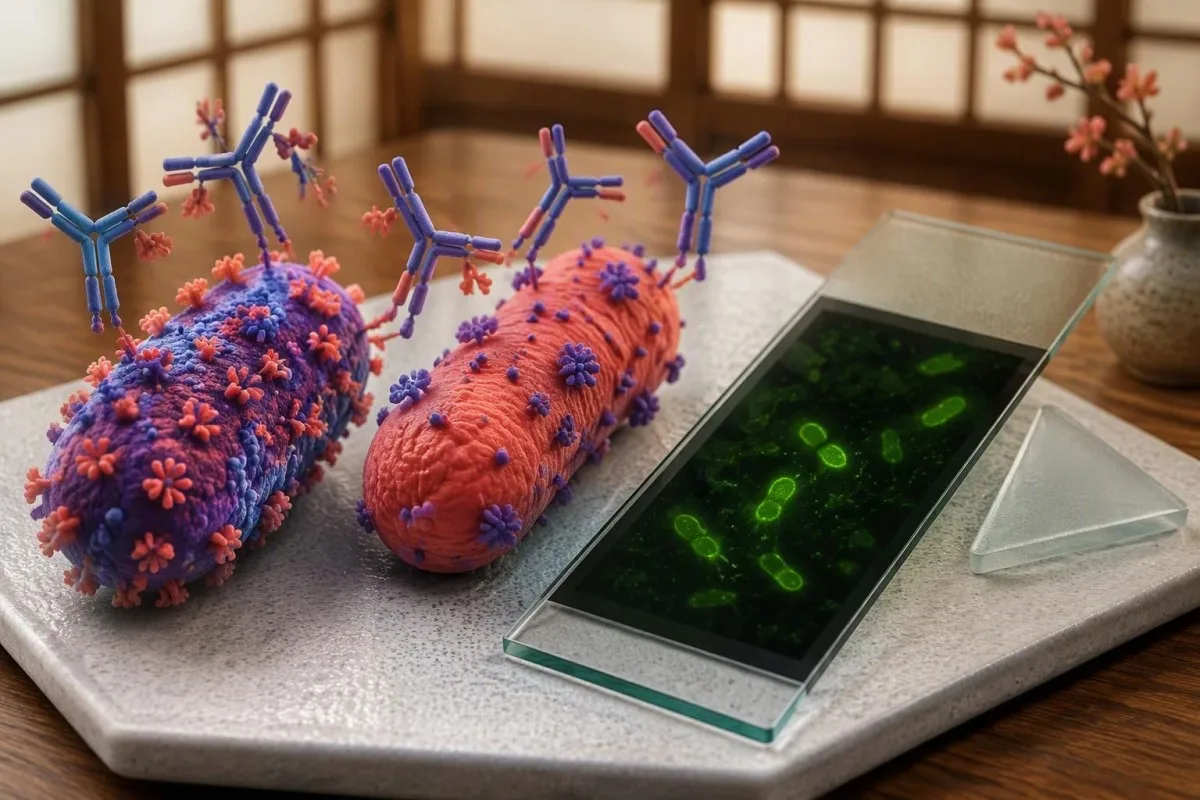
IgG ระยะที่ II มักจะบ่งชี้การติดเชื้อในระยะหลัง; IgG ระยะที่ I ที่สูงขึ้นนั้นจะทำให้เกิดคำถามที่แตกต่างออกไป การกำหนดเวลาเก็บตัวอย่าง วิธีการของห้องปฏิบัติการ และหลักฐานของการติดเชื้อเฉพาะที่ที่คงอยู่ จะเป็นตัวกำหนดว่าผลลัพธ์แต่ละอย่างมีความหมายอย่างไร.


- Q fever antibodies บ่งชี้การตอบสนองทางภูมิคุ้มกันต่อ Coxiella burnetii; ผลบวกเพียงครั้งเดียวไม่สามารถยืนยันการติดเชื้อที่กำลังดำเนินอยู่ได้ด้วยตนเอง.
- Phase II IgG มักจะเด่นใน Q fever ระยะเฉียบพลัน แต่ค่า titer บวกเพียงค่าเดียวก็อาจสะท้อนถึงการสัมผัสในอดีตได้เช่นกัน.
- Paired samples ที่เก็บห่างกันประมาณ 3–6 สัปดาห์ สามารถแสดงให้เห็นถึงการเพิ่มขึ้นสี่เท่าของ IgG ระยะที่ II ซึ่งสนับสนุนการติดเชื้อในระยะหลัง.
- Fourfold rise หมายถึง 1:64 ถึง 1:256 ไม่ใช่ 1:64 ถึง 1:128; การเปรียบเทียบที่เชื่อถือได้ต้องใช้วิธีการเดียวกัน โดยเฉพาะอย่างยิ่งห้องปฏิบัติการเดียวกัน.
- Phase I IgG ที่ 1:1024 หรือสูงกว่าโดย IFA เป็นตัวกระตุ้นที่เกี่ยวข้องกับ CDC สำหรับการประเมินการติดเชื้อที่คงอยู่ ไม่ใช่การวินิจฉัยด้วยตัวเอง.
- ຄ່າຂີດຈຳກັດຂອງຫ້ອງທົດລອງ ແຕກຕ່າງກັນ: ເກณฑ์ endocarditis ຂອງ ESC ໃຊ້ IgG ໄລยะ I ຫຼາຍກວ່າ 1:800, ເຊິ່ງບໍ່ສາມາດປ່ຽນແທນໄດ້ກັບລະດັບການລາຍງານຂອງຫ້ອງທົດລອງທຸກແຫ່ງ.
- PCR ໄລยะຕົ້ນ ມີປະໂຫຍດຫຼາຍທີ່ສຸດໃນປະມານ 2 ອາທິດທໍາອິດຂອງການເຈັບປ່ວຍ, ໂດຍສະເພາະກ່ອນຫຼືຫຼັງຈາກເລີ່ມຕົ້ນການໃຊ້ຢາຕ້ານເຊື້ອ.
- ຄົນເຈັບທີ່ມີຄວາມສ່ຽງສູງ ປະກອບມີຜູ້ທີ່ມີພະຍາດຫົວໃຈ, ປ່ຽນຖ່າຍເສັ້ນເລືອດຫຼືເສັ້ນເລືອດແດງ, ຖືພາ, ແລະພູມຕ້ານທານທີ່ຖືກກົດຂີ່ບາງຊະນິດ.
- ອາການດ່ວນ ເຊັ່ນ ອາການຫາຍໃຈຝືດຮຸນແຮງ, ສັບສົນ, ເປັນລົມ, ຫຼືອາການທາງລະບົບປະສາດໃໝ່ ຕ້ອງການການປະເມີນຜົນທັນທີ ແທນທີ່ຈະລໍຖ້າພູມຕ້ານທານຊໍ້າ.
ผล Q fever antibody ที่เป็นบวกหมายความว่าอย่างไร?
Q fever antibodies ສະແດງການຕອບສະໜອງທາງພູມຕ້ານທານຕໍ່ Coxiella burnetii. Phase II IgG ປົກກະຕິຈະໂດດເດັ່ນໃນການຕິດເຊື້ອສ້ວຍແຫຼມເມື່ອບໍ່ດົນມານີ້; Phase I IgG ທີ່ສູງຂຶ້ນຢ່າງຫຼວງຫຼາຍ ຮຽກຮ້ອງໃຫ້ມີການປະເມີນການຕິດເຊື້ອໃນຈຸດທີ່ຕິດຄົງຕົວ. ບໍ່ມີຮູບແບບໃດໜຶ່ງ ຖືກວິນິດໄສວ່າເປັນພະຍາດທີ່ກຳລັງດຳເນີນຢູ່ຄົນດຽວ: ອາການ, ຄ່າຂີດຈຳກັດສະເພາະຫ້ອງທົດລອງ, ຕົວຢ່າງຄູ່, ແລະບາງຄັ້ງ PCR ຫຼືການຖ່າຍຮູບແມ່ນມີຄວາມຈຳເປັນ.
ລາຍງານກ່າວວ່າ Coxiella burnetii IgG ຜົນເປັນບວກ ຕອບຄຳຖາມທີ່ແຄບກວ່າທີ່ຄົນເຈັບສ່ວນຫຼາຍຄາດຫວັງ: ໄດ້ກວດພົບພູມຕ້ານທານໂດຍການວິເຄາະນັ້ນ. ມັນບໍ່ໄດ້ກຳນົດວ່າການສຳຜັດເກີດຂຶ້ນເມື່ອໃດ, ເຊື້ອມີການເຄື່ອນໄຫວຢູ່ຫຼືບໍ່, ຫຼືຕ້ອງການການປິ່ນປົວຫຼືບໍ່; ນີ້ແມ່ນ 3 ຄຳຖາມທາງຄລີນິກທີ່ແຍກອອກຈາກກັນ, ແລະການເອົາລວມກັນເປັນອັນດຽວເຮັດໃຫ້ເກີດຄວາມກັງວົນທີ່ບໍ່ຈຳເປັນ.
ຂ້າພະເຈົ້າຂຽນໃນນາມ Thomas Klein, ຫົວໜ້າເຈົ້າໜ້າທີ່ການແພດ, ແລະຄຳຖາມທຳອິດຂອງຂ້າພະເຈົ້າໃນການອ່ານລາຍງານແມ່ນວ່າຜົນໄດ້ຮັບແມ່ນຄຸນນະພາບຫຼືລວມເອົາຫົວໜ່ວຍການເຈືອຈາງທີ່ແທ້ຈິງ. ຄວາມແຕກຕ່າງມີຄວາມສຳຄັນຫຼາຍກວ່າທຸງສີແດງຂ້າງຜົນໄດ້ຮັບ: 1:64 ແລະ 1:1024 ສາມາດຖືກເອີ້ນວ່າເປັນບວກໄດ້, ແຕ່ພວກມັນກະຕຸ້ນການສົນທະນາທີ່ແຕກຕ່າງກັນ. ຄຳອະທິບາຍຂອງພວກເຮົາກ່ຽວກັບ ຜົນການກວດແອນຕິບອດທີ່ເປັນບວກ ກວມເອົາຄວາມແຕກຕ່າງທີ່ກວ້າງຂວາງນັ້ນ.
Kantesti ເປັນເຄື່ອງວິເຄາະເລືອດ AI ທີ່ຊ່ວຍຈັດວາງຜົນໄດ້ຮັບສະເພາະໄລຍະພ້ອມກັບວັນທີຂອງຕົວຢ່າງ ແລະການຄົ້ນພົບໃນຫ້ອງທົດລອງອື່ນໆ, ແທນທີ່ຈະຖືວ່າ 1 ຜົນໄດ້ຮັບທີ່ຖືກໝາຍເປັນການວິນິດໄສ. ຂໍ້ມູນກ່ຽວກັບ ອົງກອນຂອງພວກເຮົາ ອະທິບາຍບໍລິບົດການບໍລິການ; Q fever ທີ່ສົງໄສຍັງຕ້ອງການທ່ານໝໍທີ່ສາມາດປະເມີນການສຳຜັດ, ຜົນການກວດ, ແລະຄວາມຈຳເປັນໃນການທົດສອບພິເສດ.
Q fever ระยะที่ 1 และระยะที่ 2 อธิบายถึงอะไร?
Q fever ໄລยะ 1 ແລະ ໄລยะ 2 ອະທິບາຍຮູບແບບແອນຕິເຈນຂອງ Coxiella burnetii, ບໍ່ແມ່ນ 2 ໄລຍະທີ່ເປັນລຳດັບທີ່ຄົນເຈັບທຸກຄົນຜ່ານ. ເຊື້ອໄລຍະ I ມີ lipopolysaccharide ຢູ່ໜ້າຜິວທີ່ເຕັມຮູບແບບ; ແອນຕິເຈນໄລຍະ II ສະທ້ອນໃຫ້ເຫັນຮູບແບບທີ່ສັ້ນລົງທີ່ຜະລິດຂຶ້ນໃນລະຫວ່າງການປັບຕົວໃນຫ້ອງທົດລອງ.
Phase II IgG ປົກກະຕິຈະເພີ່ມຂຶ້ນຢ່າງເດັ່ນຊັດກວ່າໃນລະຫວ່າງ Q fever ສ້ວຍແຫຼມ, ໃນຂະນະທີ່ Phase I IgG ສູງ ສາມາດມາພ້ອມກັບການຕິດເຊື້ອໃນຈຸດທີ່ຕິດຄົງຕົວ. ຄວາມສຳພັນເຫຼົ່ານັ້ນມີປະໂຫຍດ, ແຕ່ບໍ່ແມ່ນສິ່ງທີ່ແນ່ນອນ: ທັງສອງກຸ່ມພູມຕ້ານທານອາດຈະສູງຂຶ້ນຫຼັງຈາກການເຈັບປ່ວຍສ້ວຍແຫຼມ, ແລະ Phase II IgG ສາມາດຍັງຄົງມີຄວາມສຳຄັນແມ້ໃນເວລາທີ່ມີຈຸດຕິດຄົງຕົວ (Anderson et al., 2013).
ຕົວເລກໂຣມັນ I ແລະ II ມັກຈະຖືກແທນທີ່ດ້ວຍຕົວເລກ 1 ແລະ 2 ໃນລາຍງານ; ພວກມັນອ້າງອີງເຖິງໝວດໝູ່ແອນຕິເຈນດຽວກັນ. ພວກມັນຕ້ອງບໍ່ສັບສົນກັບ IgG ທຽບກັບ IgM, ເຊິ່ງອະທິບາຍຊັ້ນພູມຕ້ານທານ. ທົ່ວໄປ ຄູ່ມືການຕີຄວາມໝາຍຂອງ immunoglobulin ອະທິບາຍວ່າເປັນຫຍັງຄວາມເຂັ້ມຂຸ້ນຂອງ IgG ທັງໝົດ ແລະ IgG ສະເພາະເຊື້ອຈຶ່ງເປັນການວັດແທກທີ່ແຕກຕ່າງກັນ.
ການປຽບທຽບໄລຍະ I ກັບໄລຍະ II ເປັນຂໍ້ບົ່ງຊີ້ ບໍ່ແມ່ນກົດເກນຈັດຈຳແນກທີ່ໃຊ້ໄດ້ໂດຍລຳພັງ ແລະບໍ່ມີອັດຕາສ່ວນ 1:1 ທີ່ໃຊ້ວິນິດໄສໄດ້ສະເໝີໄປ. ແຕກຕ່າງຈາກຊຸດແອນຕິເຈນທີ່ມີຊື່ສະເພາະ ຊຶ່ງນຳໃຊ້ໃນ EBV antibody patterns, ການແປຜົນ Q fever ຂຶ້ນກັບຄ່າໄຕເຕີສຸດທ້າຍ, ການປ່ຽນແປງຂອງຄ່ານັ້ນຕາມເວລາ ແລະການມີ ຫຼື ບໍ່ມີບັນຫາທາງຄລີນິກທີ່ຈຳເພາະຈຸດໜຶ່ງ.
จะอ่านค่า Q fever antibody titers และเกณฑ์ของห้องปฏิบัติการได้อย่างไร?
ຄ່າໄຕເຕີແອນຕິບໍດີ Q fever ໂດຍປົກກະຕິລາຍງານຄ່າການເຈືອຈາງສູງສຸດຂອງເຊຣັມທີ່ຍັງໃຫ້ປະຕິກິລິຍາໄດ້ ໂດຍໃຊ້ວິທີອິມມູໂນຟລູອໍເຣສເຊນທາງອ້ອມ ຫຼື IFA. ຜົນ 1:256 ສູງກວ່າ 1:64; ຄ່າເຫຼົ່ານີ້ແມ່ນຄ່າສຸດທ້າຍຂອງການເຈືອຈາງ ບໍ່ແມ່ນຄວາມເຂັ້ມຂຸ້ນຂອງແອນຕິບໍດີໃນ mg/dL ຫຼື IU/mL.
ຕົວສ່ວນລຸ່ມສະແດງວ່າເຊຣັມຖືກເຈືອຈາງໄປຫຼາຍປານໃດ: 1:256 ຍັງໃຫ້ປະຕິກິລິຍາໄດ້ເມື່ອເຈືອຈາງຫຼາຍກວ່າ 1:64. ຫ້ອງປະຕິບັດການອາດເລີ່ມກວດດ້ວຍລະດັບການເຈືອຈາງທີ່ຕ່າງກັນ ແລະໃຊ້ການກະກຽມແອນຕິເຈນ ຫຼືຂັ້ນຕອນການອ່ານຜົນທີ່ຕ່າງກັນ, ດັ່ງນັ້ນຈຶ່ງບໍ່ສາມາດກຳນົດຄວາມໝາຍທາງຄລີນິກຂອງເຄື່ອງໝາຍຜົນບວກຈາກຫ້ອງປະຕິບັດການໜຶ່ງ ໃຫ້ເປັນຄວາມໝາຍທີ່ອີກຫ້ອງໜຶ່ງໃຊ້ໂດຍອັດຕະໂນມັດບໍ່ໄດ້.
ການກວດຄັດກອງໄດ້ຜົນບວກ ແລະເກນຕັດສິນທາງຄລີນິກ ບໍ່ແມ່ນສິ່ງດຽວກັນ. ຫ້ອງປະຕິບັດການອາດຈັດຄ່າສຸດທ້າຍຕ່ຳໆວ່າກວດພົບໄດ້, ໃນຂະນະທີ່ແນວທາງປະຕິບັດໃຊ້ຄ່າໄລຍະ I ທີ່ສູງກວ່າຫຼາຍເພື່ອເລີ່ມສືບສວນການຕິດເຊື້ອທີ່ຄົງຢູ່; ດັດຊະນີ ELISA 2.4 ບໍ່ສາມາດປ່ຽນເປັນຄ່າໄຕເຕີ IFA 1:1024 ໄດ້ໂດຍກົງ.
ຂໍລາຍລະອຽດ 3 ຢ່າງ: ວິທີການກວດ, ຄ່າສຸດທ້າຍແຍກຕາມໄລຍະ ແລະຄຳເຫັນການແປຜົນຂອງຫ້ອງປະຕິບັດການ. ການອະທິບາຍຂອງພວກເຮົາກ່ຽວກັບ ການທົດສອບຄຸນນະພາບທຽບກັບການທົດສອບປະລິມານ ອະທິບາຍເຫດຜົນທີ່ລາຍງານຜົນບວກ/ຜົນລົບບໍ່ສາມາດສະແດງການເພີ່ມຂຶ້ນສີ່ເທົ່າ; ຖ້າຈຳເປັນຕ້ອງປຽບທຽບ, ແພດອາດຂໍກວດ IFA ແບບປະລິມານຜ່ານຫ້ອງປະຕິບັດການອ້າງອີງ ແທນການກວດຊ້ຳດ້ວຍການກວດຄັດກອງທີ່ບໍ່ເໝາະສົມ.
ทำไมตัวอย่างที่เก็บสองครั้งจึงช่วยระบุ Q fever ในระยะหลังได้?
ຕົວຢ່າງ serum ຄູ່ ສາມາດລະບຸ Q fever ໃນຊ່ວງໄວໆນີ້ໄດ້ ໂດຍສະແດງວ່າ IgG ໄລຍະ II ເພີ່ມຂຶ້ນສີ່ເທົ່າຂຶ້ນໄປ. ວິທີວິນິດໄສຂອງ CDC ປຽບທຽບຕົວຢ່າງໃນໄລຍະສ້ວຍແຫຼມ, ໂດຍຄວນເກັບໃນຊ່ວງຕົ້ນຂອງການເຈັບປ່ວຍ, ກັບຕົວຢ່າງໃນໄລຍະຟື້ນຕົວທີ່ເກັບປະມານ 3–6 ອາທິດຕໍ່ມາ (Anderson et al., 2013).
ການເພີ່ມຈາກ 1:64 ເປັນ 1:256 ແມ່ນເພີ່ມສີ່ເທົ່າ; ຈາກ 1:64 ເປັນ 1:128 ແມ່ນເພີ່ມພຽງສອງເທົ່າ. ວິທີ IFA ຫຼາຍວິທີໃຊ້ການເຈືອຈາງແບບເພີ່ມເທົ່າສອງ, ດັ່ງນັ້ນການເພີ່ມສີ່ເທົ່າຈຶ່ງກວມເອົາ 2 ຂັ້ນຂອງການເຈືອຈາງ. ເງື່ອນໄຂນີ້ຊ່ວຍແຍກການເພີ່ມຂຶ້ນຂອງແອນຕິບອດທີ່ມີຄວາມໝາຍ ອອກຈາກຄວາມແຕກຕ່າງເລັກນ້ອຍໃນການອ່ານຜົນກວດ, ແຕ່ກໍຍັງຕ້ອງພິຈາລະນາພາບລວມທາງຄລີນິກທັງໝົດ.
ຄວນສົ່ງຕົວຢ່າງທັງສອງໄປຫ້ອງປະຕິບັດການດຽວກັນ, ໃຊ້ວິທີການດຽວກັນ ແລະອາດກວດພ້ອມກັນເພື່ອຫຼຸດຄວາມແປປວນລະຫວ່າງຮອບການກວດ. ຜົນ 1:128 ຈາກຫ້ອງປະຕິບັດການໜຶ່ງ ຕາມດ້ວຍ 1:256 ຈາກອີກແຫ່ງ ບໍ່ແມ່ນຫຼັກຖານທີ່ໜ້າເຊື່ອຖືຂອງການດຳເນີນໄປຂອງພະຍາດທາງຊີວະວິທະຍາ; ບົດຄວາມຂອງພວກເຮົາ คู่มือไทม์ไลน์การตรวจเลือด ອະທິບາຍວ່າເຫດໃດຈຶ່ງຕ້ອງລະບຸເງື່ອນໄຂການວັດແທກໃນທຸກການປຽບທຽບ.
ຕົວຢ່າງທຳອິດທີ່ເກັບໃນໄລຍະ 4 ອາທິດຫຼັງເລີ່ມປ່ວຍ ອາດສະແດງຄ່າໄຕເຕີ phase II ສູງຢູ່ແລ້ວ, ເຮັດໃຫ້ມີຊ່ອງຫວ່າງໜ້ອຍສຳລັບການເພີ່ມຂຶ້ນອີກສີ່ເທົ່າ. ການບໍ່ພົບການເພີ່ມຂຶ້ນໃນສະຖານະການນັ້ນ ບໍ່ໄດ້ຕັດຄວາມເປັນໄປໄດ້ຂອງການຕິດເຊື້ອໃໝ່ໆອອກ; ຕົວຢ່າງເບື້ອງຕົ້ນພຽງແຕ່ເກັບຊ້າເກີນໄປຈົນບໍ່ທັນຈັບການຕອບສະໜອງທີ່ກຳລັງພັດທະນາ, ແລະ PCR ກໍອາດມີຄວາມໄວລົດລົງເຊັ່ນກັນ.
ທີ່ Kantesti, ການປຽບທຽບທີ່ເປັນປະໂຫຍດບໍ່ແມ່ນພຽງກ່ອນກັບຫຼັງ: ແຕ່ແມ່ນ ວັນທີເກັບຕົວຢ່າງທຽບກັບວັນທີເລີ່ມມີອາການ, ໂດຍເພີ່ມເວລາໃຫ້ຢາຕ້ານເຊື້ອເປັນຈຸດອ້າງອີງທີສາມ. ແຈ້ງແພດວ່າຕົວຢ່າງທຳອິດເກັບໃນວັນທີ 3 ຫຼືວັນທີ 23, ແລະຖາມວ່າສາມາດນຳຕົວຢ່າງເດີມທີ່ເກັບຮັກສາໄວ້ມາກວດຊ້ຳພ້ອມກັບຕົວຢ່າງທີສອງໄດ້ຫຼືບໍ່.
ผล IgG ที่เป็นบวกเพียงครั้งเดียวสามารถบ่งชี้การติดเชื้อเก่าได้หรือไม่?
ຜົນ Coxiella burnetii IgG ບວກພຽງຄັ້ງດຽວ ອາດສະແດງເຖິງການສຳຜັດເຊື້ອໃນອະດີດ, ເພາະແອນຕິບອດອາດຄົງຢູ່ໄດ້ຫຼາຍເດືອນ ຫຼືຫຼາຍປີ. ຜົນ IgG phase II ບວກພຽງຄັ້ງດຽວ ບໍ່ສາມາດແຍກການຕິດເຊື້ອທີ່ເກີດຂຶ້ນໃໝ່ໆອອກຈາກການຕິດເຊື້ອໃນອະດີດໄດ້ຢ່າງໜ້າເຊື່ອຖື, ເຖິງແມ່ນຄ່າໄຕເຕີຕົວເລກຈະສູງຫຼາຍກໍຕາມ.
ຄວາມເປັນໄປໄດ້ຂອງໄຂ້ Q ທີ່ກຳລັງດຳເນີນຢູ່ ປ່ຽນແປງຕາມເຫດຜົນທີ່ສັ່ງກວດ. ຄ່າໄຕເຕີ phase II 1:256 ໃນຄົນທີ່ຫາກໍມີໄຂ້ຫຼັງສຳຜັດກັບສັດລ້ຽງປະສິດ ມີຄວາມໝາຍແຕກຕ່າງຈາກຜົນດຽວກັນໃນຄົນສະບາຍດີທີ່ຖືກກວດໃນການສືບຫາສາເຫດຂອງອາການເມື່ອຍລ້າແບບກວ້າງຂວາງ; ຕົວເລກຈາກຫ້ອງປະຕິບັດການບໍ່ໄດ້ປ່ຽນ, ແຕ່ຄວາມເປັນໄປໄດ້ກ່ອນກວດປ່ຽນໄປແລ້ວ.
IgM ບໍ່ສາມາດຕອບຄຳຖາມໄດ້ດ້ວຍຕົວມັນເອງ: ມັນອາດຄົງຢູ່ໄດ້ ແລະໂດຍທົ່ວໄປຈຳເພາະໜ້ອຍກວ່າ IgG ໃນການກວດໄຂ້ Q. ຜົນ IgM ບວກບວກກັບ IgG ບວກພຽງຄັ້ງດຽວ ຍັງຕ້ອງພິຈາລະນາເວລາແລະຕີຄວາມໝາຍທາງຄລີນິກ, ແທນທີ່ຈະຕິດປ້າຍວ່າເປັນການຕິດເຊື້ອໃໝ່ຢ່າງງ່າຍໆ; ຄວາມແຕກຕ່າງນີ້ຄ້າຍກັບບາງປະເດັນທີ່ກ່າວເຖິງໃນ ການຕີຄວາມໝາຍ CMV IgG, ແມ່ນແຕ່ເຊື້ອກໍ່ໂລກຕ້ອງໃຊ້ກົດການວິນິດໄສທີ່ແຕກຕ່າງກັນ.
ຄ່າໄຕເຕີ IgG phase II ສູງພຽງຄັ້ງດຽວ ອາດຊ່ວຍສະໜັບສະໜູນການວິນິດໄສວ່າໜ້າຈະເປັນພະຍາດໄລຍະສ້ວຍ ໃນກໍລະນີທີ່ອາການທາງຄລີນິກສອດຄ່ອງ, ແຕ່ຕ້ອງກວດສອບກອບການລາຍງານຜົນຂອງຫ້ອງປະຕິບັດການ. ຄຳແນະນຳຂອງ CDC ປີ 2013 ກ່າວເຖິງຄ່າໄຕເຕີ IgG phase II ຢ່າງໜ້ອຍ 1:128 ວ່າເປັນຜົນທີ່ສະໜັບສະໜູນໃນສະຖານະການນັ້ນ; ບໍ່ໄດ້ກຳນົດໃຫ້ 1:128 ເປັນເກນວິນິດໄສສາກົນສຳລັບທຸກວິທີກວດ ຫຼືຜູ້ປ່ວຍທຸກຄົນທີ່ບໍ່ມີອາການ.
PCR และการทดสอบซ้ำมีประโยชน์เมื่อใดหลังจากผลเป็นลบ?
PCR ສຳລັບ Coxiella burnetii ມີປະໂຫຍດຫຼາຍທີ່ສຸດໃນຊ່ວງຕົ້ນຂອງການເຈັບປ່ວຍໄລຍະສ້ວຍ, ໂດຍມັກແມ່ນປະມານ 2 ອາທິດທຳອິດ ແລະກ່ອນ ຫຼືບໍ່ດົນຫຼັງເລີ່ມໃຊ້ຢາຕ້ານເຊື້ອ. ມັກກວດບໍ່ພົບແອນຕິບອດໃນອາທິດທຳອິດ, ດັ່ງນັ້ນຜົນກວດຊີໂຣໂລຈີໃນຊ່ວງຕົ້ນທີ່ເປັນລົບ ບໍ່ສາມາດຕັດໄຂ້ Q ອອກໄດ້.
PCR ກວດຫາສານພັນທຸກຳຂອງແບັກທີເຣຍ, ໃນຂະນະທີ່ IFA ວັດການຕອບສະໜອງຂອງແອນຕິບອດຈາກຮ່າງກາຍ; ດັ່ງນັ້ນການກວດເຫຼົ່ານີ້ຈຶ່ງມີຊ່ວງເວລາທີ່ໃຫ້ປະໂຫຍດແຕກຕ່າງກັນ. ຜົນກວດແອນຕິບອດເປັນລົບໃນວັນທີ 5 ແລະ PCR ເປັນບວກ ສາມາດສອດຄ່ອງກັບໄຂ້ Q ໄລຍະສ້ວຍໄດ້ຢ່າງສົມບູນ, ແລະຜົນ IgG ບວກໃນພາຍຫຼັງ ບໍ່ໄດ້ໝາຍຄວາມວ່າການກວດແອນຕິບອດກ່ອນໜ້ານັ້ນຂັດຂ້ອງ.
ການໄດ້ຮັບຢາຕ້ານເຊື້ອອາດຫຼຸດຄວາມໄວຂອງ PCR, ແລະປະລິມານສານຈາກແບັກທີເຣຍທີ່ໄຫຼວຽນໃນເລືອດຫຼຸດລົງ ກໍຈຳກັດການກວດພົບໃນໄລຍະທ້າຍຂອງການເຈັບປ່ວຍເຊັ່ນກັນ. ຜົນ PCR ເປັນລົບໃນວັນທີ 18, ໂດຍສະເພາະຫຼັງໄດ້ຮັບການຮັກສາ, ບໍ່ມີນ້ຳໜັກໃນການຕັດພະຍາດອອກເທົ່າກັບຕົວຢ່າງທີ່ເກັບໃນເວລາຕົ້ນທີ່ເໝາະສົມ; ເວລາກໍເປັນປັດໃຈສຳຄັນເຊັ່ນກັນໃນ ການກວດໄຂ້ເດັງກີໃນຊ່ວງຕົ້ນ, although the actual tests and organisms differ.
Clinicians should not delay appropriate treatment for strongly suspected acute Q fever while waiting 3–6 weeks for convalescent antibodies. Record the exact antibiotic start date and ask which specimen type the reference laboratory accepts; serum, whole blood, and tissue testing have different uses, and a late negative blood PCR cannot by itself exclude a persistent focal infection.
การสัมผัสและอาการใดบ้างที่ทำให้ Q fever มีความเป็นไปได้มากขึ้น?
Q fever becomes more plausible after exposure to aerosolized material from infected livestock, particularly sheep, goats, or cattle and their birth products. Acute illness commonly begins about 2–3 weeks after exposure and may cause fever, severe headache, pneumonia, or hepatitis without a distinctive rash.
Directly touching an animal is not required for Q fever exposure, because contaminated dust can be inhaled. A useful 3-week history includes farm visits, veterinary work, abattoir work, lambing or kidding environments, and nearby livestock activity; a patient who says they never handled animals may still have an exposure worth discussing.
Fever after animal contact has more than 1 possible explanation, and broadening the differential can prevent a positive Q fever antibody result from becoming an anchor. Unpasteurized dairy exposure or occupational animal contact may also prompt brucellosis testing interpretation. Freshwater or floodwater exposure may instead strengthen the case for leptospirosis test timing.
An ALT of 180 IU/L with fever may support a hepatitis presentation, but it does not identify Coxiella as the cause; acute Q fever can also occur with a normal white-cell count. Our ຄູ່ມືຫຍໍ້ການທົດສອບຕັບ helps separate markers of liver injury from organism-specific testing, and normal routine results should not override a convincing exposure history.
เมื่อใดที่ IgG ระยะที่ I สูงขึ้นจึงควรได้รับการประเมินจากผู้เชี่ยวชาญ?
Elevated phase I IgG warrants evaluation when it reaches a method-specific concerning threshold, rises over time, or accompanies relevant symptoms or risk factors. CDC guidance uses phase I IgG at least 1:1024 by IFA together with an identifiable persistent focus to diagnose chronic Q fever (Anderson et al., 2013).
Phase I IgG at or above 1:1024 is not a treatment order. Some patients develop substantial phase I responses after acute infection without demonstrable persistent disease, so clinicians look for endocarditis, vascular infection, or another focal site rather than prescribing prolonged therapy based on the number alone; a stable patient usually needs coordinated assessment, not panic.
ໄດ້ 2023 ESC endocarditis guideline lists a phase I IgG titer greater than 1:800 as a major microbiological criterion for infective endocarditis (Delgado et al., 2023). That criterion belongs within a complete endocarditis evaluation; different dilution series and reference-laboratory methods help explain why 1:800 and 1:1024 appear in different documents without being universally interchangeable.
The Dutch consensus separates proven, probable, and possible chronic Q fever using combinations of serology, PCR, imaging, and clinical risk factors, rather than one antibody result alone (Wegdam-Blans et al., 2012). This is a useful corrective to the idea that antibodies directly measure disease severity, an issue also discussed in high ASO titer interpretation.
A Kantesti explanation should frame a phase I result of 1:2048 as a follow-up trigger, not a declaration of endocarditis. Fever, weight loss, night sweats, a known valve problem, an aortic aneurysm, or a vascular graft changes the urgency; conversely, a result below 1:1024 should not dismiss persistent focal infection when clinical suspicion is substantial.
แพทย์จะตรวจสอบ Q fever เฉพาะที่ที่คงอยู่อย่างไร?
Persistent focal Q fever requires a search for an anatomical site of ongoing infection, most often a heart valve or an abnormal vascular site. Evaluation may combine phase-specific IFA, PCR, echocardiography, vascular imaging, and specialist-directed FDG-PET/CT; no single test answers all of these questions.
A normal transthoracic echocardiogram does not exclude Q fever endocarditis, because vegetations may be small or absent and prosthetic material can complicate imaging. Transoesophageal echocardiography may be needed when suspicion remains; 1 reassuring scan should not automatically close the investigation in someone with compatible serology and a high-risk cardiac history.
Vascular grafts and aneurysms create a second major diagnostic pathway, sometimes without obvious cardiac findings. CT angiography or FDG-PET/CT may help identify the relevant focus, while tissue PCR can be informative if material becomes available for clinical reasons; clinicians should not obtain invasive tissue solely because an isolated phase I titer is 1:1024.
Routine blood cultures often remain negative in Q fever endocarditis, so culture-negative disease does not mean there is no infection. Bartonella is another organism considered in this setting, and our Bartonella titer explanation describes why targeted serology differs from ordinary culture; the selected tests depend on exposure history and the broader endocarditis differential.
Tell the laboratory when Q fever is suspected, because Coxiella culture requires specialized facilities and should not be requested casually. Our culture result interpretation guide explains what standard cultures can establish; a negative blood PCR, negative routine cultures, and 1 normal echocardiogram still need to be weighed against the patient's overall risk and imaging findings.
ใครบ้างที่ต้องได้รับการติดตามอย่างใกล้ชิดหลังจากการสัมผัสหรือการติดเชื้อ Q fever?
People with valve disease, prosthetic valves, vascular grafts, aneurysms, pregnancy, or certain forms of immunosuppression need closer clinical attention after Q fever. Persistent infection develops in a minority of patients—commonly described as fewer than 5% overall—but that average does not describe an individual high-risk patient.
A valve history can matter more than a twofold titer difference, so the clinician needs to know about prosthetic valves, congenital valve abnormalities, and previously documented valve disease. Not every incidental murmur carries the same risk; the practical question is whether a structural abnormality has been established and whether baseline cardiac assessment is appropriate.
Pregnancy changes both risk assessment and treatment choices, and compatible symptoms or newly concerning serology deserve prompt discussion with obstetric and infectious-disease teams. Q fever serology is not the same test as the routine pregnancy antibody screen, which assesses antibodies against red-cell antigens; 1 positive result on either report needs its own distinct clinical pathway.
Immunosuppression can alter symptoms and antibody responses, so a low titer should not be used as automatic reassurance in a symptomatic transplant recipient or someone receiving substantial immune-suppressing treatment. A person with no recalled acute illness can still have persistent focal Q fever; missing a memorable fever 6 months earlier does not exclude the diagnosis.
รูปแบบ IgG ระยะที่ I และระยะที่ II ทั่วไปบ่งชี้อะไร?
Common Q fever antibody patterns suggest possibilities rather than establish diagnoses. Low phase I IgG with rising phase II IgG supports recent infection; stable phase II IgG may represent past exposure; high phase I IgG raises concern for a persistent focus when the clinical circumstances fit.
Consider an illustrative—not an actual patient—case: a 34-year-old veterinary worker has fever for 5 days, phase I IgG below detection, and phase II IgG of 1:64. If phase II IgG reaches 1:256 after 4 weeks using the same IFA method, that fourfold rise supports recent Q fever; the first low result did not mean the illness was mild.
Another hypothetical patient feels well and has phase II IgG of 1:256 with phase I IgG of 1:32, unchanged on a properly timed repeat. That pattern is compatible with prior exposure, but stability alone cannot establish the precise date of infection; 2 results obtained after antibodies have plateaued may miss the earlier rise entirely.
A third example is a 68-year-old with an aortic graft, weight loss, phase I IgG of 1:2048, and phase II IgG of 1:4096. Phase II remaining higher does not exclude persistent infection; the graft history and high phase I endpoint warrant specialist evaluation, and this is exactly where a simplistic phase-I-greater-than-phase-II rule can fail.
Kantesti is an AI blood test interpretation platform that can help organize these phase-specific values and their dates, but the report should never substitute a pattern label for a clinical diagnosis. Our ມາດຕະຖານທາງການແພດແລະການກວດສອບຄວາມຖືກຕ້ອງ page describes the broader evaluation framework; an automated explanation cannot locate a vascular focus or independently exclude endocarditis.
ควรทำการตรวจ Q fever antibodies ซ้ำเมื่อใด?
Repeat testing depends on the question being asked: approximately 3–6 weeks helps confirm recent infection, while longer follow-up checks for persistent disease in selected patients. CDC guidance describes assessment around 6 months for lower-risk patients and closer monitoring at 3, 6, 12, 18, and 24 months for higher-risk groups.
Weekly antibody testing usually adds little, because IgG changes more slowly than symptoms and ordinary inflammatory markers. A repeat after 7 days may show a small fluctuation without settling whether infection is recent or persistent; worsening clinical symptoms require reassessment now, not a decision to wait until the next planned serology date.
Follow-up should preserve the original report, laboratory method, phase I and phase II values, symptom dates, and antibiotic history. Our การเปรียบเทียบทางห้องปฏิบัติการที่มีความหมาย guide explains the value of consistent measurement conditions; a switch from a 1:800-based reference scale to a 1:1024-based scale should be identified before anyone labels the difference a true rise.
Kantesti can help organize a result timeline, but a 60-second explanation is not equivalent to 24 months of specialist surveillance. Our ຄູ່ມືເທັກໂນໂລຍີ AI explains the report-processing context; the clinician should individualize follow-up for pregnancy, cardiac or vascular risk, persistent symptoms, and the exact national or reference-laboratory protocol being used.
ผล antibody ที่เป็นบวกหมายความว่าคุณต้องได้รับยาปฏิชีวนะหรือไม่?
Positive Q fever antibodies do not automatically justify antibiotics. Symptomatic acute Q fever is commonly treated with doxycycline 100 mg twice daily for 14 days in appropriate adults, while confirmed persistent focal infection requires specialist-directed prolonged treatment; pregnancy and pediatric care require separate decisions.
Q fever endocarditis treatment is measured in months, not days, and commonly combines doxycycline with hydroxychloroquine under specialist supervision. CDC guidance describes at least 18 months for native-valve infection and 24 months for prosthetic-valve infection; drug interactions, retinal monitoring, clinical response, and serological trends matter, so these durations are not instructions for self-treatment.
Persistent fatigue after acute Q fever is not synonymous with persistent focal infection, and a positive IgG result does not prove that ongoing bacterial activity explains fatigue. A separate evaluation may uncover anemia or another contributor; our คู่มือการศึกษาธาตุเหล็กที่สมบูรณ์ is relevant when hemoglobin or iron markers are abnormal, but iron results neither confirm nor exclude Coxiella infection.
There is no established supplement, dietary plan, or detox regimen that reliably clears Q fever IgG, and a titer of 1:256 does not create a nutrient prescription. New dark urine, jaundice, or renal abnormalities need their own assessment; our การตรวจปัสสาวะ (urinalysis) แบบครอบคลุม explains complementary findings without implying that urinary markers can diagnose Q fever.
คุณควรนำอะไรไปในการนัดหมายและอ่านอะไรต่อไป?
Bring the full laboratory report, not just the word positive, plus symptom-onset dates, exposure history, medication dates, and relevant cardiac or vascular records. As of October 11, 2026, the central distinction remains unchanged: antibodies support an assessment, but 1 result cannot independently establish recent or persistent focal Q fever.
Write down 4 questions: Which method was used? Are the phase I and phase II values actual endpoints? Is a paired sample needed? Does my history warrant a search for a persistent focus? Include the exact laboratory cutoff, because saying the result was high omits the detail that distinguishes a low screening positive from a phase I titer of 1:2048.
Kantesti is an AI-powered blood test analysis tool that can support preparation for that discussion, but urgent symptoms should bypass report upload and routine interpretation. Severe breathlessness, confusion, fainting, new neurological symptoms, or serious chest pain require immediate medical assessment; even 1 of these symptoms matters more than waiting for a second antibody sample.
My checklist as Thomas Klein, Chief Medical Officer at Kantesti LTD, separates report explanation from diagnosis and treatment. Information about our ໂຄງສ້າງການໃຫ້ຄຳປຶກສາທາງການແພດ describes the organization of clinical input; this article does not represent a personal consultation, and any claim of independent review should be confirmed through the publication's actual editorial records.
The following 2 associated publications concern complementary laboratory interpretation, not the evidence for Q fever thresholds or treatment. Their DOI identifiers are supplied below for traceability; they should not be read as substitutes for the CDC, ESC, and Dutch consensus references cited in this article.
Kantesti. (n.d.). Urobilinogen in urine test: Complete urinalysis guide 2026. Zenodo. DOI: 10.5281/zenodo.18226379. A ຄົ້ນຫາບັນທຶກ ResearchGate ແລະ ຄົ້ນຫາສິ່ງພິມຂອງ Academia are discovery links, not verified hosted copies. Kantesti. (n.d.). Iron studies guide: TIBC, iron saturation & binding capacity. Zenodo. DOI: 10.5281/zenodo.18248745. The corresponding ຄົ້ນຫາສິ່ງພິມຂອງ ResearchGate ແລະ Academia record search likewise do not establish independent peer review.
ຄໍາຖາມທີ່ຖາມເລື້ອຍໆ
Coxiella burnetii IgG positive ໝາຍຄວາມວ່າແນວໃດ?
Coxiella burnetii IgG positive ໝາຍຄວາມວ່າການທົດສອບໄດ້ກວດພົບພູມຕ້ານທານຕໍ່ກັບເຊື້ອຈຸລິນຊີທີ່ເຮັດໃຫ້ເກີດໄຂ້ Q. ຜົນໄດ້ຮັບທີ່ເປັນບວກໜຶ່ງຢ່າງສາມາດສະທ້ອນເຖິງການຕິດເຊື້ອເມື່ອບໍ່ດົນມານີ້ຫຼືການສຳຜັດເມື່ອຫຼາຍເດືອນຫາຫຼາຍປີກ່ອນໜ້ານີ້ ແລະບໍ່ໄດ້ກຳນົດພະຍາດທີ່ກຳລັງລຸກລາມຢ່າງເປັນເອກລາດ. ການຕີຄວາມຮຽກຮ້ອງໃຫ້ມີຜົນໄດ້ຮັບໄລຍະ I ແລະ II, ວິທີການຫ້ອງທົດລອງ, ອາການ, ແລະເວລາຂອງຕົວຢ່າງ. ຕົວຢ່າງຄູ່ທີ່ມີໄລຍະຫ່າງປະມານ 3–6 ອາທິດສາມາດຊ່ວຍລະບຸການຕິດເຊື້ອເມື່ອບໍ່ດົນມານີ້.
ຄວາມແຕກຕ່າງລະຫວ່າງ Q fever phase I ແລະ phase II IgG ແມ່ນຫຍັງ?
Q fever phase I ແລະ phase II IgG ຈຳແນກຮູບແບບແອນຕິເຈນ 2 ຮູບແບບຂອງ Coxiella burnetii ແທນທີ່ຈະເປັນ 2 ໄລຍະຕາຍຕົວຂອງອາການເຈັບປ່ວຍຂອງຄົນເຈັບ. Phase II IgG ມັກມີລະດັບເດັ່ນໃນຊ່ວງຕິດເຊື້ອແບບສະເພາະໜ້າ, ໃນຂະນະທີ່ phase I IgG ທີ່ສູງຂຶ້ນຢ່າງຫຼາຍບົ່ງຊີ້ເຖິງຄວາມກັງວົນວ່າອາດມີການຕິດເຊື້ອສະເພາະຈຸດທີ່ຄົງຢູ່. ແນວທາງຂອງ CDC ໃຊ້ phase I IgG ຢ່າງໜ້ອຍ 1:1024 ໂດຍວິທີ IFA ຮ່ວມກັບຫຼັກຖານວ່າມີຈຸດຕິດເຊື້ອທີ່ຄົງຢູ່. ຂະໜາດສຳພັນຂອງຄ່າໄຕເຕີ 2 ຄ່ານີ້ບໍ່ສາມາດໃຊ້ວິນິດໄສ ຫຼື ຕັດອອກວ່າມີການຕິດເຊື້ອທີ່ຄົງຢູ່ໄດ້ດ້ວຍຕົນເອງ.
4 เท่าของ titres แอนติบอดี Q fever นับเป็นเท่าใด?
ການເພີ່ມຂຶ້ນສີ່ເທົ່າຂອງຄ່າ titer ຂອງພູມຕ້ານທານ Q fever ໝາຍຄວາມວ່າຄ່ານ້ອຍສຸດເພີ່ມຂຶ້ນ 4 ເທົ່າ, ເຊັ່ນ IgG ໄລຍະ II ຈາກ 1:64 ຫາ 1:256. ການປ່ຽນແປງຈາກ 1:64 ຫາ 1:128 ແມ່ນພຽງແຕ່ສອງເທົ່າແລະບໍ່ເປັນໄປຕາມເງື່ອນໄຂນັ້ນ. ໂດຍປົກກະຕິແລ້ວ, ຕົວຢ່າງສ້ວຍແຫຼມແລະ convalescent ທີ່ຈັບຄູ່ກັນຈະຖືກເກັບໃນໄລຍະຫ່າງປະມານ 3–6 ອາທິດ. ການປຽບທຽບແມ່ນເຊື່ອຖືໄດ້ຫຼາຍທີ່ສຸດດ້ວຍວິທີ IFA ດຽວກັນ ແລະ ດີທີ່ສຸດແມ່ນຫ້ອງທົດລອງດຽວກັນ.
ລະດັບ IgG ຂອງໄລຍະ I 1:1024 ໝາຍຄວາມວ່າເປັນໄຂ້ Q ຊໍາເຮື້ອບໍ?
ລະດັບ IgG ໄລຍະ I 1:1024 ໂດຍ IFA ເປັນສາເຫດທີ່ຈະປະເມີນຫາ Q fever ທີ່ຕິດຕໍ່ເນື່ອງ, ບໍ່ແມ່ນການວິນິດໄສໃນຕົວມັນເອງ. ຄຳແນະນຳຂອງ CDC ໄດ້ລວມເອົາລະດັບ IgG ໄລຍະ I ຢ່າງໜ້ອຍ 1:1024 ພ້ອມດ້ວຍຈຸດທີ່ຕິດຕໍ່ເນື່ອງທີ່ສາມາດລະບຸໄດ້, ເຊັ່ນ endocarditis ຫລື ການຕິດເຊື້ອເສັ້ນເລືອດ. ບາງຄັ້ງໃນປີ 2023 ຂອງຄຳແນະນຳກ່ຽວກັບ endocarditis ໃຊ້ລະດັບ IgG ໄລຍະ I ຫຼາຍກວ່າ 1:800 ເປັນຄຸນລັກສະນະທາງຈຸລຊີວະວິທະຍາຫລັກພາຍໃນການປະເມີນ endocarditis ທີ່ສົມບູນ. ຫ້ອງທົດລອງມີຄວາມແຕກຕ່າງກັນ, ສະນັ້ນຕ້ອງໄດ້ກວດສອບວິທີການລາຍງານແລະການຕີຄວາມກ່ອນທີ່ຈະນຳໃຊ້ຂີດຈຳກັດໃດໜຶ່ງ.
แอนติบอดี Q fever สามารถเป็นลบในช่วงแรกของการติดเชื้อได้หรือไม่?
Q fever antibodies ທີ່ເປັນລົບອາດພົບໄດ້ໃນອາທິດທຳອິດຂອງອາການແບບສ້ວຍແຫຼມ. Coxiella burnetii PCR ມັກຈະມີປະໂຫຍດຫຼາຍທີ່ສຸດໃນຊ່ວງປະມານ 2 ອາທິດທຳອິດ, ໂດຍສະເພາະກ່ອນຫຼືຫຼັງການເລີ່ມການປິ່ນປົວດ້ວຍຢາຕ້ານເຊື້ອບໍ່ດົນ. ຕົວຢ່າງການຫາຍດີໃນໄລຍະຫຼັງທີ່ເກັບໄດ້ປະມານ 3–6 ອາທິດຫຼັງຈາກຕົວຢ່າງແບບສ້ວຍແຫຼມສາມາດສະແດງໃຫ້ເຫັນການເພີ່ມຂຶ້ນຂອງພູມຕ້ານທານທີ່ສາມາດວິນิจໄສໄດ້. ນັກແພດບໍ່ຄວນຊັກຊ້າໃນການປິ່ນປົວທີ່ເໝາະສົມສຳລັບອາການ Q fever ແບບສ້ວຍແຫຼມທີ່ສົງໄສຢ່າງແຮງໃນຂະນະທີ່ລໍຖ້າຕົວຢ່າງທີສອງນັ້ນ.
แอนติบอดี Q fever จะมีผลเป็นบวกนานเท่าใด และจำเป็นต้องได้รับการรักษาหรือไม่?
ພູມຕ້ານທານຕໍ່ໄຂ້ Q ອາດຍັງມີຜົນບວກຢູ່ໄດ້ເປັນເດືອນ ຫຼືເປັນປີຫຼັງຈາກສຳຜັດເຊື້ອ, ດັ່ງນັ້ນ IgG ທີ່ຍັງຄົງຢູ່ບໍ່ໄດ້ໝາຍຄວາມວ່າຕ້ອງໃຊ້ຢາຕ້ານເຊື້ອສະເໝີໄປ. ໃນຜູ້ໃຫຍ່ທີ່ເໝາະສົມ, ໄຂ້ Q ໄລຍະສ້ວຍແບບມີອາການມັກຮັກສາດ້ວຍ doxycycline 100 mg ມື້ລະສອງຄັ້ງ ເປັນເວລາ 14 ມື້, ສ່ວນການຕິດເຊື້ອສະເພາະຈຸດທີ່ຄົງຢູ່ແລະຢືນຢັນແລ້ວ ຕ້ອງຮັກສາໄລຍະຍາວຕາມຄຳແນະນຳຂອງຜູ້ຊ່ຽວຊານ. ແນວໂນ້ມຂອງລະດັບພູມຕ້ານທານເປັນພຽງສ່ວນໜຶ່ງຂອງການຕິດຕາມອາການ ແລະບໍ່ສາມາດໃຊ້ຢືນຢັນການຫາຍດີ ຫຼືການຮັກສາລົ້ມເຫຼວໄດ້ໂດຍລຳພັງ. ການຖືພາ, ໄວເດັກ, ປະຕິສຳພັນລະຫວ່າງຢາ ແລະພະຍາດຫົວໃຈ ຫຼືຫຼອດເລືອດ ຕ້ອງການການດູແລແບບສະເພາະບຸກຄົນ.
ຮັບການວິເຄາະຜົນກວດເລືອດດ້ວຍ AI ທັນທີ
ເຂົ້າຮ່ວມຜູ້ໃຊ້ຫຼາຍກວ່າ 2 ລ້ານຄົນທົ່ວໂລກ ທີ່ໄວ້ໃຈ Kantesti ສຳລັບການວິເຄາະການກວດເລືອດທີ່ທັນທີ ແລະຖືກຕ້ອງ. ອັບໂຫຼດຜົນກວດເລືອດຂອງທ່ານ ແລະຮັບການຕີຄວາມໝາຍຢ່າງຄົບຖ້ວນຂອງ biomarker 15,000+ ໃນວິນາທີ.
📚 ບົດຄວາມວິຈັຍທີ່ອ້າງອີງ
Klein, T., Mitchell, S., & Weber, H. (2026). Urobilinogen ໃນການກວດຍ່ຽວ: ຄູ່ມືການກວດຍ່ຽວຄົບຖ້ວນ 2026. ການຄົ້ນຄວ້າທາງການແພດຂອງ AI Kantesti.
Klein, T., Mitchell, S., & Weber, H. (2026). ຄູ່ມືການສຶກສາທາດເຫຼັກ: TIBC, ຄວາມອີ່ມຕົວຂອງທາດເຫຼັກ ແລະ ຄວາມສາມາດໃນການຜູກມັດ. ການຄົ້ນຄວ້າທາງການແພດຂອງ AI Kantesti.
📖 ເອກະສານອ້າງອີງທາງການແພດພາຍນອກ
Anderson A et al. (2013). Diagnosis and Management of Q Fever—United States, 2013: Recommendations from CDC and the Q Fever Working Group. MMWR Recommendations and Reports.
Delgado V et al. (2023). 2023 ESC Guidelines for the management of endocarditis. European Heart Journal.
Wegdam-Blans MCA et al. (2012). Chronic Q fever: review of the literature and a proposal of new diagnostic criteria. ວາລະສານການຕິດເຊື້ອ.
⚕️ ຂໍ້ສັງເກດທາງການແພດ
ບົດຄວາມນີ້ມີຈຸດປະສົງເພື່ອການສຶກສາເທົ່ານັ້ນ ແລະບໍ່ແມ່ນຄຳແນະນຳທາງການແພດ. ຄວນປຶກສາຜູ້ໃຫ້ບໍລິການດ້ານສຸຂະພາບທີ່ມີຄຸນວຸດທິສະເໝີ ສຳລັບການວິນິດໄຊ ແລະ ການຕັດສິນໃຈດ້ານການຮັກສາ.
ສັນຍານຄວາມໄວ້ໃຈ E-E-A-T
ປະສົບການ
ການທົບທວນຄລີນິກຂອງແພດຜູ້ນຳພາ ກ່ຽວກັບຂັ້ນຕອນການຕີຄວາມໝາຍຜົນການກວດໃນຫ້ອງທົດລອງ.
ຄວາມຊ່ຽວຊານ
ວິຊາການແພດທົດລອງ (ການແພດທາງຫ້ອງທົດລອງ) ເນັ້ນໃສ່ວ່າຕົວຊີ້ວັດ (biomarkers) ມີພຶດຕິກຳແນວໃດໃນບັນບົດທາງຄລີນິກ.
ຄວາມເປັນອຳນາດ
ຂຽນໂດຍທ່ານດຣ. Thomas Klein ໂດຍມີການກວດທານໂດຍທ່ານດຣ. Sarah Mitchell ແລະ ສາດສະດາຈານດຣ. Hans Weber.
ຄວາມໜ້າເຊື່ອຖື
ການຕີຄວາມໝາຍອີງຕາມຫຼັກຖານດ້ວຍເສັ້ນທາງຕິດຕາມທີ່ຊັດເຈນ ເພື່ອຫຼຸດການຕົກໃຈ.