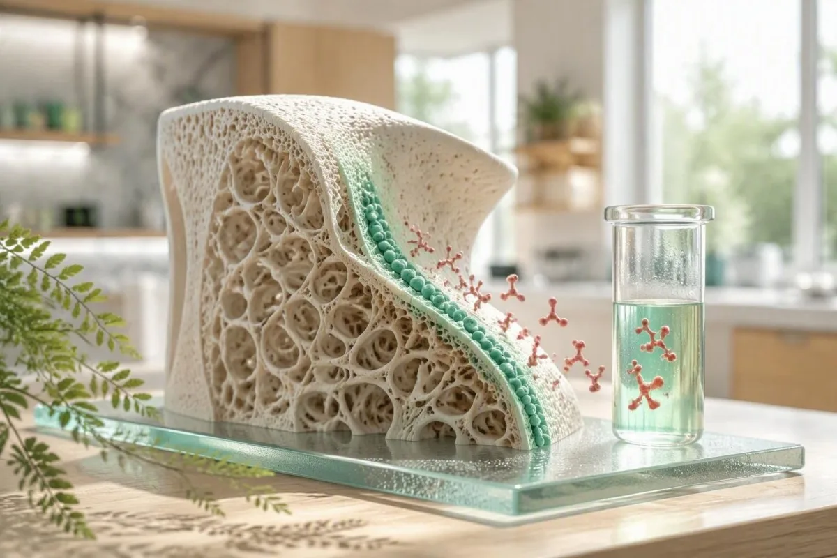
An osteocalcin result is a snapshot of bone-building activity, not a stand-alone osteoporosis diagnosis. The useful question is whether the value fits your age, kidney function, glucose status, recent fracture, and treatment history.


- Bone formation marker: Osteocalcin is released by osteoblasts during bone formation, but it can also rise when overall bone turnover is high.
- No universal range: Adult osteocalcin normal values vary by assay; many N-MID assays report roughly 11-43 ng/mL before menopause, but the laboratory range controls.
- 높은 결과: A high osteocalcin level can occur with growth, menopause, fracture repair, hyperthyroidism, vitamin D deficiency with secondary hyperparathyroidism, or high-turnover bone disease.
- 낮은 결과: A low osteocalcin level may reflect suppressed bone turnover from antiresorptive treatment, glucocorticoids, diabetes, low energy availability, or hypothyroidism.
- Kidney caveat: Reduced eGFR can elevate circulating osteocalcin because renal clearance falls, so chronic kidney disease changes interpretation.
- Best companions: P1NP, CTX, calcium, phosphate, alkaline phosphatase, 25-hydroxyvitamin D, PTH, creatinine/eGFR, TSH, and HbA1c give osteocalcin clinical meaning.
- Treatment monitoring: Bone-turnover markers can shift within 3-6 months of therapy, whereas a DXA scan usually changes more slowly.
- Repeat wisely: Use the same laboratory, assay, collection time, and fasting routine for serial osteocalcin measurements whenever possible.
What an osteocalcin blood test actually measures
An osteocalcin blood test measures a protein made chiefly by osteoblasts, the cells that lay down new bone matrix. A high result usually means bone turnover is active, while a low result often means bone formation is suppressed; neither result alone proves that bone is strong or weak.
Osteocalcin becomes incorporated into bone after vitamin K-dependent carboxylation, and a smaller fraction enters circulation. Laboratories may measure total, intact, N-terminal mid-fragment, or undercarboxylated osteocalcin; those are related measurements, not interchangeable numbers. For that reason, a value of 25 ng/mL can be routine in one assay and impossible to compare directly with another.
In my clinical practice, patients often assume osteocalcin is a calcium test. It is not: calcium is tightly regulated in serum, while osteocalcin is a dynamic marker of osteoblast activity that can move noticeably after a fracture, a new medicine, or menopause.
칸테스티는 AI 혈액검사 분석기 that reads osteocalcin alongside mineral, kidney, thyroid, and metabolic markers rather than attaching a diagnosis to one flagged line. This matters because a normal calcium result does not rule out accelerated skeletal turnover; our 바이오마커 참조 가이드 explains why markers answer different biological questions.
Why osteocalcin is not a screening test
Osteocalcin is not recommended as a population screening test for osteoporosis. DXA bone-density testing and fracture-risk assessment remain the tools that estimate fracture risk; osteocalcin is mainly useful when a clinician needs a biochemical view of turnover or response to treatment.
Osteocalcin normal values depend on the assay and life stage
Osteocalcin normal values are assay-specific and must be read against the reference interval printed on your report. Many adult N-MID osteocalcin assays use approximate intervals near 11-43 ng/mL in premenopausal women and 15-46 ng/mL after menopause, but those figures are not universal cutoffs.
Reference intervals differ because assays recognize different fragments and because osteocalcin is unstable enough that sample handling matters. Some European laboratories report results in micrograms per litre, which is numerically equivalent to ng/mL, while others report pmol/L; unit conversion is only safe when the exact analyte and calibrator are known.
Children and adolescents commonly have substantially higher osteocalcin than adults because longitudinal growth accelerates bone formation. A pubertal value above 70 ng/mL may be physiologically expected, whereas the same result in a 68-year-old needs context such as a recent fracture, PTH level, and thyroid status.
Dr. Thomas Klein advises patients to save the laboratory name, assay name, collection time, and menopause status with every result. A result that is 20% different may reflect real turnover, but it can also reflect a different assay or pre-analytical handling; our guide to 혈액검사 간의 실제 차이 그 구분을 다룹니다.
What a high osteocalcin level can mean
A high osteocalcin level most often reflects increased bone turnover, not automatically bone gain. Menopause, adolescence, healing fractures, hyperthyroidism, hyperparathyroidism, osteomalacia, and some high-turnover bone disorders can all raise it.
The distinction between formation and turnover is the trap. When osteoclast-driven resorption and osteoblast-driven formation both accelerate, osteocalcin can be high while net bone density falls—particularly in untreated hyperthyroidism or early postmenopause.
A healing fracture can raise bone markers for months. I have seen a 59-year-old whose osteocalcin remained near twice her laboratory upper limit 10 weeks after wrist fixation; her calcium, PTH, and TSH were unremarkable, and the timing was far more informative than the isolated number.
A high osteocalcin paired with high alkaline phosphatase may support active skeletal remodeling, but liver disease can also raise total alkaline phosphatase. Review the pattern through our explanation of alkaline phosphatase after fracture before assuming the source is bone.
When high turnover deserves prompt review
Unexplained high osteocalcin with bone pain, recurrent low-trauma fractures, calcium above 10.5 mg/dL, phosphate abnormalities, or a suppressed TSH deserves clinician review. These combinations can point toward endocrine or mineral disorders that require targeted testing rather than supplements alone.
What a low osteocalcin level can mean
A low osteocalcin level usually indicates low bone formation activity or pharmacologic suppression of turnover. Common explanations include antiresorptive medicines, glucocorticoids, type 2 diabetes, hypothyroidism, low energy availability, and prolonged immobility.
Bisphosphonates and denosumab reduce bone resorption and, secondarily, formation markers; a lower osteocalcin after treatment may show the medicine is biologically active. It is not a reason to stop treatment unless the prescribing clinician sees a broader safety concern.
Type 2 diabetes is associated with lower bone-turnover markers despite a DXA result that can look normal or even high. This helps explain the so-called diabetic bone paradox: density does not capture all aspects of bone material quality and fall risk.
Severe calorie restriction, amenorrhea, and endurance training can suppress formation markers before a DXA scan changes. Athletes with fatigue, recurrent stress injury, or altered periods should consider a RED-S laboratory review rather than treating a low marker as an isolated nutrition problem.
Low is not always safer
Very low turnover is not automatically protective because bone needs ongoing remodeling to repair microdamage. The evidence does not support a single osteocalcin threshold for “over-suppression,” which is why symptoms, fracture history, duration of therapy, and DXA trends still lead clinical decisions.
Why age, puberty, and menopause shift osteocalcin
Osteocalcin is highest during periods of rapid skeletal change, especially puberty and the first years after menopause. Estrogen decline removes a brake on bone remodeling, so formation and resorption markers often rise together after the final menstrual period.
Peak osteocalcin values often occur during adolescence, when growth plates and cortical expansion demand active matrix production. Adult reference ranges should never be applied to a 13-year-old without paediatric, age- and sex-specific intervals.
After menopause, marker changes can precede a measurable DXA decline. The 2020 Endocrine Society osteoporosis guideline recommends treatment decisions based on fracture risk and bone density rather than osteocalcin alone, while acknowledging that bone-turnover markers can help monitor adherence in selected patients (Eastell et al., 2019).
Menstrual timing has a smaller effect than menopause status, illness, and treatment, but consistency still helps when trending a result. For related hormone context, our article on estradiol results in menopause explains why one hormone measurement rarely dates the transition precisely.
Men receiving androgen deprivation therapy
Men receiving androgen deprivation therapy can develop rapid turnover and bone loss, often before symptoms arise. A baseline DXA, vitamin D measurement, calcium intake review, and fracture-risk assessment are more actionable than an osteocalcin value by itself.
Kidney function can make osteocalcin look higher
Reduced kidney function can elevate osteocalcin because the kidneys contribute to clearance of circulating osteocalcin fragments. A high value in chronic kidney disease may therefore reflect retention, altered turnover, or both—not simply faster bone formation.
This is clinically significant once eGFR is persistently below 60 mL/min/1.73 m², and interpretation becomes especially unreliable in advanced CKD. The 2017 KDIGO CKD-MBD guideline favors serial PTH and bone-specific alkaline phosphatase to assess turnover because extremes of either marker help predict underlying turnover state (KDIGO CKD-MBD Update Work Group, 2017).
A high osteocalcin with eGFR 32 mL/min/1.73 m² and normal calcium is a different clinical problem from the same osteocalcin result with eGFR 95. Phosphate, corrected calcium, PTH, bicarbonate, 25-hydroxyvitamin D, and medication exposure often explain more than osteocalcin does in this setting.
칸테스티는 AI 검사 결과 해석 서비스는 that treats creatinine and eGFR as essential context before flagging a bone marker pattern. Patients can compare this with our practical CKD 병기 가이드 and the kidney-focused BUN-to-creatinine review.
Diabetes can lower osteocalcin while fracture risk rises
Type 2 diabetes often lowers osteocalcin and other formation markers even when DXA bone density is not low. Chronic hyperglycaemia may impair osteoblast function and alter collagen cross-linking, contributing to fracture risk that density alone can underestimate.
HbA1c above 6.5% confirms diabetes in appropriate clinical circumstances, but the osteocalcin relationship is associative rather than a treatment target. An unexpectedly low osteocalcin in someone with HbA1c 8.4% should prompt a wider review of diabetes control, falls, kidney function, vitamin D, and medications—not an attempt to raise osteocalcin directly.
Advanced glycation end-products stiffen collagen and may reduce the bone's ability to absorb energy. That is one reason a person with diabetes and a “reassuring” DXA T-score can still warrant careful fracture-risk assessment after a low-trauma fracture.
When fasting insulin, triglycerides, and glucose move together, metabolic risk may be developing before diabetes is formally diagnosed. Our review of borderline fasting insulin shows which companion results make that pattern more convincing.
Undercarboxylated osteocalcin and glucose
Undercarboxylated osteocalcin has been studied as a possible metabolic signal, but results in humans remain inconsistent and assays are not standardized enough for diabetes management. I would not use it to diagnose insulin resistance or choose a glucose-lowering medicine.
Bone medicines and common drugs can change osteocalcin
Antiresorptive osteoporosis medicines generally lower osteocalcin, while anabolic therapies usually raise formation markers early in treatment. The direction and size of change depend on the medicine, baseline turnover, adherence, renal function, and assay.
Oral bisphosphonates commonly reduce formation and resorption markers by roughly 30-70% within 3-6 months in adherent patients, although there is wide individual variation. Denosumab can suppress resorption markers more rapidly, so timing relative to the injection matters when interpreting a panel.
Teriparatide and abaloparatide usually increase formation markers within 1-3 months; romosozumab produces an early formation rise followed by a return toward baseline. The IOF and IFCC recommend P1NP as the reference formation marker and serum CTX as the reference resorption marker for many osteoporosis studies, not osteocalcin, because standardization is better (Vasikaran et al., 2011).
Prednisone at doses of 5 mg daily or more for 3 months can suppress osteoblast activity and increase fracture risk. Do not self-adjust calcium, vitamin D, or osteoporosis medication based on one result; our medication trend safety guide explains what clinicians look for over time.
Other medications to disclose
Thyroid hormone excess, anticonvulsants, aromatase inhibitors, androgen deprivation therapy, proton-pump inhibitors in selected patients, and loop diuretics can affect bone risk through different mechanisms. Bring a complete medicines and supplement list, including injections and intermittent steroid courses, to the appointment.
The companion tests that give osteocalcin clinical context
Osteocalcin is most useful when paired with P1NP or CTX, calcium, phosphate, alkaline phosphatase, 25-hydroxyvitamin D, PTH, creatinine/eGFR, TSH, and HbA1c. Each companion test answers a different question about formation, resorption, mineral supply, endocrine drive, or clearance.
P1NP reflects type I collagen formation and CTX reflects type I collagen breakdown; both are usually collected fasting in the morning for serial comparison, especially CTX because it has pronounced day-to-day and circadian variation. A raised CTX plus raised osteocalcin supports high turnover more clearly than osteocalcin alone.
Calcium should be interpreted with albumin or measured as ionized calcium when clinically necessary. Phosphate below 2.5 mg/dL, elevated PTH, and low 25-hydroxyvitamin D can suggest mineral stress or secondary hyperparathyroidism, while a suppressed TSH can uncover an overlooked driver of bone loss.
Kantesti AI interprets osteocalcin results by mapping these companion markers into a time-based pattern, not a simplistic high-or-low verdict. Our vitamin D retest schedule 그리고 인산염 범위 가이드에서 are useful preparation for that discussion.
How to prepare for an osteocalcin test and repeat it fairly
Use the same laboratory and, when possible, collect osteocalcin at the same morning time for repeat testing. Fasting is not always mandatory for osteocalcin, but fasting morning collection improves comparability when CTX or other bone-turnover markers are measured at the same visit.
Avoid scheduling a trend test immediately after a major fracture, orthopedic procedure, acute illness, or an unusually hard endurance event unless the clinician specifically wants that information. These states can temporarily change turnover markers enough to obscure the baseline signal.
Tell the laboratory and clinician about biotin, because high-dose supplements can interfere with some immunoassays. Biotin doses of 5-10 mg daily, common in hair and nail products, are more relevant than the 30 micrograms found in a typical multivitamin; laboratory-specific instructions should guide any pause.
Record the date of your last denosumab injection, anabolic dose, menstruation changes, and steroid use. A useful baseline blood-test checklist can prevent the common problem of having numbers without the clinical timeline needed to read them.
Can exercise change the number?
A single workout has less predictable effect on osteocalcin than on CK or lactate, but strenuous training, low energy availability, and recent injury can shift turnover biology. For a planned serial result, keeping exercise ordinary for 24 hours beforehand is a reasonable pragmatic approach unless your own laboratory instructs otherwise.
When clinicians use osteocalcin and when they usually do not
Clinicians may order osteocalcin to investigate unusual bone turnover or monitor a selected treatment response, but they do not use it to diagnose osteoporosis by itself. A DXA scan, vertebral imaging when indicated, fracture history, and medication review remain more decisive.
I most often find bone markers helpful when a patient has an unexpected fracture, a strikingly abnormal PTH or TSH result, uncertain adherence to therapy, or a mismatch between symptoms and DXA. They can also help distinguish an actively remodeling state from a lower-turnover state, though bone biopsy remains the definitive test in rare difficult CKD cases.
Routine testing in a well adult without fracture risk factors rarely changes management. A 2026 panel with a modestly high osteocalcin but no symptoms, normal kidney function, normal calcium, normal TSH, and recent menopause generally calls for risk assessment and follow-up—not panic.
Dr. Thomas Klein has seen the greatest value when the request includes a specific clinical question: “Is turnover suppressed after six months of therapy?” is far better than “Is my bone marker normal?” Patients can prepare with our blood tests for women over 60 개요.
DXA and FRAX remain central
DXA measures areal bone mineral density, whereas osteocalcin reflects current biology; one cannot substitute for the other. A low-trauma hip or vertebral fracture warrants medical assessment even if a turnover marker appears unremarkable.
Four osteocalcin result patterns clinicians recognize
The most informative osteocalcin results are patterns: high with high CTX, high with reduced eGFR, low after antiresorptive therapy, or low with diabetes and low energy availability. The same numerical result can mean different things in each pattern.
High osteocalcin plus high CTX, normal eGFR, low 25-hydroxyvitamin D, and high PTH suggests a high-turnover, secondary hyperparathyroid pattern. In contrast, high osteocalcin plus eGFR 28 mL/min/1.73 m² should make renal retention and CKD-mineral bone disorder central considerations.
Low osteocalcin with a 50% fall in CTX after six months of alendronate may support adherence and expected antiresorptive effect. Low osteocalcin with HbA1c 9.1%, low activity, and no bone medication points in a very different direction: metabolic and mechanical bone-quality risk.
A borderline number should not be upgraded to disease merely because it is marked red. Our explanation of 범위를 벗어난 검사 경고 is particularly relevant for assays with wide biological variation.
Why trends beat single values
A personal baseline is often more clinically useful than a population interval for a dynamic marker. A 35% sustained change on the same method, aligned with a medicine start or corrected vitamin D deficiency, is generally more persuasive than a one-time mild elevation.
Practical next steps after high or low osteocalcin
A high or low osteocalcin result should lead to a focused review of fracture history, medicines, kidney function, calcium-phosphate balance, vitamin D, PTH, thyroid status, and DXA timing. Seek urgent care for acute severe bone pain with confusion, marked weakness, dehydration, or symptoms of significant calcium disturbance—not for osteocalcin alone.
Bring prior results rather than only the newest report. A clinician can interpret whether an osteocalcin shift followed a fracture, menopause, a 20 mg prednisone course, a denosumab injection, reduced eGFR, or a major change in training volume.
If results are mildly abnormal but you feel well, repeat testing is commonly planned after 3-6 months under comparable conditions, alongside whichever companion markers were abnormal. If you have a fragility fracture, new height loss, persistent bone pain, kidney disease, or calcium above the reference range, do not wait for a routine repeat.
칸테스티는 AI 기반 혈액검사 분석 도구 used by more than 2 million people across 127+ countries to organize longitudinal laboratory context for a clinician conversation. Our medical review standards are described in the 임상적 타당성 개요, 그리고 저희는 의료 자문 위원회 helps keep patient-facing interpretations appropriately cautious.
Questions to ask at your appointment
Ask which osteocalcin assay was used, whether a repeat should use the same laboratory, whether P1NP and CTX would change management, and whether you need DXA or vertebral fracture assessment. That short list usually produces a more useful consultation than asking whether the number is simply “good” or “bad.”
Limits of osteocalcin testing and what the evidence supports
Osteocalcin has biological value, but evidence does not support a universal diagnostic cutoff for osteoporosis, diabetes, or “bone age.” Its strongest role is as a contextual turnover marker, while treatment and screening decisions still rest on validated fracture-risk tools.
The evidence for osteocalcin as a metabolic hormone in humans is honestly mixed. Associations with insulin sensitivity and fat mass are interesting, but they are not sufficient to make osteocalcin a diabetes screening or nutrition-targeting biomarker.
International work has focused on standardizing P1NP and CTX because their analytical and clinical use is more mature for osteoporosis trials. Osteocalcin remains useful in specific endocrine and research settings, but its fragment diversity makes comparison across laboratories difficult.
As of September 22, 2026, Dr. Thomas Klein's practical advice is simple: use osteocalcin to ask a better question about turnover, then verify the answer with the right companion tests and fracture-risk assessment. Readers who want to understand how our reasoning system handles such uncertainty can review the AI 해석 기술 가이드는.
자주 묻는 질문
골칼신 혈액 검사의 정상 범위는 얼마인가요?
Osteocalcin normal values depend on the exact assay, age, sex, menopause status, and units used by the laboratory. Many N-MID osteocalcin assays report approximate adult intervals of 11-43 ng/mL before menopause and 15-46 ng/mL after menopause, but your report's interval takes priority. Children and teenagers can have values above 70 ng/mL during rapid growth without bone disease. A result should be compared only with previous tests performed by the same method whenever possible.
높은 골카르복실화 수준은 무엇을 의미합니까?
A high osteocalcin level usually means bone turnover is increased, with osteoblasts actively producing bone matrix. Common causes include puberty, the first years after menopause, fracture healing, hyperthyroidism, vitamin D deficiency with raised PTH, and some high-turnover bone disorders. Chronic kidney disease can also raise osteocalcin because renal clearance falls, particularly when eGFR is below 60 mL/min/1.73 m². High osteocalcin alone does not diagnose osteoporosis or prove that bones are gaining density.
낮은 골칼신 수치는 무엇을 의미하나요?
A low osteocalcin level usually reflects reduced bone formation activity or successful suppression of turnover by an antiresorptive medicine. Bisphosphonates and denosumab may reduce bone-turnover markers by roughly 30-70% within 3-6 months, depending on the marker and treatment. Low osteocalcin also occurs with type 2 diabetes, glucocorticoid exposure, hypothyroidism, immobility, and low energy availability. There is no validated single low cutoff that proves dangerous over-suppression of bone turnover.
신장 질환이 높은 골교질 수치의 원인이 될 수 있습니까?
Yes, kidney disease can cause a high osteocalcin result because circulating osteocalcin fragments are partly cleared by the kidneys. When eGFR is persistently below 60 mL/min/1.73 m², the result becomes less specific for bone formation alone. In chronic kidney disease, clinicians usually interpret PTH, calcium, phosphate, vitamin D, and bone-specific alkaline phosphatase together. Advanced CKD may require nephrology or endocrine input when mineral markers are abnormal or fractures occur.
골칼신 혈액 검사 전에 금식해야 하나요?
Fasting is not universally required for osteocalcin, but a fasting morning sample is sensible when osteocalcin is measured with CTX or used for serial monitoring. CTX has substantial circadian variation and is commonly collected in the morning after an overnight fast. Use the same collection time, laboratory, and assay for repeat tests to reduce noise. Avoid major training sessions and report recent fractures, acute illness, high-dose biotin, and osteoporosis medicine timing.
Is osteocalcin better than a DEXA scan for osteoporosis?
No, osteocalcin is not better than DXA for diagnosing osteoporosis or estimating fracture risk. DXA measures bone mineral density, while osteocalcin measures current bone formation activity and can change without a meaningful density change. A DXA T-score of -2.5 or lower at accepted sites meets the densitometric definition of osteoporosis in appropriate adults, whereas no universal osteocalcin cutoff does. Osteocalcin can complement DXA when clinicians are assessing turnover, endocrine causes, or treatment response.
오늘 AI 기반 혈액검사 분석 받기
즉시 정확한 검사 분석을 위해 Kantesti를 신뢰하는 전 세계 200만 명 이상의 사용자와 함께하세요. 혈액검사 결과를 업로드하고 몇 초 만에 15,000+ 바이오마커에 대한 포괄적인 해석을 받아보세요.
📚 참고된 연구 출판물
📖 외부 의학 참고문헌
⚕️ 의료 면책 조항
이 글은 교육 목적만을 위한 것이며 의학적 조언을 구성하지 않습니다. 진단 및 치료 결정에는 항상 자격을 갖춘 의료 전문가와 상담하세요.
E-E-A-T 신뢰 신호
경험
의사가 주도하는 검사 해석 워크플로 임상 검토.
전문적 지식
임상 맥락에서 바이오마커가 어떻게 거동하는지에 대한 검사실 의학 중심.
권위
Dr. Thomas Klein이 작성했으며 Dr. Sarah Mitchell과 Prof. Dr. Hans Weber가 검토했습니다.
신뢰성
경고를 줄이기 위한 명확한 후속 경로가 포함된 근거 기반 해석.