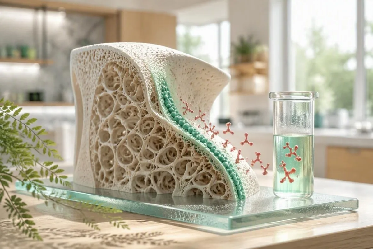
Остеокальциндин жыйынтыгы - бул сөөк түзүүчү активдүүлүктүн сүрөтү, өз алдынча остеопороз диагнозу эмес. Пайдалуу суроо - бул көрсөткүч сиздин жашыңызга, бөйрөк функцияңызга, глюкоза статусуңузга, жакында болгон сыныкка жана дарылоо тарыхыңызга туура келеби.


- Сөөк пайда болуу маркеры: Остеокальцин сөөк пайда болгондо остеобласттар тарабынан бөлүнүп чыгат, бирок ал жалпы сөөк айлануусу жогору болгондо да көбөйүшү мүмкүн.
- Универсалдуу диапазон жок: Чоңдордогу остеокальциндин нормалдуу көрсөткүчтөрү анализ ыкмасына жараша өзгөрөт; көптөгөн N-MID анализи менопаузага чейин болжол менен 11-43 нг/мл көрсөтөт, бирок лабораториялык диапазон аны көзөмөлдөйт.
- Жогору натыйжа: Остеокальциндин жогорку деңгээли өсүү, менопауза, сыныктын айыгышы, гипертиреоз, экинчилик гиперпаратиреоз менен коштолгон D витамининин жетишсиздиги же жогорку айлануучу сөөк оорусу менен пайда болушу мүмкүн.
- Төмөн натыйжа: Остеокальциндин төмөнкү деңгээли антирезорбтивдик дарылоо, глюкокортикоиддер, диабет, энергиянын аз болушу же гипотиреоздон улам сөөк айлануусунун басылышын чагылдырышы мүмкүн.
- Бөйрөк эскертүүсү: eGFRдин төмөндөшү циркуляциядагы остеокальциндди жогорулатышы мүмкүн, анткени бөйрөк клиренси төмөндөйт, ошондуктан өнөкөт бөйрөк оорусу чечмелөөнү өзгөртөт.
- Негизги жардамчылар: P1NP, CTX, кальций, фосфат, ишкер фосфатаза, 25-гидроксивитамин D, PTH, креатинин/eGFR, TSH, жана HbA1c остеокальциндин клиникалык маанисин берет.
- Дарылоону мониторингдоо: Сөөктүн алмашуу маркерлери терапиядан кийин 3-6 айдын ичинде өзгөрүшү мүмкүн, ал эми DXA сканери, адатта, жайыраак өзгөрөт.
- Акылдуулук менен кайталаъыз: Мүмкүн болгондо, сериялык остеокальцинди өлчөө үчүн бир эле лабораторияны, анализди, чогултуу убактысын жана ачка отуруу режимин колдонуңуз.
Остеокальцинди кан анализи чындап эмнени өлчөйт
Остеокальцин кан анализи негизинен остеобласттар, башкача айтканда, жаңы сөөк матриксин түзгөн клеткалар тарабынан чыгарылган белокту өлчөйт. Жогорку натыйжа, адатта, сөөктүн алмашуусу активдүү дегенди билдирет, ал эми төмөнкү натыйжа көбүнчө сөөктүн пайда болушу басылганын билдирет; эч бир натыйжа сөөк күчтүү же алсыз экенин далилдебейт.
Остеокальцин витамин К-көз каранды карбоксилдешүүдөн кийин сөөккө биригет, ал эми кичинекей бөлүгү циркуляцияга кирет. Лабораториялар жалпы, бүтүн, N-терминалдык ортоңку фрагмент же карбоксилдешпеген остеокальцинди өлчөй алышат; алар байланышкан өлчөөлөр, алмаштырылгыс сандар эмес. Ошол себептен, 25 нг/мл мааниси бир анализде кадимки болуп, башкасына түздөн-түз салыштырууга мүмкүн болбой калышы мүмкүн.
Менин клиникалык тажрыйбамда, пациенттер көбүнчө остеокальцинди кальций тести деп ойлошот. Ал эмес: кальций сывороткасында так жөнгө салынат, ал эми остеокальцин остеобласттардын активдүүлүгүнүн динамикалык маркери болуп саналат, ал сыныктан, жаңы дарыдан же менопаузадан кийин байкаларлык жылышы мүмкүн.
Кантести - бул AI кан анализи анализатору минералдык, бөйрөк, калкан бези жана метаболикалык маркерлер менен бирге остеокальцинин окуйт, бир белгиленген сызыкка диагноз койбостон. Бул маанилүү, анткени нормалдуу кальций натыйжасы сөөк алмашуусунун тездешин четке какпайт; биздин биомаркер маалымдамасы маркерлердин ар кандай биологиялык суроолорго жооп берерин түшүндүрөт.
Эмне үчүн остеокальцин скринингдик тест эмес
Остеокальцин остеопороз үчүн популяциялык скринингдик тест катары сунушталбайт. DXA сөөк тыгыздыгын текшерүү жана сынык тобокелдигин баалоо сынык тобокелдигин баалоо үчүн куралдар бойдон калууда; остеокальцин негизинен клиницист алмашуунун биохимиялык көрүнүшүн же дарылоого жооп берүүсүн кааласа пайдалуу.
Остеокальциндин нормалдуу көрсөткүчтөрү анализ ыкмасына жана жашоо баскычына жараша болот
Остеокальциндин нормалдуу маанилери анализге жараша болот жана сиздин отчетуңузда басылган шилтеме интервалына каршы окулушу керек. Көптөгөн чоңдордун N-MID остеокальцин анализи пременопаузалдуу аялдарда 11-43 нг/мл, ал эми менопаузадан кийин 15-46 нг/мл жакынкы интервалдарды колдонот, бирок бул көрсөткүчтөр универсалдуу чектөөлөр эмес.
Шилтеме интервалдары ар түрдүү, анткени анализдер ар кандай фрагменттерди тааныйт жана остеокальцин жетиштүү туруксуз болгондуктан, үлгүлөрдү иштетүү маанилүү. Кээ бир европалык лабораториялар натыйжаларды микрограмм/литр менен билдиришет, бул сандык жактан нг/мл менен барабар, ал эми башкалары সংখ/л менен билдиришет; бир гана белгилүү болгондо гана бирдик конверсиясы коопсуз.
Балдарда жана өспүрүмдөрдө чоңдорго караганда остеокальцин бир кыйла жогору, анткени узунунан өсүү сөөк пайда болушун тездетет. 70 нг/мл жогору болгон пубертеттик маани физиологиялык жактан күтүлүүдө, ал эми 68 жаштагы адамдагы ушундай натыйжа контекстти талап кылат, мисалы, акыркы сынык, PTH деңгээли жана калкан безинин абалы.
Доктор Томас Клейн пациенттерге ар бир жыйынтык менен лабораториянын атын, анализдин атын, чогултуу убактысын жана менопауза статусун сактап коюуну сунуштайт. 20% башкача болгон натыйжа чыныгы алмашууну чагылдырышы мүмкүн, бирок ал ошондой эле башка анализди же анализге чейинки иштетүүнү чагылдырышы мүмкүн; биздин колдонмо кан анализдериндеги чыныгы айырмачылыктар жөнүндө бул айырманы түшүндүрөт.
Остеокальциндин жогорку деңгээли эмнени билдириши мүмкүн
A high osteocalcin level most often reflects increased bone turnover, not automatically bone gain. Menopause, adolescence, healing fractures, hyperthyroidism, hyperparathyroidism, osteomalacia, and some high-turnover bone disorders can all raise it.
The distinction between formation and turnover is the trap. When osteoclast-driven resorption and osteoblast-driven formation both accelerate, osteocalcin can be high while net bone density falls—particularly in untreated hyperthyroidism or early postmenopause.
A healing fracture can raise bone markers for months. I have seen a 59-year-old whose osteocalcin remained near twice her laboratory upper limit 10 weeks after wrist fixation; her calcium, PTH, and TSH were unremarkable, and the timing was far more informative than the isolated number.
A high osteocalcin paired with high alkaline phosphatase may support active skeletal remodeling, but liver disease can also raise total alkaline phosphatase. Review the pattern through our explanation of alkaline phosphatase after fracture before assuming the source is bone.
When high turnover deserves prompt review
Unexplained high osteocalcin with bone pain, recurrent low-trauma fractures, calcium above 10.5 mg/dL, phosphate abnormalities, or a suppressed TSH deserves clinician review. These combinations can point toward endocrine or mineral disorders that require targeted testing rather than supplements alone.
Остеокальциндин төмөнкү деңгээли эмнени билдириши мүмкүн
A low osteocalcin level usually indicates low bone formation activity or pharmacologic suppression of turnover. Common explanations include antiresorptive medicines, glucocorticoids, type 2 diabetes, hypothyroidism, low energy availability, and prolonged immobility.
Bisphosphonates and denosumab reduce bone resorption and, secondarily, formation markers; a lower osteocalcin after treatment may show the medicine is biologically active. It is not a reason to stop treatment unless the prescribing clinician sees a broader safety concern.
Type 2 diabetes is associated with lower bone-turnover markers despite a DXA result that can look normal or even high. This helps explain the so-called diabetic bone paradox: density does not capture all aspects of bone material quality and fall risk.
Severe calorie restriction, amenorrhea, and endurance training can suppress formation markers before a DXA scan changes. Athletes with fatigue, recurrent stress injury, or altered periods should consider a RED-S laboratory review rather than treating a low marker as an isolated nutrition problem.
Low is not always safer
Very low turnover is not automatically protective because bone needs ongoing remodeling to repair microdamage. The evidence does not support a single osteocalcin threshold for “over-suppression,” which is why symptoms, fracture history, duration of therapy, and DXA trends still lead clinical decisions.
Эмне үчүн жаш курак, жыныстык жетилүү жана менопауза остеокальциндди өзгөртөт
Osteocalcin is highest during periods of rapid skeletal change, especially puberty and the first years after menopause. Estrogen decline removes a brake on bone remodeling, so formation and resorption markers often rise together after the final menstrual period.
Peak osteocalcin values often occur during adolescence, when growth plates and cortical expansion demand active matrix production. Adult reference ranges should never be applied to a 13-year-old without paediatric, age- and sex-specific intervals.
After menopause, marker changes can precede a measurable DXA decline. The 2020 Endocrine Society osteoporosis guideline recommends treatment decisions based on fracture risk and bone density rather than osteocalcin alone, while acknowledging that bone-turnover markers can help monitor adherence in selected patients (Eastell et al., 2019).
Menstrual timing has a smaller effect than menopause status, illness, and treatment, but consistency still helps when trending a result. For related hormone context, our article on estradiol results in menopause explains why one hormone measurement rarely dates the transition precisely.
Men receiving androgen deprivation therapy
Men receiving androgen deprivation therapy can develop rapid turnover and bone loss, often before symptoms arise. A baseline DXA, vitamin D measurement, calcium intake review, and fracture-risk assessment are more actionable than an osteocalcin value by itself.
Бөйрөк функциясы остеокальцинди жогорураак көрсөтүшү мүмкүн
Reduced kidney function can elevate osteocalcin because the kidneys contribute to clearance of circulating osteocalcin fragments. A high value in chronic kidney disease may therefore reflect retention, altered turnover, or both—not simply faster bone formation.
This is clinically significant once eGFR is persistently below 60 mL/min/1.73 m², and interpretation becomes especially unreliable in advanced CKD. The 2017 KDIGO CKD-MBD guideline favors serial PTH and bone-specific alkaline phosphatase to assess turnover because extremes of either marker help predict underlying turnover state (KDIGO CKD-MBD Update Work Group, 2017).
A high osteocalcin with eGFR 32 mL/min/1.73 m² and normal calcium is a different clinical problem from the same osteocalcin result with eGFR 95. Phosphate, corrected calcium, PTH, bicarbonate, 25-hydroxyvitamin D, and medication exposure often explain more than osteocalcin does in this setting.
Кантести - бул AI лабораториялык тест чечмелөө кызматы аркылуу жарыяланат that treats creatinine and eGFR as essential context before flagging a bone marker pattern. Patients can compare this with our practical CKD стадиялары боюнча колдонмону караңыз and the kidney-focused BUN-to-creatinine review.
Диабет остеокальциндди төмөндөтүп, сынык тобокелдиги жогорулайт
Type 2 diabetes often lowers osteocalcin and other formation markers even when DXA bone density is not low. Chronic hyperglycaemia may impair osteoblast function and alter collagen cross-linking, contributing to fracture risk that density alone can underestimate.
HbA1c above 6.5% confirms diabetes in appropriate clinical circumstances, but the osteocalcin relationship is associative rather than a treatment target. An unexpectedly low osteocalcin in someone with HbA1c 8.4% should prompt a wider review of diabetes control, falls, kidney function, vitamin D, and medications—not an attempt to raise osteocalcin directly.
Advanced glycation end-products stiffen collagen and may reduce the bone's ability to absorb energy. That is one reason a person with diabetes and a “reassuring” DXA T-score can still warrant careful fracture-risk assessment after a low-trauma fracture.
When fasting insulin, triglycerides, and glucose move together, metabolic risk may be developing before diabetes is formally diagnosed. Our review of borderline fasting insulin shows which companion results make that pattern more convincing.
Undercarboxylated osteocalcin and glucose
Undercarboxylated osteocalcin has been studied as a possible metabolic signal, but results in humans remain inconsistent and assays are not standardized enough for diabetes management. I would not use it to diagnose insulin resistance or choose a glucose-lowering medicine.
Сөөк дарылары жана кеңири таралган дарылар остеокальциндди өзгөртө алат
Antiresorptive osteoporosis medicines generally lower osteocalcin, while anabolic therapies usually raise formation markers early in treatment. The direction and size of change depend on the medicine, baseline turnover, adherence, renal function, and assay.
Oral bisphosphonates commonly reduce formation and resorption markers by roughly 30-70% within 3-6 months in adherent patients, although there is wide individual variation. Denosumab can suppress resorption markers more rapidly, so timing relative to the injection matters when interpreting a panel.
Teriparatide and abaloparatide usually increase formation markers within 1-3 months; romosozumab produces an early formation rise followed by a return toward baseline. The IOF and IFCC recommend P1NP as the reference formation marker and serum CTX as the reference resorption marker for many osteoporosis studies, not osteocalcin, because standardization is better (Vasikaran et al., 2011).
Prednisone at doses of 5 mg daily or more for 3 months can suppress osteoblast activity and increase fracture risk. Do not self-adjust calcium, vitamin D, or osteoporosis medication based on one result; our medication trend safety guide explains what clinicians look for over time.
Other medications to disclose
Thyroid hormone excess, anticonvulsants, aromatase inhibitors, androgen deprivation therapy, proton-pump inhibitors in selected patients, and loop diuretics can affect bone risk through different mechanisms. Bring a complete medicines and supplement list, including injections and intermittent steroid courses, to the appointment.
Остеокальцинге клиникалык контекст берген коштомо тесттер
Osteocalcin is most useful when paired with P1NP or CTX, calcium, phosphate, alkaline phosphatase, 25-hydroxyvitamin D, PTH, creatinine/eGFR, TSH, and HbA1c. Each companion test answers a different question about formation, resorption, mineral supply, endocrine drive, or clearance.
P1NP reflects type I collagen formation and CTX reflects type I collagen breakdown; both are usually collected fasting in the morning for serial comparison, especially CTX because it has pronounced day-to-day and circadian variation. A raised CTX plus raised osteocalcin supports high turnover more clearly than osteocalcin alone.
Calcium should be interpreted with albumin or measured as ionized calcium when clinically necessary. Phosphate below 2.5 mg/dL, elevated PTH, and low 25-hydroxyvitamin D can suggest mineral stress or secondary hyperparathyroidism, while a suppressed TSH can uncover an overlooked driver of bone loss.
Kantesti AI interprets osteocalcin results by mapping these companion markers into a time-based pattern, not a simplistic high-or-low verdict. Our vitamin D retest schedule жана фосфат диапазону боюнча колдонмодон are useful preparation for that discussion.
Остеокальцин тестине кантип даярдануу жана аны адилеттүү кайталоо керек
Use the same laboratory and, when possible, collect osteocalcin at the same morning time for repeat testing. Fasting is not always mandatory for osteocalcin, but fasting morning collection improves comparability when CTX or other bone-turnover markers are measured at the same visit.
Avoid scheduling a trend test immediately after a major fracture, orthopedic procedure, acute illness, or an unusually hard endurance event unless the clinician specifically wants that information. These states can temporarily change turnover markers enough to obscure the baseline signal.
Tell the laboratory and clinician about biotin, because high-dose supplements can interfere with some immunoassays. Biotin doses of 5-10 mg daily, common in hair and nail products, are more relevant than the 30 micrograms found in a typical multivitamin; laboratory-specific instructions should guide any pause.
Record the date of your last denosumab injection, anabolic dose, menstruation changes, and steroid use. A useful baseline blood-test checklist can prevent the common problem of having numbers without the clinical timeline needed to read them.
Can exercise change the number?
A single workout has less predictable effect on osteocalcin than on CK or lactate, but strenuous training, low energy availability, and recent injury can shift turnover biology. For a planned serial result, keeping exercise ordinary for 24 hours beforehand is a reasonable pragmatic approach unless your own laboratory instructs otherwise.
Дарыгерлер качан остеокальциндди колдонушат жана качан көбүнчө колдонушпайт
Clinicians may order osteocalcin to investigate unusual bone turnover or monitor a selected treatment response, but they do not use it to diagnose osteoporosis by itself. A DXA scan, vertebral imaging when indicated, fracture history, and medication review remain more decisive.
I most often find bone markers helpful when a patient has an unexpected fracture, a strikingly abnormal PTH or TSH result, uncertain adherence to therapy, or a mismatch between symptoms and DXA. They can also help distinguish an actively remodeling state from a lower-turnover state, though bone biopsy remains the definitive test in rare difficult CKD cases.
Routine testing in a well adult without fracture risk factors rarely changes management. A 2026 panel with a modestly high osteocalcin but no symptoms, normal kidney function, normal calcium, normal TSH, and recent menopause generally calls for risk assessment and follow-up—not panic.
Dr. Thomas Klein has seen the greatest value when the request includes a specific clinical question: “Is turnover suppressed after six months of therapy?” is far better than “Is my bone marker normal?” Patients can prepare with our blood tests for women over 60 жалпы көрүнүш.
DXA and FRAX remain central
DXA measures areal bone mineral density, whereas osteocalcin reflects current biology; one cannot substitute for the other. A low-trauma hip or vertebral fracture warrants medical assessment even if a turnover marker appears unremarkable.
Дарыгерлер тааныган төрт остеокальцин жыйынтыгынын үлгүлөрү
The most informative osteocalcin results are patterns: high with high CTX, high with reduced eGFR, low after antiresorptive therapy, or low with diabetes and low energy availability. The same numerical result can mean different things in each pattern.
High osteocalcin plus high CTX, normal eGFR, low 25-hydroxyvitamin D, and high PTH suggests a high-turnover, secondary hyperparathyroid pattern. In contrast, high osteocalcin plus eGFR 28 mL/min/1.73 m² should make renal retention and CKD-mineral bone disorder central considerations.
Low osteocalcin with a 50% fall in CTX after six months of alendronate may support adherence and expected antiresorptive effect. Low osteocalcin with HbA1c 9.1%, low activity, and no bone medication points in a very different direction: metabolic and mechanical bone-quality risk.
A borderline number should not be upgraded to disease merely because it is marked red. Our explanation of лабораториялык белгилер диапазондон тышкары болгондо is particularly relevant for assays with wide biological variation.
Why trends beat single values
A personal baseline is often more clinically useful than a population interval for a dynamic marker. A 35% sustained change on the same method, aligned with a medicine start or corrected vitamin D deficiency, is generally more persuasive than a one-time mild elevation.
Остеокальциндин жогору же төмөн болушунан кийинки практикалык кийинки кадамдар
A high or low osteocalcin result should lead to a focused review of fracture history, medicines, kidney function, calcium-phosphate balance, vitamin D, PTH, thyroid status, and DXA timing. Seek urgent care for acute severe bone pain with confusion, marked weakness, dehydration, or symptoms of significant calcium disturbance—not for osteocalcin alone.
Bring prior results rather than only the newest report. A clinician can interpret whether an osteocalcin shift followed a fracture, menopause, a 20 mg prednisone course, a denosumab injection, reduced eGFR, or a major change in training volume.
If results are mildly abnormal but you feel well, repeat testing is commonly planned after 3-6 months under comparable conditions, alongside whichever companion markers were abnormal. If you have a fragility fracture, new height loss, persistent bone pain, kidney disease, or calcium above the reference range, do not wait for a routine repeat.
Кантести - бул AI менен иштеген кан анализин талдоо куралы used by more than 2 million people across 127+ countries to organize longitudinal laboratory context for a clinician conversation. Our medical review standards are described in the клиникалык валидацияга сереп, жана биздин Медициналык кеңеш helps keep patient-facing interpretations appropriately cautious.
Questions to ask at your appointment
Ask which osteocalcin assay was used, whether a repeat should use the same laboratory, whether P1NP and CTX would change management, and whether you need DXA or vertebral fracture assessment. That short list usually produces a more useful consultation than asking whether the number is simply “good” or “bad.”
Остеокальцин тестирлөөсүнүн чектөөлөрү жана далилдер эмнени колдойт
Osteocalcin has biological value, but evidence does not support a universal diagnostic cutoff for osteoporosis, diabetes, or “bone age.” Its strongest role is as a contextual turnover marker, while treatment and screening decisions still rest on validated fracture-risk tools.
The evidence for osteocalcin as a metabolic hormone in humans is honestly mixed. Associations with insulin sensitivity and fat mass are interesting, but they are not sufficient to make osteocalcin a diabetes screening or nutrition-targeting biomarker.
International work has focused on standardizing P1NP and CTX because their analytical and clinical use is more mature for osteoporosis trials. Osteocalcin remains useful in specific endocrine and research settings, but its fragment diversity makes comparison across laboratories difficult.
As of September 22, 2026, Dr. Thomas Klein's practical advice is simple: use osteocalcin to ask a better question about turnover, then verify the answer with the right companion tests and fracture-risk assessment. Readers who want to understand how our reasoning system handles such uncertainty can review the AI интерпретация технологиясы боюнча колдонмо.
Көп берилүүчү суроолор
Сөөк кальцийинин кандагы нормалдуу көрсөткүчү кандай?
Osteocalcin normal values depend on the exact assay, age, sex, menopause status, and units used by the laboratory. Many N-MID osteocalcin assays report approximate adult intervals of 11-43 ng/mL before menopause and 15-46 ng/mL after menopause, but your report's interval takes priority. Children and teenagers can have values above 70 ng/mL during rapid growth without bone disease. A result should be compared only with previous tests performed by the same method whenever possible.
Сөөк кальцийинин жогорку деңгээли эмнени билдирет?
A high osteocalcin level usually means bone turnover is increased, with osteoblasts actively producing bone matrix. Common causes include puberty, the first years after menopause, fracture healing, hyperthyroidism, vitamin D deficiency with raised PTH, and some high-turnover bone disorders. Chronic kidney disease can also raise osteocalcin because renal clearance falls, particularly when eGFR is below 60 mL/min/1.73 m². High osteocalcin alone does not diagnose osteoporosis or prove that bones are gaining density.
Сөөк кальцинин төмөн деңгээли эмнени билдирет?
A low osteocalcin level usually reflects reduced bone formation activity or successful suppression of turnover by an antiresorptive medicine. Bisphosphonates and denosumab may reduce bone-turnover markers by roughly 30-70% within 3-6 months, depending on the marker and treatment. Low osteocalcin also occurs with type 2 diabetes, glucocorticoid exposure, hypothyroidism, immobility, and low energy availability. There is no validated single low cutoff that proves dangerous over-suppression of bone turnover.
Бөйрөк оорусу кальцийдин деңгээлин жогорулатабы?
Yes, kidney disease can cause a high osteocalcin result because circulating osteocalcin fragments are partly cleared by the kidneys. When eGFR is persistently below 60 mL/min/1.73 m², the result becomes less specific for bone formation alone. In chronic kidney disease, clinicians usually interpret PTH, calcium, phosphate, vitamin D, and bone-specific alkaline phosphatase together. Advanced CKD may require nephrology or endocrine input when mineral markers are abnormal or fractures occur.
Остеокальцин кан анализин тапшырардан мурун орозо тутушум керекпи?
Fasting is not universally required for osteocalcin, but a fasting morning sample is sensible when osteocalcin is measured with CTX or used for serial monitoring. CTX has substantial circadian variation and is commonly collected in the morning after an overnight fast. Use the same collection time, laboratory, and assay for repeat tests to reduce noise. Avoid major training sessions and report recent fractures, acute illness, high-dose biotin, and osteoporosis medicine timing.
Остеокальцин DEXA сканосунан остеопороз үчүн жакшыбы?
No, osteocalcin is not better than DXA for diagnosing osteoporosis or estimating fracture risk. DXA measures bone mineral density, while osteocalcin measures current bone formation activity and can change without a meaningful density change. A DXA T-score of -2.5 or lower at accepted sites meets the densitometric definition of osteoporosis in appropriate adults, whereas no universal osteocalcin cutoff does. Osteocalcin can complement DXA when clinicians are assessing turnover, endocrine causes, or treatment response.
Бүгүн AI менен күчөтүлгөн кан анализин талдоону алыңыз
Дүйнө жүзү боюнча 2 миллиондон ашык колдонуучу Kantestiти заматта, так лабораториялык анализ талдоосу үчүн ишенет. Кан анализиңизди жүктөп, бир нече секунд ичинде 15,000+ биомаркерлеринин комплекстүү чечмелөөсүн алыңыз.
📚 Шилтемеленген изилдөө басылмалары
Кляйн, Т., Митчелл, С., & Вебер, Х. (2026). Kantesti LTD. (2026). BUN/Креатинин катышы түшүндүрмөсү: Бөйрөк функциясынын анализи боюнча колдонмо. Zenodo.. Kantesti AI медициналык изилдөө.
Кляйн, Т., Митчелл, С., & Вебер, Х. (2026). Kantesti LTD. (2026). Уробилиноген в моче тест: Толук уриналитикалык нускам 2026. Zenodo.. Kantesti AI медициналык изилдөө.
📖 Тышкы медициналык шилтемелер
⚕️ Медициналык эскертүү
Бул макала билим берүү максатында гана жана медициналык кеңеш болуп саналбайт. Диагноз коюу жана дарылоо чечимдери үчүн дайыма квалификациялуу саламаттыкты сактоо адисине кайрылыңыз.
E-E-A-T ишеним сигналдары
Тажрыйба
Дарыгер жетектеген лабораториялык чечмелөө иш процесстерин клиникалык кароо.
Экспертиза
Биомаркерлер клиникалык контекстте кандай жүрөрүнө лабораториялык медицина багыты.
Авторитеттүүлүк
Доктор Томас Клейн жазган, доктор Сара Митчелл жана профессор доктор Ханс Вебер тарабынан каралган.
Ишенимдүүлүк
Тынчсызданууну азайтуу үчүн так кийинки кадамдар менен далилге негизделген чечмелөө.