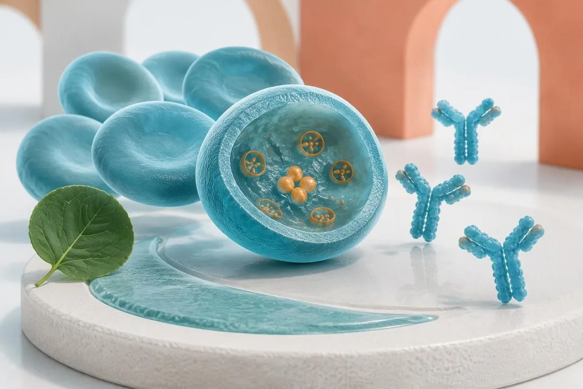
A positive antibody result can reflect an earlier encounter with Babesia rather than illness today. The deciding evidence usually comes from parasite testing, symptoms, and your individual risk.


- Positive Babesia IgG does not establish active infection; antibodies can remain detectable for more than 1 year after infection clears.
- A Babesia PCR test detects parasite DNA and is particularly useful when parasite levels are too low for a blood smear to identify reliably.
- Blood smear parasitemia measures the percentage of red blood cells containing parasites; 1% means approximately 1 infected cell per 100 red blood cells examined.
- IgG titers of 1:1024 or higher may suggest recent or active Babesia microti infection, but do not replace PCR or smear confirmation.
- A 4-fold antibody rise between appropriately timed samples supports recent infection; a single positive result cannot provide that comparison.
- 응급 증상 include confusion, breathlessness at rest, fainting, chest pain, or markedly reduced urine output, regardless of the antibody titer.
- Fever of 38°C or higher without a spleen warrants immediate medical contact and urgent assessment, even before Babesia testing is complete.
- 치료 기간 is usually 7–10 days for uncomplicated symptomatic disease in immunocompetent patients, but selected highly immunocompromised patients need at least 6 weeks.
- Persistent antibodies after treatment are not a test of cure and do not, by themselves, justify another antibiotic course.
Do positive Babesia test results mean active infection?
Positive Babesia antibodies alone do not establish active infection. IgG can remain positive for more than 1 year after infection clears; a Babesia PCR test or blood smear, interpreted alongside symptoms, helps determine whether parasites are present now. Persistent antibodies alone do not indicate treatment failure.
The word positive describes the test, not necessarily your current health. An antibody assay detects your immune response, whereas PCR detects parasite DNA and microscopy looks for parasites inside red blood cells; these 3 methods provide different kinds of evidence rather than interchangeable diagnoses.
Kantesti is an AI blood test analyzer that helps explain why 1 positive antibody result needs clinical context. I’m Thomas Klein, Chief Medical Officer at Kantesti; my approach is to separate evidence of exposure from evidence of present parasitemia before discussing what a result could mean for treatment.
A symptom-free person and a feverish person can have the same IgG titer but need different next steps. In an illustrative comparison, a well 45-year-old with an old positive result may need clarification, while a patient with fever, falling hemoglobin, and recent tick exposure needs prompt direct testing.
The first practical step is to identify exactly what was positive. Bring the complete report, including the organism, method, collection date, and reference interval; our explanation of 양성 항체 결과 distinguishes the 2 broad questions of immune recognition and active disease, while our organization overview explains our role.
What does a positive Babesia IgG actually measure?
Positive Babesia IgG measures immune recognition of Babesia antigens, not the number of living parasites. Antibodies can persist for more than 1 year, so a positive result may reflect prior infection, recent infection, or occasionally a misleading reaction that needs clarification.
Antibody persistence is a normal property of immune memory. Long-lived antibody-producing cells can continue making IgG after detectable parasites disappear; the IDSA babesiosis guideline specifically warns that antibodies may persist for a year or more and recommends smear or PCR confirmation before treating an antibody-positive patient (Krause et al., 2021).
IgM is not an automatic proof of new babesiosis. Although IgM can support a recent immune response, false-positive reactions and persistence complicate interpretation; 1 isolated IgM result should not override a negative diagnostic evaluation or become the sole reason for a prolonged treatment course.
A negative antibody test does not reliably exclude very early infection. If testing occurs during the first days of illness, the antibody response may not yet be detectable; this is why clinicians may order 2 direct tests—PCR and a smear—instead of waiting for serology to become positive.
The meaning of IgG is organism-specific. A Babesia antibody result should not be interpreted as proof of protective immunity or as a vaccination-style target; our discussion of CMV IgG patterns illustrates why 2 infections can produce similar-looking antibody reports yet require different clinical decisions.
How should Babesia antibody titers be interpreted?
Babesia antibody test interpretation depends on the assay, species, and timing—not a universal positive cutoff. For Babesia microti, an IgG titer of 1:1024 or higher can suggest recent or active infection, but even a high titer does not replace direct parasite testing.
A titer reports how far a sample can be diluted while remaining reactive. A result of 1:256 means reactivity remains detectable at that dilution under the laboratory’s method; it does not mean 256 parasites, 256 days of illness, or a particular percentage of affected red blood cells.
A 4-fold rise is more informative than a small fluctuation. For example, a change from 1:64 to 1:256 in appropriately timed paired samples supports recent infection, whereas 1:128 to 1:256 is only a 2-fold change and may reflect biological or assay variation.
Comparing different laboratories can create a false trend. If a result changes from 1:128 to 1:256 after switching methods, the apparent increase may not represent a genuine immune change; paired testing is easiest to interpret when the same laboratory, species antigen, and assay are used.
High titers are supporting evidence, not a severity scale. The IDSA guideline describes a Babesia microti IgG titer of at least 1:1024 as suggestive of recent or active infection, while still prioritizing smear or PCR confirmation (Krause et al., 2021); Q열 항체 해석 shows why infectious antibody cutoffs cannot be transferred between organisms.
What can a Babesia PCR test confirm?
A Babesia PCR test detects parasite DNA in a laboratory sample and can confirm babesiosis when symptoms and exposure fit. PCR is especially useful at low parasite levels, but 1 positive result after treatment does not necessarily prove that viable parasites are causing ongoing illness.
PCR often detects lower parasite burdens than routine microscopy. A patient can therefore have positive PCR and a negative smear without either result being an error; the 2 tests have different detection limits, and a negative smear alone cannot settle a strongly suspected low-level infection.
A negative PCR only addresses the organisms covered by that assay. A Babesia microti-specific assay may not detect Babesia duncani, Babesia divergens, or Babesia venatorum; these 3 examples matter when travel, geography, transfusion exposure, or immune status raises suspicion beyond the species named on the report.
Collection timing can change the result. Treatment before sampling, a very low parasite burden, or specimen problems can reduce detection; when symptoms remain convincing, the clinician may request a repeat sample and expert microscopy rather than treating 1 negative PCR as an absolute exclusion.
PCR positivity is not the same as a parasite viability test. DNA may remain detectable after clinical improvement, particularly with sensitive assays, so repeat testing needs a defined clinical purpose; our guide to PCR 검사 시점 및 음성 결과 explains the general timing problem without implying that 2 different infections share identical detection windows.
How do blood smears identify Babesia parasites?
A blood smear can confirm Babesia by showing parasites inside red blood cells and can quantify parasitemia. Parasitemia of 1% means approximately 1 affected red blood cell per 100 examined; that percentage helps assess burden, but symptoms and organ function still determine urgency.
Babesia often appears as ring forms within red blood cells. The characteristic 4-part Maltese cross tetrad is strongly suggestive when present, but it is uncommon enough that its absence cannot rule out babesiosis; an experienced microscopist must consider the complete pattern rather than hunt for one famous image.
Smear examination requires an explicit clinical request when suspicion is high. An automated complete blood count does not substitute for a manual parasite search, and the IDSA guideline recommends examining at least 200–300 oil-immersion fields before considering a smear negative when parasites are sparse.
A small parasitemia percentage can coexist with significant illness. Severe anemia, kidney dysfunction, respiratory compromise, or immune suppression can make a result below 10% clinically serious; conversely, the percentage alone does not describe blood pressure, oxygenation, or the speed at which hemoglobin is falling.
Malaria and Babesia can look similar on an initial smear. Travel history, parasite morphology, and molecular testing help separate them, and fever after travel should never be dismissed because a Babesia antibody test is positive; our travel-fever smear guide explains why these 2 possibilities sometimes need parallel evaluation.
What if IgG, PCR, and smear results disagree?
Disagreeing Babesia test results usually reflect differences in timing, sensitivity, or what each method measures. Positive IgG with negative PCR and smear is different from positive PCR with a negative smear; interpreting all 3 results together prevents both missed infection and unnecessary treatment.
IgG-positive, PCR-negative, smear-negative results often favor past exposure when the patient is well. They do not absolutely exclude infection under every circumstance, but 1 isolated antibody result is insufficient evidence for active babesiosis; current fever, immune suppression, assay coverage, and prior treatment determine whether more testing is reasonable.
PCR-positive, smear-negative results can indicate low-level parasitemia. In an untreated symptomatic patient, that combination can support diagnosis; after treatment in a clinically recovered immunocompetent patient, the same 2-result pattern may not warrant further medication, because molecular detection can outlast symptoms.
Smear-positive, PCR-negative results deserve laboratory clarification. Possible explanations include a species not covered by PCR, a specimen problem, or misidentified cellular material; the next step may be expert slide review and broader molecular testing rather than choosing whichever of the 2 reports seems more reassuring.
Kantesti helps organize results, but cannot independently certify parasite identification from an uploaded report. The distinction between a yes/no PCR and a percentage parasitemia is explained in our 정성 대 정량 가이드; 입니다. 저희 임상 검사 방법 정보는 explains why 2 contradictory results require clinical and laboratory oversight.
Which other blood results support suspected babesiosis?
Babesiosis often produces anemia, low platelets, and laboratory evidence of red blood cell breakdown. No single CBC or chemistry value confirms Babesia, but a falling hemoglobin combined with rising LDH and indirect bilirubin can make a positive antibody result more clinically concerning than IgG alone.
The hemoglobin trend matters more than one unfamiliar flag. In an illustrative case, a decline from 13.5 to 9.0 g/dL over several days, accompanied by fever and new thrombocytopenia, warrants prompt evaluation for hemolysis and other serious causes; the antibody titer cannot explain that deterioration on its own.
LDH is sensitive to cellular injury but not specific to Babesia. An illustrative LDH of 600 U/L may support hemolysis if the laboratory upper limit is 250 U/L, yet liver, muscle, and specimen-handling problems can also raise it; our LDH result explanation separates these possibilities.
Low haptoglobin supports hemolysis only in context. A result of 10 mg/dL may be below a laboratory’s range, but liver function and baseline variation affect interpretation; our guide to 낮은 하플토글로빈 원인 explains why 2 patients with the same number may have different underlying problems.
Kidney function, bilirubin, and oxygenation help assess severity rather than establish species. A creatinine increase from 0.8 to 1.6 mg/dL over a short interval deserves urgent attention even with modest parasitemia, and a hemolyzed laboratory specimen must not be confused with hemolysis occurring inside the patient.
Which symptoms need urgent or emergency assessment?
Suspected babesiosis needs emergency assessment when there is confusion, fainting, chest pain, breathlessness at rest, or markedly reduced urine output. A person without a spleen who develops a fever of 38°C 이상 should seek urgent assessment rather than wait for antibody or PCR confirmation.
New jaundice or dark urine with fever needs same-day medical assessment. These findings can signal hemolysis, liver disease, or another urgent condition rather than harmless dehydration; having 1 positive Babesia IgG result does not identify the cause, so clinicians usually assess the broader pattern.
Dark urine is a clue, not a home diagnostic test. Tea-colored urine with weakness and a falling hemoglobin is more concerning than an isolated color change after reduced fluid intake; our 소변이 어두울 때의 경고 신호에 대한 안내는 explains why the combination of 2 or more new symptoms changes the response.
Do not use the parasite percentage as permission to stay home when you feel seriously unwell. Respiratory difficulty, confusion, or poor circulation can require hospital care even below 10% parasitemia; if you are breathless at rest or fainting, call your local emergency number rather than arranging routine repeat testing.
Urgent treatment decisions may precede the final PCR report. In severe illness, clinicians can collect diagnostic samples and begin management based on the clinical picture and smear findings; tell the team about spleen removal, immune-suppressing medicines, recent transfusion, and symptom onset during the first assessment.
Why are people without a spleen or with weak immunity at higher risk?
People without a functioning spleen and those with substantial immune suppression have greater risks of severe or persistent babesiosis. The spleen helps remove affected red blood cells, while impaired antibody responses can make 1 negative serology result less reassuring than it would be in an otherwise healthy patient.
Spleen removal changes the safety threshold for fever. After splenectomy, a temperature of 38°C or higher warrants urgent assessment for serious infection, including bacterial causes unrelated to Babesia; do not assume the fever is explained by an old antibody result, and follow any individualized emergency plan already provided.
B-cell-depleting treatment can complicate both diagnosis and recovery. Rituximab, certain blood cancers, transplantation, and other major immune-suppressing conditions can increase persistence or relapse risk; documenting the last treatment date and current medicines may be more useful than ordering a second antibody titer.
Selected highly immunocompromised patients may need at least 6 weeks of treatment. The prolonged approach includes at least the final 2 weeks with no parasites detected on smears, according to evidence discussed in the IDSA guideline and the persistent-babesiosis series by Krause et al. (2008).
Kantesti can help organize the relevant reports, but immune risk must come from your history as well as laboratory values. A total IgG measurement and a Babesia-specific IgG result answer 2 different questions; our frequent-infection testing guide explains why normal routine blood counts do not rule out clinically significant immune suppression.
Do persistent Babesia antibodies mean treatment failed?
Persistent Babesia antibodies do not, by themselves, indicate treatment failure. IgG can remain detectable for more than 1년, and a stable or slowly declining titer is not a reliable test of cure; recovery is assessed through symptoms and, when indicated, direct parasite testing.
Repeated antibody testing can create anxiety without changing management. A patient may feel well after a standard 7–10-day course and still receive a positive IgG result months later; that finding alone is not a reason to restart treatment or continue medication until the antibody becomes negative.
PCR can also stay positive after symptoms resolve. The IDSA guideline notes that molecular positivity can persist for months after standard treatment in immunocompetent patients, which is why routine ongoing PCR monitoring after symptom resolution is generally discouraged in this group (Krause et al., 2021).
Persistent fatigue does not automatically equal persistent parasitemia. A hemoglobin that is recovering from 8.5 to 10.5 g/dL can still leave someone tired, and sleep disruption or another diagnosis may contribute; recurrent fever, new hemolysis, or worsening organ function deserves a fresh assessment rather than an assumption.
Hemolysis after parasite clearance may occasionally have another mechanism. Delayed autoimmune hemolytic anemia has been described after babesiosis, particularly in people without a spleen; a direct Coombs result can help investigate immune-mediated red cell destruction, but neither 1 positive Coombs test nor persistent IgG independently proves Babesia relapse.
When is treatment needed, and how is recovery monitored?
Treatment is generally directed at confirmed symptomatic babesiosis, not isolated antibody positivity. Uncomplicated illness in an immunocompetent patient is usually treated for 7–10일; severe disease, persistent parasitemia, and major immune suppression require individualized specialist management and a different follow-up plan.
Atovaquone plus azithromycin is the preferred regimen for many patients with babesiosis. Clindamycin plus quinine is an alternative in selected circumstances, but drug choice depends on severity, tolerability, interactions, and patient factors; the 7–10-day standard course must not be extended solely because an antibody remains positive.
An asymptomatic positive PCR or smear is different from isolated positive IgG. IDSA guidance generally does not recommend treatment for asymptomatic patients unless parasites continue to be seen on thin smears for more than 1 month; immune suppression or other high-risk circumstances still require an individualized specialist decision.
Exchange transfusion is considered for selected severe cases, not triggered by a titer. Parasitemia above 10%, severe hemolytic anemia, or major pulmonary, kidney, or liver compromise can prompt discussion with infectious-disease and transfusion specialists; the decision incorporates the whole clinical picture and is not an automatic numerical rule.
Monitoring should have a purpose and a stopping point. Smears are followed during treatment in immunocompetent patients, but usually not after symptoms resolve; highly immunocompromised patients need closer surveillance, and our 혈액검사 타임라인 가이드 helps separate 2 issues: biological recovery and the slower disappearance of laboratory signals.
How do exposure, geography, and species change interpretation?
Exposure history changes how plausible a positive Babesia result is and which confirmatory assay is appropriate. Babesia can be transmitted through ticks and transfusion, and more than 1 species causes human disease; a Babesia microti-only test should not be treated as a universal Babesia screen.
No remembered tick bite does not exclude babesiosis. Small ticks can go unnoticed, and transfusion provides a separate transmission route; the review by Vannier and Krause (2012) describes both routes, so clinicians should ask about transfusion as well as outdoor exposure when evaluating 1 unexplained febrile illness.
European and North American testing needs are not identical. Babesia microti predominates in many recognized United States cases, whereas Babesia divergens and Babesia venatorum are relevant in Europe; these 3 examples explain a difference in assay coverage, not a different principle for distinguishing antibodies from active parasitemia.
Symptoms often appear weeks after exposure rather than immediately after a tick is removed. Reconstructing the previous 1–2 months of outdoor activities, travel, and transfusions can therefore be more helpful than focusing only on yesterday; transfusion-associated timing can be longer, so provide dates rather than a guessed source.
Tick-related conditions do not share the same diagnostic pathway. Tick bites can also precede Lyme disease, anaplasmosis, or alpha-gal allergy, but 1 diagnosis does not establish the others; prevention includes appropriate repellents, clothing protection, and tick checks, while unexplained symptoms still need condition-specific testing.
What should you bring to a Babesia result review?
A useful Babesia review needs the complete report, symptom dates, exposure history, and immune-risk information. Bring all 3 test categories if available—antibody, PCR, and smear—because a cropped positive flag can hide the species, method, or timing that changes the interpretation.
Record units and result formats exactly. A titer of 1:256, parasitemia of 2%, and a PCR marked detected describe 3 different measurements; copying them into one generic positive-result field removes information the clinician needs, especially when comparing pretreatment and post-treatment samples.
An uploaded image can introduce transcription errors. A faint colon may turn 1:256 into 1256, and a decimal can change 0.2% into 2%; our PDF 전사 체크리스트 recommends checking the extracted value against the source before interpreting a trend or acting on an apparent change.
Kantesti is an AI blood test interpretation platform that can place antibody reports beside CBC and chemistry trends. The useful output is a clearer set of questions—not a substitute for parasite microscopy, prescribing, or urgent assessment; our AI 기술 가이드 explains the distinction between interpreting 1 report and establishing a clinical diagnosis.
My review checklist is deliberately short. I, Thomas Klein, would ask: Which species was tested, were direct tests collected before treatment, are symptoms improving, and is there spleen or immune risk? Those 4 questions usually provide more actionable information than repeating an antibody panel without a reason.
Research sources and the limits of related laboratory guides
Babesia management should rest on disease-specific evidence, not unrelated biomarker publications. , 두 개의 공개 DOI 기록은 Kantesti의 혈액검사 결과 해석 작업과 대규모 사용 데이터의 출처 자료를 원하는 독자에게 가장 좋은 출발점입니다. 저희가 발행한 2026년 10월 15일, this article draws its diagnostic and follow-up recommendations from the cited IDSA guideline and peer-reviewed Babesia literature; the 2 Zenodo guides below provide background on associated laboratory questions only.
The IDSA 2020 guideline was published in Clinical Infectious Diseases in 2021. Its central distinction remains useful: confirm suspected acute babesiosis with smear or PCR rather than a single antibody result; the 2012 New England Journal of Medicine review provides additional context on transmission, diagnosis, and severe-disease risk.
Kantesti is an AI-powered blood test analysis tool, not a source of independent Babesia treatment-trial evidence. Our background 철분 검사 가이드 helps explain anemia-related measurements, but iron deficiency and hemolysis are 2 different processes; a low hemoglobin should not prompt iron supplementation without evidence that iron replacement is appropriate.
Coagulation testing may become relevant in severe systemic illness. The related coagulation laboratory guide describes aPTT and other tests, but neither an abnormal aPTT nor a D-dimer result confirms babesiosis; the 2 listed Zenodo deposits are supporting educational material rather than Babesia diagnostic guidelines.
Source labels should not imply a review or indexing status that has not been verified. The publication-search links supplied with the 2 deposits are discovery links, not proof of ResearchGate or Academia.edu hosting; our 의료 콘텐츠 표준 describe the editorial boundaries, including why persistent antibodies must not be presented as treatment failure.
자주 묻는 질문
바베시아 IgG 양성이면 현재 감염 상태라는 뜻인가요?
Babesia IgG 양성만으로는 활동성 감염이 확립되지 않습니다. 감염이 해소된 후에도 항체가 1년 넘게 검출될 수 있으며, 단일 검사 결과로는 과거 노출과 현재의 기생충혈증을 구분할 수 없습니다. 임상의는 일반적으로 증상 및 노출 이력과 함께 Babesia PCR 검사 또는 혈액 도말 검사를 이용해 의심되는 급성 바베시아증을 확인합니다. 발열, 혈색소 수치 저하 또는 상당한 면역억제가 있는 경우 신속한 평가가 더욱 중요합니다.
Babesia IgG 역가 1:256은 무엇을 의미하나요?
Babesia IgG 역가 1:256은 검사실의 분석법에서 해당 희석배율에서도 검체가 반응성을 유지했음을 의미합니다. 기생충 수나 질병 중증도를 측정하는 것은 아니며, 양성 여부는 검사실의 기준치와 검사한 종에 따라 달라집니다. 검체 채취 시기가 적절하고 서로 비교 가능한 경우, 1:64에서 1:256으로의 상승은 4배 증가이며 최근 감염을 뒷받침할 수 있습니다. 단일 1:256 결과도 여전히 임상적 맥락을 고려해야 하며, 활동성 감염이 의심될 때는 직접적인 기생충 검사가 필요합니다.
Can Babesia PCR be positive when the blood smear is negative?
A Babesia PCR test can be positive when a blood smear is negative because PCR can detect parasite DNA at lower burdens than routine microscopy. In an untreated symptomatic patient, these 2 results can be compatible with low-level infection rather than a laboratory contradiction. After treatment, molecular positivity may persist despite clinical recovery, so a positive PCR does not automatically justify more medication. Immune status, symptom recurrence, and smear findings determine the follow-up plan.
How long can Babesia antibodies stay positive after treatment?
Babesia antibodies can remain positive for more than 1 year after infection clears. Persistent IgG is not a reliable test of cure and does not, by itself, indicate treatment failure. For an immunocompetent patient whose symptoms have resolved after the usual 7–10-day treatment course, routine repeated antibody testing generally does not help determine recovery. New fever, worsening anemia, or major immune suppression requires a separate clinical assessment.
Do I need treatment if Babesia antibodies are positive but I feel well?
A person who feels well generally should not receive Babesia treatment solely because an antibody test is positive. The IDSA guideline recommends direct confirmation with smear or PCR before considering treatment based on positive antibodies. For asymptomatic patients with confirmed parasitemia, treatment is generally not recommended unless parasites continue to be seen on thin smears for more than 1 month. People without a spleen or with substantial immune suppression need individualized specialist advice.
When should someone without a spleen seek urgent help for possible Babesia?
A person without a functioning spleen who develops a fever of 38°C or higher should seek immediate medical contact and urgent assessment, without waiting for Babesia confirmation. Confusion, fainting, chest pain, breathlessness at rest, or markedly reduced urine output requires emergency care. Severe babesiosis can occur even when parasitemia is below 10%, so the percentage alone cannot establish safety. Fever after splenectomy can also reflect dangerous bacterial infections unrelated to Babesia.
오늘 AI 기반 혈액검사 분석 받기
즉시 정확한 검사 분석을 위해 Kantesti를 신뢰하는 전 세계 200만 명 이상의 사용자와 함께하세요. 혈액검사 결과를 업로드하고 몇 초 만에 15,000+ 바이오마커에 대한 포괄적인 해석을 받아보세요.
📚 참고된 연구 출판물
📖 외부 의학 참고문헌
Krause PJ et al. (2021). Clinical Practice Guidelines by the Infectious Diseases Society of America (IDSA): 2020 Guideline on Diagnosis and Management of Babesiosis. 임상 감염병 학회지.
Vannier E, Krause PJ. (2012). Human Babesiosis. 뉴잉글랜드 의학저널(NEJM).
Krause PJ et al. (2008). Persistent and Relapsing Babesiosis in Immunocompromised Patients. 임상 감염병 학회지.
⚕️ 의료 면책 조항
이 글은 교육 목적만을 위한 것이며 의학적 조언을 구성하지 않습니다. 진단 및 치료 결정에는 항상 자격을 갖춘 의료 전문가와 상담하세요.
E-E-A-T 신뢰 신호
경험
의사가 주도하는 검사 해석 워크플로 임상 검토.
전문적 지식
임상 맥락에서 바이오마커가 어떻게 거동하는지에 대한 검사실 의학 중심.
권위
Dr. Thomas Klein이 작성했으며 Dr. Sarah Mitchell과 Prof. Dr. Hans Weber가 검토했습니다.
신뢰성
경고를 줄이기 위한 명확한 후속 경로가 포함된 근거 기반 해석.