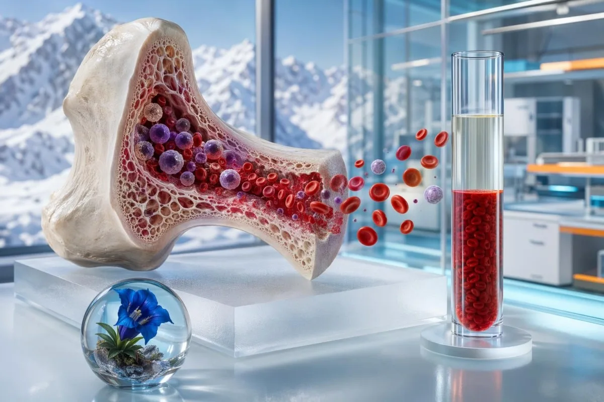
ಕಡಿಮೆ ಹೀಮಾಟೋಕ್ರಿಟ್ ಒಂದು ಮಾತ್ರ ರೋಗನಿರ್ಣಯವಲ್ಲ. ಅದರ ಸುತ್ತಲಿನ ಮಾದರಿ — ಹೀಮೋಗ್ಲೋಬಿನ್, RBC ಎಣಿಕೆ, MCV, ರೆಟಿಕ್ಯುಲೋಸೈಟ್ಸ್, ಫೆರಿಟಿನ್, ಕಿಡ್ನಿ ಸೂಚಕಗಳು ಮತ್ತು ದೇಹದ ನೀರಿನ ಸ್ಥಿತಿಯ ಸುಳಿವುಗಳು — ಕೆಂಪು ರಕ್ತಕಣಗಳು ನಿಜವಾಗಿಯೂ ಕಡಿಮೆಯೇ ಅಥವಾ ಕೇವಲ ದ್ರವದಿಂದ ದಟ್ಟಣೆ ಕಡಿಮೆಯಾಗಿದೆ ಎಂಬುದನ್ನು ತಿಳಿಸುತ್ತದೆ.


- ಕಡಿಮೆ ಹೆಮಾಟೋಕ್ರಿಟ್ ಅಂದರೆ ಕೆಂಪು ರಕ್ತಕಣಗಳು ರಕ್ತದ ಒಟ್ಟು ಪರಿಮಾಣದಲ್ಲಿ ಕಡಿಮೆ ಶೇಕಡಾವನ್ನು ಆಕ್ರಮಿಸುತ್ತವೆ; ವಯಸ್ಕರ ಶ್ರೇಣಿಗಳು ಸುಮಾರು 40-52% ಪುರುಷರಿಗೆ ಮತ್ತು 36-46% ಗರ್ಭಿಣಿಯಲ್ಲದ ಮಹಿಳೆಯರಿಗೆ.
- ನಿಜವಾದ ರಕ್ತಹೀನತೆ ಹೀಮಾಟೋಕ್ರಿಟ್, ಹೀಮೋಗ್ಲೋಬಿನ್ ಮತ್ತು ಕೆಂಪು ರಕ್ತಕಣ ಎಣಿಕೆ—all ಒಂದೇ ಸಮಯದಲ್ಲಿ ಕಡಿಮೆಯಾಗಿದ್ದಾಗ, ವಿಶೇಷವಾಗಿ ಲಕ್ಷಣಗಳು ಇದ್ದರೆ, ಬಹುಶಃ ಸಂಭವಿಸುತ್ತದೆ.
- ದ್ರವದಿಂದ ಉಂಟಾಗುವ ಕಡಿಮೆ ಹೀಮಾಟೋಕ್ರಿಟ್ IV ದ್ರವಗಳ ನಂತರ, ಹೆಚ್ಚು ದ್ರವ ಸೇವನೆ, ಗರ್ಭಧಾರಣೆ ಅಥವಾ ಅತಿಹೈಡ್ರೇಶನ್ನ ನಂತರ ಸಂಭವಿಸಬಹುದು; ಅಲ್ಬ್ಯುಮಿನ್ ಮತ್ತು ಸೋಡಿಯಂ ಕೂಡ ಕಡಿಮೆಯಾಗಿರಬಹುದು.
- ಇತ್ತೀಚಿನ ರಕ್ತಸ್ರಾವ ತಕ್ಷಣವೇ ಹೀಮಾಟೋಕ್ರಿಟ್ ಕಡಿಮೆಯಾಗದೇ ಇರಬಹುದು; ದ್ರವ ಬದಲಾವಣೆಗಳ ನಂತರ 6-24 ಗಂಟೆಗಳ ಬಳಿಕ ಕುಸಿತ ಹೆಚ್ಚು ಸ್ಪಷ್ಟವಾಗುತ್ತದೆ.
- ಕಬ್ಬಿಣದ ಕೊರತೆ ಸಾಮಾನ್ಯವಾಗಿ MCV 80 fL ಕ್ಕಿಂತ ಕಡಿಮೆ, ಫೆರಿಟಿನ್ 30 ng/mL ಕ್ಕಿಂತ ಕಡಿಮೆ, ಅಥವಾ ಟ್ರಾನ್ಸ್ಫೆರಿನ್ ಸ್ಯಾಚುರೇಶನ್ 20% ಕ್ಕಿಂತ ಕಡಿಮೆ ಎಂದು ತೋರಿಸುತ್ತದೆ.
- B12 ಅಥವಾ ಫೋಲೇಟ್ ಕೊರತೆ ಸಾಮಾನ್ಯವಾಗಿ MCV 100 fL ಕ್ಕಿಂತ ಹೆಚ್ಚು ಆಗುತ್ತದೆ; B12 200 pg/mL ಕ್ಕಿಂತ ಕಡಿಮೆ ಅಥವಾ MMA 0.40 µmol/L ಕ್ಕಿಂತ ಹೆಚ್ಚು ಇದ್ದರೆ ಕೊರತೆಯನ್ನು ಬೆಂಬಲಿಸುತ್ತದೆ.
- ರೆಟಿಕ್ಯುಲೋಸೈಟ್ಗಳು ಉತ್ಪಾದನೆ ಕಡಿಮೆಯಾಗುವುದನ್ನು ನಷ್ಟದಿಂದ ಬೇರ್ಪಡಿಸಲು ಸಹಾಯ ಮಾಡುತ್ತದೆ; ರೆಟಿಕ್ಯುಲೋಸೈಟ್ ಉತ್ಪಾದನಾ ಸೂಚ್ಯಂಕ 2 ಕ್ಕಿಂತ ಕಡಿಮೆ ಇದ್ದರೆ ದುರ್ಬಲ ಮಜ್ಜೆ ಪ್ರತಿಕ್ರಿಯೆಯನ್ನು ಸೂಚಿಸುತ್ತದೆ.
- ಗರ್ಭಧಾರಣೆ ಪ್ಲಾಸ್ಮಾ ವಿಸ್ತರಣೆಯ ಮೂಲಕ ಹೀಮಾಟೋಕ್ರಿಟ್ ಕಡಿಮೆಯಾಗುತ್ತದೆ; 1ನೇ ಅಥವಾ 3ನೇ ತ್ರೈಮಾಸಿಕದಲ್ಲಿ ಹೀಮೋಗ್ಲೋಬಿನ್ 11.0 g/dL ಕ್ಕಿಂತ ಕಡಿಮೆ ಇದ್ದರೆ ಸಾಮಾನ್ಯವಾಗಿ ಪರಿಶೀಲನೆ ಅಗತ್ಯ.
- ತುರ್ತು ಲಕ್ಷಣಗಳು ಎದೆನೋವು, ಮೂರ್ಚೆ ಹೋಗುವುದು, ವಿಶ್ರಾಂತಿಯಲ್ಲಿ ಉಸಿರಾಟದ ತೊಂದರೆ, ಕಪ್ಪು ಮಲ, ಅಥವಾ ನಿರಂತರವಾಗಿ ಹೆಚ್ಚು ರಕ್ತಸ್ರಾವ ಅಥವಾ ಹಿಮೋಗ್ಲೋಬಿನ್ ಸುಮಾರು 7-8 g/dL ಸಮೀಪದಲ್ಲಿರುವುದು.
ಕಡಿಮೆ ಹೀಮಾಟೋಕ್ರಿಟ್ ಫಲಿತಾಂಶದ ನಿಜವಾದ ಅರ್ಥ
ಕಡಿಮೆ ಹೆಮಾಟೋಕ್ರಿಟ್ ಸಾಮಾನ್ಯವಾಗಿ ಮೂರು ವಿಷಯಗಳಲ್ಲಿ ಒಂದನ್ನು ಸೂಚಿಸುತ್ತದೆ: ಕೆಂಪು ರಕ್ತಕಣಗಳು ಕಡಿಮೆ ಇರುವುದು, ಇತ್ತೀಚಿನ ಕೆಂಪು ರಕ್ತಕಣಗಳ ನಷ್ಟ, ಅಥವಾ ಹೆಚ್ಚುವರಿ ಪ್ಲಾಸ್ಮಾ ಮಾದರಿಯನ್ನು ದಪ್ಪಗೊಳಿಸಿ (dilute) ಪ್ರಮಾಣವನ್ನು ಕಡಿಮೆ ತೋರಿಸುವುದು. ಪ್ರಾಯೋಗಿಕವಾಗಿ, ನಾನು ಹೀಮಾಟೋಕ್ರಿಟ್ ಅನ್ನು ಮಾತ್ರ ಅರ್ಥೈಸುವುದಿಲ್ಲ; ಅದನ್ನು ಹಿಮೋಗ್ಲೋಬಿನ್, RBC count, MCV, RDW, ರೆಟಿಕ್ಯುಲೋಸೈಟ್ಸ್ ಮತ್ತು ರಕ್ತ ತೆಗೆದ ಸಂದರ್ಭದ ಕಥೆಯೊಂದಿಗೆ ಹೋಲಿಸುತ್ತೇನೆ. 2 ಲೀಟರ್ IV ದ್ರವದ ನಂತರ ಕಡಿಮೆ ಮೌಲ್ಯವು, ಫೆರಿಟಿನ್ 9 ng/mL ಇರುವುದರಿಂದ ಬಂದ ಕಡಿಮೆ ಮೌಲ್ಯಕ್ಕಿಂತ ಬೇರೆ ಸಮಸ್ಯೆ. ಆ ವ್ಯತ್ಯಾಸ ಮುಂದಿನ ಪರೀಕ್ಷೆ, ತುರ್ತುತೆ ಮತ್ತು ಚಿಕಿತ್ಸೆ ಬದಲಿಸುತ್ತದೆ.
ಹೀಮಾಟೋಕ್ರಿಟ್ ಎಂದರೆ ಸಂಪೂರ್ಣ ರಕ್ತದ ಪರಿಮಾಣದಲ್ಲಿ ಕೆಂಪು ಕೋಶೀಯ ಅಂಶಗಳು ಆಕ್ರಮಿಸುವ ಶೇಕಡಾವಾರು. 33% ಹೀಮಾಟೋಕ್ರಿಟ್ ಎಂದರೆ ಮಾದರಿಯ ಸುಮಾರು ಮೂರನೇ ಭಾಗ ಕೋಶೀಯ ಅಂಶಗಳು ಮತ್ತು ಸುಮಾರು ಎರಡು ಮೂರನೇ ಭಾಗ ಪ್ಲಾಸ್ಮಾ; ಆದ್ದರಿಂದ ಆ ಸಮೀಕರಣದ ಎರಡೂ ಬದಿಗಳಲ್ಲಿನ ಬದಲಾವಣೆಗಳು ಫಲಿತಾಂಶವನ್ನು ಸರಿಸಬಹುದು.
Kantesti ಒಂದು AI blood test interpretation ವೇದಿಕೆ; ಇದು ಶೇಕಡಾವಾರನ್ನು ಸ್ವತಂತ್ರ ರೋಗನಿರ್ಣಯವಾಗಿ ಪರಿಗಣಿಸುವ ಬದಲು, CBC ಉಳಿದ ಭಾಗಗಳ ಜೊತೆಗೆ ಹೀಮಾಟೋಕ್ರಿಟ್ ಅನ್ನು ಓದುತ್ತದೆ. ನಮ್ಮ ಕ್ಲಿನಿಕಲ್ ತಂಡ, ವಿವರಿಸಿರುವ ನಮ್ಮ ಬಗ್ಗೆ, ಪ್ರತೀ ವಾರವೂ ಒಂದೇ ರೀತಿಯ ಪ್ರಾಯೋಗಿಕ ಸಮಸ್ಯೆಯನ್ನು ನೋಡುತ್ತದೆ: ಕಡಿಮೆ ಎಂದು ಗುರುತಿಸಲಾದ ಹೀಮಾಟೋಕ್ರಿಟ್ ಎಂದರೆ ಐರನ್ ಕೊರತೆ, ತುರ್ತು ದ್ರವ ದಪ್ಪಗೊಳಿಕೆ (acute fluid dilution), ಗರ್ಭಧಾರಣೆಯ ದೇಹಶಾಸ್ತ್ರ, ಕಿಡ್ನಿ ಸಂಬಂಧಿತ ಕಡಿಮೆ ಉತ್ಪಾದನೆ ಅಥವಾ ಲ್ಯಾಬ್ ಸಮಯದ ಸಮಸ್ಯೆ ಎಂದಾಗಬಹುದು.
WHO 2011 ರ ರಕ್ತಹೀನತೆ (anemia) ಮಾರ್ಗಸೂಚಿಗಳು ಹೀಮಾಟೋಕ್ರಿಟ್ ಬದಲು ಹಿಮೋಗ್ಲೋಬಿನ್ ಮೂಲಕ ರಕ್ತಹೀನತೆಯನ್ನು ವ್ಯಾಖ್ಯಾನಿಸುತ್ತವೆ: ಪುರುಷರಲ್ಲಿ 13.0 g/dL ಕ್ಕಿಂತ ಕಡಿಮೆ, ಗರ್ಭಿಣಿಯಲ್ಲದ ಮಹಿಳೆಯರಲ್ಲಿ 12.0 g/dL ಕ್ಕಿಂತ ಕಡಿಮೆ ಮತ್ತು ಗರ್ಭಧಾರಣೆಯಲ್ಲಿ 11.0 g/dL ಕ್ಕಿಂತ ಕಡಿಮೆ ಇದ್ದರೆ ರಕ್ತಹೀನತೆ ಮಾನದಂಡಗಳನ್ನು ಪೂರೈಸುತ್ತದೆ (WHO, 2011). ನೀವು ಪ್ಯಾನೆಲ್ನ ಉಳಿದ ಭಾಗವನ್ನು ಅರ್ಥಮಾಡಿಕೊಳ್ಳಲು ಪ್ರಯತ್ನಿಸುತ್ತಿದ್ದರೆ, ನಮ್ಮ ಮಾರ್ಗದರ್ಶಿ CBC ಘಟಕಗಳು ಸಂಪೂರ್ಣ ರಕ್ತ ಎಣಿಕೆ (complete blood count) ಒಳಗೆ ಹೀಮಾಟೋಕ್ರಿಟ್ ಎಲ್ಲಿದೆ ಎಂಬುದನ್ನು ವಿವರಿಸುತ್ತದೆ.
ಸಾಮಾನ್ಯ ಹೀಮಾಟೋಕ್ರಿಟ್ ಶ್ರೇಣಿಗಳು ಲಿಂಗ, ವಯಸ್ಸು ಮತ್ತು ಗರ್ಭಧಾರಣೆಯ ಮೇಲೆ ಅವಲಂಬಿತವಾಗಿರುತ್ತವೆ
ವಯಸ್ಕರ ಹೀಮಾಟೋಕ್ರಿಟ್ ಸಾಮಾನ್ಯವಾಗಿ ಪುರುಷರಲ್ಲಿ ಸುಮಾರು 40-52% ಮತ್ತು ಗರ್ಭಿಣಿಯಲ್ಲದ ಮಹಿಳೆಯರಲ್ಲಿ 36-46% ಇರುತ್ತದೆ, ಆದರೂ ಪ್ರತಿಯೊಂದು ಲ್ಯಾಬ್ ತನ್ನದೇ ಮಧ್ಯಂತರವನ್ನು ನಿಗದಿಪಡಿಸುತ್ತದೆ. ಸಾಮಾನ್ಯ ಶ್ರೇಣಿಗಿಂತ ಕೇವಲ 1-2 ಶೇಕಡಾ ಪಾಯಿಂಟ್ಗಳಷ್ಟು ಕೆಳಗಿರುವ ಫಲಿತಾಂಶವು, ನಿಮ್ಮ ಸಾಮಾನ್ಯ ಮೂಲಮಟ್ಟದಿಂದ 6-10 ಪಾಯಿಂಟ್ಗಳಷ್ಟು ಏಕಾಏಕಿ ಕುಸಿತವಾಗುವುದಕ್ಕಿಂತ ಸಾಮಾನ್ಯವಾಗಿ ಕಡಿಮೆ ಚಿಂತಾಜನಕ.
ಕೆಲವು ಯುರೋಪಿಯನ್ ಲ್ಯಾಬ್ಗಳು ಸ್ವಲ್ಪ ಕಡಿಮೆ ಮಧ್ಯಂತರಗಳನ್ನು ಬಳಸುತ್ತವೆ, ಮತ್ತು ಹೆಚ್ಚಿನ ಎತ್ತರದ ಪ್ರದೇಶಗಳು ನಿರೀಕ್ಷಿತ ಹೀಮಾಟೋಕ್ರಿಟ್ ಅನ್ನು ಹೆಚ್ಚಾಗಿ ವರದಿ ಮಾಡಬಹುದು, ಏಕೆಂದರೆ ದೀರ್ಘಕಾಲದ ಕಡಿಮೆ ಆಮ್ಲಜನಕ ಒತ್ತಡವು ಕೆಂಪು ರಕ್ತಕಣಗಳ ಉತ್ಪಾದನೆಯನ್ನು ಉತ್ತೇಜಿಸುತ್ತದೆ. ನನ್ನ ಕ್ಲಿನಿಕಲ್ ಅನುಭವದಲ್ಲಿ, ಅತ್ಯಂತ ಉಪಯುಕ್ತ ಹೋಲಿಕೆ ಬಹುಶಃ ನಿಮ್ಮ ಇಂದಿನ ಸಂಖ್ಯೆಯ ಪಕ್ಕದಲ್ಲಿರುವ ಮುದ್ರಿತ ಶ್ರೇಣಿಗಿಂತ ನಿಮ್ಮ ಹಿಂದಿನ ಫಲಿತಾಂಶವೇ ಆಗಿರುತ್ತದೆ.
ಲ್ಯಾಬ್ ಶ್ರೇಣಿಗಳು ಭಿನ್ನವಾಗುತ್ತವೆ, ಏಕೆಂದರೆ ಹೀಮಾಟೋಕ್ರಿಟ್ ಅನ್ನು ಲಿಂಗ ಹಾರ್ಮೋನುಗಳು, ಎತ್ತರ, ಧೂಮಪಾನ, ಗರ್ಭಧಾರಣೆ, ಹೈಡ್ರೇಶನ್ ಮತ್ತು ಅನಲೈಸರ್ ವಿಧಾನ ಪ್ರಭಾವಿಸುತ್ತವೆ. ನಮ್ಮ ಲೇಖನ ಲಿಂಗ-ನಿರ್ದಿಷ್ಟ ಲ್ಯಾಬ್ ಶ್ರೇಣಿಗಳ ಕುರಿತು ಇರುವ ಹೆಚ್ಚಿನ ಮಾರ್ಗದರ್ಶಿಯನ್ನು ನೋಡಿ ಒಂದು ವಯಸ್ಕನಿಗೆ ಕಡಿಮೆ ಎಂದು ಕಾಣುವ ಮೌಲ್ಯ ಮತ್ತೊಬ್ಬನಿಗೆ ಸಾಮಾನ್ಯವಾಗಿರುವುದಕ್ಕೆ ಕಾರಣವನ್ನು ವಿವರಿಸುತ್ತದೆ.
ನಾನು ಬಳಸುವ ಪ್ರಾಯೋಗಿಕ ನಿಯಮ: ಹೀಮಾಟೋಕ್ರಿಟ್ ಅನ್ನು ಎರಡೂ conventional units ನಲ್ಲಿ ವರದಿ ಮಾಡಿದಾಗ ಅದು ಹಿಮೋಗ್ಲೋಬಿನ್ನ ಸುಮಾರು ಮೂರು ಪಟ್ಟು. ಹಿಮೋಗ್ಲೋಬಿನ್ 10.5 g/dL ಇದ್ದರೆ, ಹೀಮಾಟೋಕ್ರಿಟ್ ಸುಮಾರು 31-32% ಇದ್ದರೆ ಅದು ಆಂತರಿಕವಾಗಿ ಹೊಂದಿಕೆಯಾಗುತ್ತದೆ; ಈ ಜೋಡಿ ಬಹಳಷ್ಟು ಹೊಂದಿಕೆಯಾಗದಿದ್ದರೆ, ರೋಗವಿದೆ ಎಂದು ಊಹಿಸುವ ಮೊದಲು ನಾನು ಯೂನಿಟ್ಗಳು, ಅನಲೈಸರ್ ಫ್ಲ್ಯಾಗ್ಗಳು ಮತ್ತು ಮಾದರಿ ಟಿಪ್ಪಣಿಗಳನ್ನು ಪರಿಶೀಲಿಸುತ್ತೇನೆ.
CBC ಮಾದರಿಯ ಸುಳಿವುಗಳು ರಕ್ತಹೀನತೆಯನ್ನು ದಟ್ಟಣೆ ಕಡಿಮೆಯಿಂದ ಬೇರ್ಪಡಿಸುತ್ತವೆ
ನಿಜವಾದ ರಕ್ತಹೀನತೆ (true anemia) ಹೆಚ್ಚು ಸಾಧ್ಯವಾಗುವುದು ಹೀಮಾಟೋಕ್ರಿಟ್, ಹಿಮೋಗ್ಲೋಬಿನ್ ಮತ್ತು ಕೆಂಪು ರಕ್ತಕಣಗಳ ಸಂಖ್ಯೆ—all ಒಂದೇ ಸಮಯದಲ್ಲಿ ಕಡಿಮೆಯಾಗಿದ್ದಾಗ. ದ್ರವಗಳ ನಂತರ ಹೀಮಾಟೋಕ್ರಿಟ್ ಕಡಿಮೆಯಾಗುವುದರಿಂದ ದಪ್ಪಗೊಳಿಕೆ (dilution) ಹೆಚ್ಚು ಸಾಧ್ಯ, ಮತ್ತು ಆ ಮಾದರಿಯ ಜೊತೆಗೆ ಕಡಿಮೆ-ಸಾಮಾನ್ಯ ಸೋಡಿಯಂ, ಕಡಿಮೆ ಆಲ್ಬ್ಯುಮಿನ್ ಅಥವಾ ಹೊಂದಿಕೆಯಾಗುವ ಲಕ್ಷಣಗಳಿಲ್ಲದೆ ಅದೇ ದಿನದ ಏಕಾಏಕಿ ಕುಸಿತ ಕಂಡುಬಂದರೆ.
ಕಡಿಮೆ RBC count ಜೊತೆಗೆ ಕಡಿಮೆ ಹಿಮೋಗ್ಲೋಬಿನ್ ಸಾಮಾನ್ಯವಾಗಿ ನಿಜವಾದ ಆಮ್ಲಜನಕ ಸಾಗಿಸುವ ಸಾಮರ್ಥ್ಯ ಕಡಿಮೆಯಾಗಿರುವುದನ್ನು ಸೂಚಿಸುತ್ತದೆ. RBC count ಮತ್ತು ಹಿಮೋಗ್ಲೋಬಿನ್ ಕೆಲವೊಮ್ಮೆ ಏಕೆ ಭಿನ್ನವಾಗುತ್ತವೆ ಎಂಬುದರ ಆಳವಾದ ವಿವರಣೆಗೆ, ನಮ್ಮ ಮಾರ್ಗದರ್ಶಿಯನ್ನು ನೋಡಿ RBC ವಿರುದ್ಧ ಹಿಮೋಗ್ಲೋಬಿನ್.
MCV ಮತ್ತು RDW ಮುಂದಿನ ಪದರವನ್ನು ಸೇರಿಸುತ್ತವೆ. MCV 80 fL ಕ್ಕಿಂತ ಕಡಿಮೆ ಇದ್ದರೆ ಮೈಕ್ರೋಸೈಟೋಸಿಸ್ ಸೂಚಿಸುತ್ತದೆ, MCV 100 fL ಕ್ಕಿಂತ ಹೆಚ್ಚು ಇದ್ದರೆ ಮ್ಯಾಕ್ರೋಸೈಟೋಸಿಸ್ ಸೂಚಿಸುತ್ತದೆ, ಮತ್ತು RDW ಸುಮಾರು 14.5% ಕ್ಕಿಂತ ಹೆಚ್ಚಿದ್ದರೆ ಕೋಶದ ಗಾತ್ರದ ವ್ಯತ್ಯಾಸವು ಇಷ್ಟು ಹೆಚ್ಚಾಗಿದೆ ಎಂದು ಅರ್ಥವಾಗುತ್ತದೆ—ಅದು ಬೆಳೆಯುತ್ತಿರುವ ಕೊರತೆ ಅಥವಾ ಮಿಶ್ರ ಕಾರಣಗಳನ್ನು ಬೆಂಬಲಿಸುತ್ತದೆ; ನಮ್ಮ RDW ಮಾರ್ಗದರ್ಶಿ ಅದನ್ನು ಇನ್ನಷ್ಟು ವಿವರವಾಗಿ ಮಾದರಿಯಲ್ಲಿ ವಿವರಿಸುತ್ತದೆ.
Kantesti AI ಹೀಮಾಟೋಕ್ರಿಟ್ ಅನ್ನು ಹೀಮೋಗ್ಲೋಬಿನ್-ಹೀಮಾಟೋಕ್ರಿಟ್ ಸಂಬಂಧವು ಜೈವಿಕವಾಗಿ ಸಾಧ್ಯವಿದೆಯೇ ಎಂದು ಪರಿಶೀಲಿಸುವ ಮೂಲಕ ವ್ಯಾಖ್ಯಾನಿಸುತ್ತದೆ, ನಂತರ ಪ್ಲೇಟ್ಲೆಟ್ ಕ್ಲಂಪಿಂಗ್, ಮಾದರಿಯ ವಯಸ್ಸು ಅಥವಾ ಕೊನೆಯ CBC ಯಿಂದ ಇದ್ದಕ್ಕಿದ್ದಂತೆ ಡೆಲ್ಟಾ ಮುಂತಾದ ಅಸಂಗತ ಸೂಚನೆಗಳನ್ನು ಹುಡುಕುತ್ತದೆ. ಇಂತಹ ಮಾದರಿ ಪರಿಶೀಲನೆ ಸಾಮಾನ್ಯವಾಗಿ ಸೌಮ್ಯ dilutional ಫಲಿತಾಂಶವನ್ನು ದೀರ್ಘಕಾಲದ ಅನೀಮಿಯಾ ಎಂದು ತಪ್ಪಾಗಿ ಲೇಬಲ್ ಆಗುವುದನ್ನು ತಡೆಯುವದೇ.
ಇತ್ತೀಚಿನ ರಕ್ತಸ್ರಾವ ಮೊದಲ ಹೀಮಾಟೋಕ್ರಿಟ್ನಲ್ಲಿ ಮರೆಮಾಡಬಹುದು
ತೀವ್ರ ರಕ್ತಸ್ರಾವವು ಆರಂಭದಲ್ಲೇ ಹೀಮಾಟೋಕ್ರಿಟ್ ಅನ್ನು ತಕ್ಷಣ ಕಡಿಮೆ ಮಾಡದೇ ಇರಬಹುದು, ಏಕೆಂದರೆ ಮೊದಲಿಗೆ ಕೆಂಪು ರಕ್ತಕಣಗಳು ಮತ್ತು ಪ್ಲಾಸ್ಮಾ ಎರಡೂ ಒಟ್ಟಿಗೆ ಕಳೆದುಹೋಗುತ್ತವೆ. ದ್ರವವು ರಕ್ತನಾಳಗಳಿಗೆ ಸರಿಯುವಾಗ ಅಥವಾ IV ದ್ರವಗಳನ್ನು ನೀಡಿದಾಗ 6-24 ಗಂಟೆಗಳ ನಂತರ ಹೀಮಾಟೋಕ್ರಿಟ್ ಹೆಚ್ಚು ಸ್ಪಷ್ಟವಾಗಿ ಇಳಿಯುತ್ತದೆ.
ಸ್ಪಷ್ಟವಾಗಿ ಭಾರೀ ಮೂಗಿನ ರಕ್ತಸ್ರಾವಗಳು ಅಥವಾ ಜೀರ್ಣಾಂಗದ ನಷ್ಟ ಹೊಂದಿರುವ ರೋಗಿಗಳು ಆಶ್ಚರ್ಯಕರವಾಗಿ ಸಾಮಾನ್ಯ ಹೀಮಾಟೋಕ್ರಿಟ್ನೊಂದಿಗೆ ಬಂದಿರುವುದನ್ನು ನಾನು ನೋಡಿದ್ದೇನೆ; ನಂತರ ಮುಂದಿನ ಬೆಳಿಗ್ಗೆ 39% ರಿಂದ 31% ಗೆ ಇಳಿದಿದೆ. ಮೊದಲ ಸಂಖ್ಯೆ ನಕಲಿ ಅಲ್ಲ; ಪ್ಲಾಸ್ಮಾ ಪುನರ್ವಿತರಣೆಯು ಕೆಂಪು ರಕ್ತಕಣಗಳ ಕೊರತೆಯನ್ನು ಬಹಿರಂಗಪಡಿಸುವ ಮೊದಲು ಅದನ್ನು ತೆಗೆದುಕೊಂಡಿದ್ದರು.
ಋತುಚಕ್ರದ ಸಮಯವೂ ಮಹತ್ವದ್ದಾಗಿದೆ. ಭಾರೀ ಪಿರಿಯಡ್ಸ್ ಇರುವ ರೋಗಿಯೊಬ್ಬರು ರಕ್ತಸ್ರಾವದ ನಂತರದ ವಾರದಲ್ಲಿ ಹೀಮೋಗ್ಲೋಬಿನ್ 11.2 g/dL ಮತ್ತು ಹೀಮಾಟೋಕ್ರಿಟ್ 34% ತೋರಿಸಬಹುದು, ಆದರೆ ಫೆರಿಟಿನ್ ಹಲವು ತಿಂಗಳುಗಳಿಂದ ಇಳಿಯುತ್ತಲೇ ಇದೆ; ನಮ್ಮ ಪಿರಿಯಡ್-ಸಂಬಂಧಿತ ಹೀಮೋಗ್ಲೋಬಿನ್ ಬದಲಾವಣೆಗಳ ಕುರಿತು ಇರುವ ಲೇಖನ ಆ ಸಮಯರೇಖೆಯನ್ನು ವಿವರಿಸುತ್ತದೆ.
ಇತ್ತೀಚಿನ ರಕ್ತಸ್ರಾವವು ಹೆಚ್ಚು ಚಿಂತಾಜನಕವಾಗುತ್ತದೆ, ಹೀಮಾಟೋಕ್ರಿಟ್ ಮೂಲಮಟ್ಟದಿಂದ 3-5 ಶೇಕಡಾ ಅಂಕಗಳಿಗಿಂತ ಹೆಚ್ಚು ಇಳಿದರೆ, ಪಲ್ಸ್ ನಿಮಿಷಕ್ಕೆ 100 ಕ್ಕಿಂತ ಹೆಚ್ಚಿದ್ದರೆ, ಮಲ ಕಪ್ಪಾಗಿದ್ದರೆ, ಮೂತ್ರವು ದೃಶ್ಯವಾಗಿ ಕೆಂಪಾಗಿದ್ದರೆ ಅಥವಾ ಮೂರ್ಚೆ ಬಂದರೆ. ಕಥೆ ಮುಂದುವರಿಯುತ್ತಿರುವ ನಷ್ಟದಂತೆ ಕೇಳಿದರೆ, ಪುನಃ CBC ಗಾಗಿ ಒಂದು ತಿಂಗಳು ಕಾಯುವುದು ಸಾಮಾನ್ಯವಾಗಿ ತಪ್ಪು ಯೋಜನೆ.
ಐರನ್ ಕೊರತೆ ಸಾಮಾನ್ಯವಾಗಿ ವೈದ್ಯರು ಹುಡುಕುವ ಕಡಿಮೆ ಹೀಮಾಟೋಕ್ರಿಟ್ ಮಾದರಿ
ಐರನ್ ಕೊರತೆ ಸಾಮಾನ್ಯವಾಗಿ ಕಡಿಮೆ ಹೀಮಾಟೋಕ್ರಿಟ್ ಅನ್ನು ಕಡಿಮೆ ಹೀಮೋಗ್ಲೋಬಿನ್, ಕಡಿಮೆ MCV ಮತ್ತು ಹೆಚ್ಚಿನ RDW ಜೊತೆಗೆ ಉಂಟುಮಾಡುತ್ತದೆ. ಫೆರಿಟಿನ್ 15 ng/mL ಕ್ಕಿಂತ ಕಡಿಮೆ ಇದ್ದರೆ ಖಾಲಿ ಐರನ್ ಸಂಗ್ರಹಗಳಿಗೆ ಅತ್ಯಂತ ನಿರ್ದಿಷ್ಟವಾಗಿದ್ದು, ಫೆರಿಟಿನ್ 30 ng/mL ಕ್ಕಿಂತ ಕಡಿಮೆ ಇದ್ದರೆ ಅನೇಕ ವಯಸ್ಕ ರೋಗಿಗಳಲ್ಲಿ ಬಲವಾಗಿ ಸೂಚಿಸುತ್ತದೆ.
British Society of Gastroenterology ಮಾರ್ಗಸೂಚಿ ಫೆರಿಟಿನ್ ಅನ್ನು ಐರನ್ ಡಿಫಿಷಿಯನ್ಸಿ ಅನೀಮಿಯಾದ ಏಕೈಕ ಅತ್ಯಂತ ಉಪಯುಕ್ತ ಸೂಚಕ ಎಂದು ಹೇಳುತ್ತದೆ, ಆದರೆ ಉರಿಯೂತ ಇದ್ದಾಗ ಅದು ತಪ್ಪಾಗಿ ಸಾಮಾನ್ಯವಾಗಿ ಕಾಣಬಹುದು (Snook et al., 2021). ಉರಿಯೂತದ ಸ್ಥಿತಿಗಳಲ್ಲಿ, ಫೆರಿಟಿನ್ 100 ng/mL ಕ್ಕಿಂತ ಕಡಿಮೆ ಇದ್ದರೂ ಟ್ರಾನ್ಸ್ಫೆರಿನ್ ಸ್ಯಾಚುರೇಶನ್ 20% ಕ್ಕಿಂತ ಕಡಿಮೆ ಇದ್ದರೆ ಕೂಡ ಐರನ್-ನಿಯಂತ್ರಿತ ಕೆಂಪು ರಕ್ತಕಣಗಳ ಉತ್ಪಾದನೆಗೆ ಹೊಂದಿಕೊಳ್ಳಬಹುದು.
ಸೀರಮ್ ಐರನ್ ದಿನದೊಳಗೆ ಮತ್ತು ಊಟದ ನಂತರ ಬದಲಾಗುತ್ತದೆ, ಆದ್ದರಿಂದ ನಾನು ಅದನ್ನು ಒಂಟಿಯಾಗಿ ಅಪರೂಪವಾಗಿ ಬಳಸುತ್ತೇನೆ. ನಿಮ್ಮ ವರದಿಯಲ್ಲಿ ಕಡಿಮೆ ಸೀರಮ್ ಐರನ್, ಹೆಚ್ಚಿನ TIBC ಮತ್ತು ಕಡಿಮೆ ಟ್ರಾನ್ಸ್ಫೆರಿನ್ ಸ್ಯಾಚುರೇಶನ್ ತೋರಿದರೆ, ನಮ್ಮ ಕಡಿಮೆ ಸೀರಮ್ ಕಬ್ಬಿಣ ಮತ್ತು ಇನ್ನಷ್ಟು ಆಳವಾದ ಕಬ್ಬಿಣ ಅಧ್ಯಯನ ಮಾರ್ಗದರ್ಶಿ ನಿಮ್ಮಿಗೆ ಆ ಮಾದರಿಯನ್ನು ಅರ್ಥಮಾಡಿಕೊಳ್ಳಲು ಸಹಾಯ ಮಾಡಬಹುದು.
ಕ್ಲಿನಿಕ್ನಲ್ಲಿ, ಹೀಮಾಟೋಕ್ರಿಟ್ ಕಡಿಮೆ ಮತ್ತು ಐರನ್ ಕೂಡ ಕಡಿಮೆ ಕಾಣಿಸಿದಾಗ ನಾನು ಮೂರು ಅಷ್ಟಾಗಿ ಆಕರ್ಷಕವಲ್ಲದ ಪ್ರಶ್ನೆಗಳನ್ನು ಕೇಳುತ್ತೇನೆ: ಭಾರೀ ಪಿರಿಯಡ್ಸ್, ರಕ್ತದಾನ ಮತ್ತು ಜೀರ್ಣಾಂಗದ ನಷ್ಟ. ಸುಮಾರು 50 ವರ್ಷಕ್ಕಿಂತ ಮೇಲ್ಪಟ್ಟ ವಯಸ್ಕರಲ್ಲಿ, ಆಹಾರವು ಸಮಂಜಸವಾಗಿ ತೋರುತ್ತಿದ್ದರೂ ಸಹ, ಹೊಸ ಐರನ್ ಡಿಫಿಷಿಯನ್ಸಿ ಅನೀಮಿಯಾ ಎಂದರೆ GI ಕಾರಣಗಳಿಗಾಗಿ ವೈದ್ಯರಿಂದಲೇ ನೇತೃತ್ವದ ಹುಡುಕಾಟಕ್ಕೆ ಅರ್ಹವಾಗಿದೆ.
ಕಡಿಮೆ ಹೀಮಾಟೋಕ್ರಿಟ್ ಜೊತೆಗೆ ಹೆಚ್ಚಿನ MCV ಇದ್ದರೆ B12 ಅಥವಾ ಫೋಲೇಟ್ ಸಮಸ್ಯೆಗಳತ್ತ ಸೂಚಿಸುತ್ತದೆ
MCV 100 fL ಕ್ಕಿಂತ ಹೆಚ್ಚಿರುವಾಗ ಕಡಿಮೆ ಹೀಮಾಟೋಕ್ರಿಟ್ ಸಾಮಾನ್ಯವಾಗಿ B12 ಕೊರತೆ, ಫೋಲೇಟ್ ಕೊರತೆ, ಮದ್ಯದ ಪರಿಣಾಮ, ಯಕೃತ್ ರೋಗ, ಹೈಪೋಥೈರಾಯ್ಡಿಸಮ್ ಅಥವಾ ಔಷಧಿ ಹಸ್ತಕ್ಷೇಪವನ್ನು ಸೂಚಿಸುತ್ತದೆ. B12 200 pg/mL ಕ್ಕಿಂತ ಕಡಿಮೆ ಇದ್ದರೆ ಸಾಮಾನ್ಯವಾಗಿ ಕೊರತೆಯನ್ನು ಬೆಂಬಲಿಸುತ್ತದೆ, ಆದರೆ 200-300 pg/mL ಒಂದು ಗಡಿ ಪ್ರದೇಶವಾಗಿದ್ದು, MMA ಅದನ್ನು ಸ್ಪಷ್ಟಪಡಿಸಬಹುದು.
ಸುಮಾರು 0.40 µmol/L ಕ್ಕಿಂತ ಹೆಚ್ಚಿನ ಮೆಥೈಲ್ಮ್ಯಾಲೋನಿಕ್ ಆಸಿಡ್ (MMA) ಕಾರ್ಯಾತ್ಮಕ B12 ಕೊರತೆಯನ್ನು ಬೆಂಬಲಿಸುತ್ತದೆ—ವಿಶೇಷವಾಗಿ ಲಕ್ಷಣಗಳಲ್ಲಿ ನಂಬ್ನೆಸ್, ಸಮತೋಲನ ತೊಂದರೆ, ಕಾಲುಗಳಲ್ಲಿ ಸುಡುವಿಕೆ ಅಥವಾ ನೆನಪಿನ ಬದಲಾವಣೆ ಇದ್ದರೆ. ಹೋಮೊಸಿಸ್ಟೀನ್ B12 ಮತ್ತು ಫೋಲೇಟ್ ಕೊರತೆ ಎರಡರಲ್ಲೂ ಹೆಚ್ಚಾಗಬಹುದು, ಆದ್ದರಿಂದ ಅದು ಸ್ವತಃ ಕಡಿಮೆ ನಿರ್ದಿಷ್ಟ.
MCV ತುಂಬಾ ಹೆಚ್ಚಾಗುವ ಮೊದಲುಲೇ ರೋಗಿಗೆ ಕಡಿಮೆ ಹೀಮೋಗ್ಲೋಬಿನ್ ಲಕ್ಷಣಗಳು ಇರಬಹುದು. ಅದೇ ಸಮಯದಲ್ಲಿ ಐರನ್ ಕೊರತೆ ಇದ್ದಾಗ, MCV ಕೇವಲ 94 fL ಆಗಿದ್ದರೂ B12 ಕೊರತೆಯನ್ನು ನಾನು ನೋಡಿದ್ದೇನೆ; ಏಕೆಂದರೆ ಸರಾಸರಿಯಲ್ಲಿ ಮೈಕ್ರೋಸೈಟೋಸಿಸ್ ಮತ್ತು ಮ್ಯಾಕ್ರೋಸೈಟೋಸಿಸ್ ಭಾಗಶಃ ಪರಸ್ಪರ ರದ್ದುಮಾಡಿಕೊಳ್ಳುತ್ತವೆ.
ನಿಮ್ಮ B12 ಗಡಿ ಮಟ್ಟದಲ್ಲಿದ್ದರೆ, ಘಟಕಗಳನ್ನು ಜಾಗರೂಕವಾಗಿ ಹೋಲಿಸಿ, ಏಕೆಂದರೆ pg/mL ಮತ್ತು pmol/L ಪರಸ್ಪರ ಬದಲಾಯಿಸಿಕೊಳ್ಳಲಾಗುವುದಿಲ್ಲ. ನಮ್ಮ B12 ಶ್ರೇಣಿ ಮಾರ್ಗದರ್ಶಿ ಆ ಕಟ್ಆಫ್ಗಳನ್ನು ಮತ್ತು MMA ಕೆಲವೊಮ್ಮೆ ಮುಖ್ಯ B12 ಸಂಖ್ಯೆಯಿಗಿಂತ ಹೆಚ್ಚು ಏಕೆ ಮಹತ್ವವಾಗುತ್ತದೆ ಎಂಬುದನ್ನು ವಿವರಿಸುತ್ತದೆ.
ಕಿಡ್ನಿ ರೋಗ ಮತ್ತು ಉರಿಯೂತ ಕೆಂಪು ರಕ್ತಕಣಗಳ ಉತ್ಪಾದನೆಯನ್ನು ಕಡಿಮೆ ಮಾಡುತ್ತದೆ
ಕಡಿಮೆ ಉತ್ಪಾದನೆಯಿಂದ ಉಂಟಾಗುವ ಕಡಿಮೆ ಹೀಮಾಟೋಕ್ರಿಟ್ ಸಾಮಾನ್ಯವಾಗಿ ಕಡಿಮೆ ರೆಟಿಕ್ಯುಲೋಸೈಟ್ ಪ್ರತಿಕ್ರಿಯೆ, ಸಾಮಾನ್ಯ ಅಥವಾ ಸಣ್ಣ MCV, ಮತ್ತು ದೀರ್ಘಕಾಲದ ಉರಿಯೂತ ಅಥವಾ ಕಿಡ್ನಿ ಸಂಬಂಧಿತ ಸೂಚಕಗಳನ್ನು ತೋರಿಸುತ್ತದೆ. eGFR 60 mL/min/1.73 m² ಕ್ಕಿಂತ ಕಡಿಮೆಯಾಗುವಾಗ CKD-ಸಂಬಂಧಿತ ಅನೀಮಿಯಾ ಹೆಚ್ಚು ಸಾಮಾನ್ಯವಾಗುತ್ತದೆ ಮತ್ತು ವಿಶೇಷವಾಗಿ 30 ಕ್ಕಿಂತ ಕಡಿಮೆ ಇದ್ದಾಗ ಹೆಚ್ಚಾಗಿ ಕಾಣಿಸುತ್ತದೆ.
ಕಿಡ್ನಿ ಎರಿತ್ರೋಪೊಯಿಟಿನ್ ಅನ್ನು ಉತ್ಪಾದಿಸುತ್ತದೆ—ಮಜ್ಜೆಗೆ ಕೆಂಪು ರಕ್ತಕಣಗಳನ್ನು ತಯಾರಿಸಲು ಹೇಳುವ ಹಾರ್ಮೋನ್ ಸಂಕೇತ. KDIGO ಯ ಅನೀಮಿಯಾ ಮಾರ್ಗಸೂಚಿ, ಅನೀಮಿಯಾವನ್ನು ಕೇವಲ ದೀರ್ಘಕಾಲದ ಕಿಡ್ನಿ ರೋಗಕ್ಕೆ ಕಾರಣವೆಂದು ಹೇಳುವ ಮೊದಲು ಐರನ್ ಸ್ಥಿತಿ, B12, ಫೋಲೇಟ್ ಮತ್ತು ಉರಿಯೂತವನ್ನು ಮೌಲ್ಯಮಾಪನ ಮಾಡಲು ಶಿಫಾರಸು ಮಾಡುತ್ತದೆ (KDIGO, 2012).
ಉರಿಯೂತವು ಹೆಪ್ಸಿಡಿನ್ ಅನ್ನು ಹೆಚ್ಚಿಸುವ ಮೂಲಕ ಕಬ್ಬಿಣದ ನಿರ್ವಹಣೆಯನ್ನು ಬದಲಿಸುತ್ತದೆ; ಒಟ್ಟು ದೇಹದ ಕಬ್ಬಿಣ ನಿಜವಾಗಿಯೂ ಇಲ್ಲದಿದ್ದರೂ ಕೂಡ ಅದು ಕಬ್ಬಿಣವನ್ನು ಸಂಗ್ರಹ ಸ್ಥಳಗಳಲ್ಲಿ ಬಂಧಿಸುತ್ತದೆ. ಆದ್ದರಿಂದ ಉರಿಯೂತದ ಅನೀಮಿಯಾದಲ್ಲಿ ಫೆರಿಟಿನ್ 150 ng/mL ಆಗಿರಬಹುದು ಮತ್ತು ಟ್ರಾನ್ಸ್ಫೆರಿನ್ ಸ್ಯಾಚುರೇಶನ್ 14% ಆಗಿರಬಹುದು — ಸಾಕಷ್ಟು ಸಂಗ್ರಹಿತ ಕಬ್ಬಿಣ, ಆದರೆ ಸರಿಯಾದ ವಿತರಣೆ ಕಡಿಮೆ.
Kantesti ಒಂದು AI-ಚಾಲಿತ ರಕ್ತ ಪರೀಕ್ಷಾ ವಿಶ್ಲೇಷಣಾ ಸಾಧನವಾಗಿದ್ದು, ಇದು eGFR, ಕ್ರಿಯಾಟಿನಿನ್, CRP, ಆಲ್ಬ್ಯುಮಿನ್ ಮತ್ತು ಕಬ್ಬಿಣ ಸೂಚಕಗಳ ಜೊತೆಗೆ ಹೆಮಾಟೋಕ್ರಿಟ್ ಅನ್ನು ಓದುತ್ತದೆ. ನಿಮ್ಮ ಮಾದರಿಯಲ್ಲಿ ಮೂತ್ರಪಿಂಡದ ಸಂಖ್ಯೆಗಳು ಭಾಗವಾಗಿದ್ದರೆ, ನಮ್ಮ CKD ಹಂತ ಮಾರ್ಗದರ್ಶಿ ರೋಗಿಗಳಿಗೆ ಸಾಮಾನ್ಯವಾಗಿ ಬೇಕಾಗುವ eGFR ಮತ್ತು ಮೂತ್ರ ACR ಸಂದರ್ಭವನ್ನು ನೀಡುತ್ತದೆ.
ರೆಟಿಕ್ಯುಲೋಸೈಟ್ಸ್ ಮಜ್ಜೆ ಪ್ರತಿಕ್ರಿಯಿಸುತ್ತಿದೆಯೇ ಎಂಬುದನ್ನು ತೋರಿಸುತ್ತವೆ
ರೆಟಿಕ್ಯುಲೋಸೈಟ್ಸ್ ಯುವ ಕೆಂಪು ರಕ್ತಕಣಗಳು; ಮಜ್ಜೆ ಪರಿಹಾರ ನೀಡಲು ಪ್ರಯತ್ನಿಸುತ್ತಿದೆಯೇ ಎಂಬುದನ್ನು ಅವು ನಮಗೆ ತಿಳಿಸುತ್ತವೆ. ಅನೀಮಿಯಾದಲ್ಲಿ, ಮಜ್ಜೆ ಆರೋಗ್ಯವಾಗಿದ್ದರೆ ರೆಟಿಕ್ಯುಲೋಸೈಟ್ ಉತ್ಪಾದನಾ ಸೂಚ್ಯಂಕ 2 ಕ್ಕಿಂತ ಕಡಿಮೆ ಇದ್ದರೆ ಕಡಿಮೆ ಉತ್ಪಾದನೆ ಸೂಚಿಸುತ್ತದೆ, ಆದರೆ 3 ಕ್ಕಿಂತ ಹೆಚ್ಚು ಇದ್ದರೆ ಇತ್ತೀಚಿನ ನಷ್ಟ ಅಥವಾ ಹಿಮೋಲಿಸಿಸ್ ಸೂಚಿಸುತ್ತದೆ.
ಹಿಮೋಲಿಸಿಸ್ ಶಂಕೆ ಇರುವುದು ಕಡಿಮೆ ಹೆಮಾಟೋಕ್ರಿಟ್ ಜೊತೆಗೆ ಹೆಚ್ಚಿನ ರೆಟಿಕ್ಯುಲೋಸೈಟ್ಸ್, ಹೆಚ್ಚಿನ LDH, ಪರೋಕ್ಷ ಬಿಲಿರುಬಿನ್ ಏರಿಕೆ ಮತ್ತು ಕಡಿಮೆ ಹ್ಯಾಪ್ಟೋಗ್ಲೋಬಿನ್ ಕಂಡುಬಂದಾಗ. ಸುಮಾರು 25 mg/dL ಕ್ಕಿಂತ ಕಡಿಮೆ ಹ್ಯಾಪ್ಟೋಗ್ಲೋಬಿನ್ ಕೆಂಪು ರಕ್ತಕಣಗಳ ವಿಗಡಣೆಯನ್ನು ಬೆಂಬಲಿಸುತ್ತದೆ; ಆದರೆ ಯಕೃತ್ ರೋಗವೂ ಹ್ಯಾಪ್ಟೋಗ್ಲೋಬಿನ್ ಅನ್ನು ಕಡಿಮೆ ಮಾಡಬಹುದು ಮತ್ತು ಓದನ್ನು ಸಂಕೀರ್ಣಗೊಳಿಸಬಹುದು.
ಡೈರೆಕ್ಟ್ ಆಂಟಿಗ್ಲೋಬುಲಿನ್ ಟೆಸ್ಟ್, ಕೆಲವೊಮ್ಮೆ DAT ಅಥವಾ ಕೂಮ್ಸ್ ಟೆಸ್ಟಿಂಗ್ ಎಂದು ಕರೆಯಲಾಗುತ್ತದೆ, ಇದು ರೋಗನಿರೋಧಕ ಹಿಮೋಲಿಸಿಸ್ ಅನ್ನು ಪತ್ತೆಹಚ್ಚಲು ಸಹಾಯ ಮಾಡುತ್ತದೆ. ರೋಗಿಗಳು ಸಾಮಾನ್ಯವಾಗಿ ಕಾಮಾಲೆ, ಕತ್ತಲೆ ಮೂತ್ರ, ಬೆನ್ನು ಅಸೌಕರ್ಯ ಅಥವಾ ಏಕಾಏಕಿ ದಣಿವು ಗಮನಿಸುತ್ತಾರೆ; ಆದರೆ ಸೌಮ್ಯ ಹಿಮೋಲಿಸಿಸ್ ಅಚ್ಚರಿಯಂತೆ ಶಾಂತವಾಗಿರಬಹುದು.
ನಾನು ಈ ಮಾದರಿಯನ್ನು ಪರಿಶೀಲಿಸಿದಾಗ, ಒಂದೇ ನಾಟಕೀಯ ಮೌಲ್ಯಕ್ಕಿಂತ ಸಂಪೂರ್ಣ ಗುಚ್ಛವನ್ನು ಹುಡುಕುತ್ತೇನೆ. ನಮ್ಮ ಹ್ಯಾಪ್ಟೋಗ್ಲೋಬಿನ್ ಫಲಿತಾಂಶಗಳು ಮತ್ತು ನಮ್ಮ ಹೆಮಟಾಲಜಿ ವಿಮರ್ಶೆ ರೆಟಿಕ್ಯುಲೋಸೈಟ್ ಮಾರ್ಕರ್ಸ್ ಈ ಸಂಯೋಜನೆ ಏಕೆ ಯಾವುದೇ ಒಂದೇ ಸೂಚಕಕ್ಕಿಂತ ಹೆಚ್ಚು ವಿಶ್ವಾಸಾರ್ಹವೆಂದು ವಿವರಿಸುತ್ತದೆ.
ಅತಿಹೈಡ್ರೇಶನ್ ಮತ್ತು IV ದ್ರವಗಳು ಹೀಮಾಟೋಕ್ರಿಟ್ ಅನ್ನು ದಟ್ಟಣೆ ಕಡಿಮೆ ಮಾಡುವಂತೆ ಮಾಡಬಹುದು
ದಿಲ್ಯೂಷನಲ್ ಕಡಿಮೆ ಹೆಮಾಟೋಕ್ರಿಟ್ ಆಗುವುದು ಪ್ಲಾಸ್ಮಾ ಪ್ರಮಾಣವು ಕೆಂಪು ರಕ್ತಕಣಗಳ ದ್ರವ್ಯರಾಶಿಗಿಂತ ವೇಗವಾಗಿ ವಿಸ್ತರಿಸಿದಾಗ. ಆಸ್ಪತ್ರೆಯ ಚಾರ್ಟ್ಗಳಲ್ಲಿ, 1-2 ಲೀಟರ್ ಕ್ರಿಸ್ಟಲಾಯ್ಡ್ ದ್ರವವು ಯಾವುದೇ ಹೊಸ ಕೆಂಪು ರಕ್ತಕಣ ನಷ್ಟವಿಲ್ಲದೆ ಹೆಮಾಟೋಕ್ರಿಟ್ ಅನ್ನು ಸುಮಾರು 2-5 ಶೇಕಡಾ ಅಂಕಗಳಿಂದ ಕಡಿಮೆ ಮಾಡಬಹುದು.
ನೀವು ಎಲ್ಲಿ ನೋಡಬೇಕು ಎಂದು ತಿಳಿದಿದ್ದರೆ ಸುಳಿವುಗಳು ಸೂಕ್ಷ್ಮವಾಗಿರುವುದಿಲ್ಲ: ಹೆಮಾಟೋಕ್ರಿಟ್ ತ್ವರಿತವಾಗಿ ಇಳಿಯುತ್ತದೆ, ಆಲ್ಬ್ಯುಮಿನ್ ಕಡಿಮೆಯಾಗಬಹುದು, ಸೋಡಿಯಂ ಕಡಿಮೆಯಾಗಬಹುದು ಮತ್ತು ರೋಗಿಗೆ ದ್ರವಗಳನ್ನು ನೀಡಿರಬಹುದು, ಗರ್ಭಧಾರಣೆಯ ಆರೈಕೆ, ಎಂಡ್ಯುರನ್ಸ್-ಈವೆಂಟ್ ರಿಹೈಡ್ರೇಶನ್ ಅಥವಾ ವಾಂತಿಗೆ ಚಿಕಿತ್ಸೆ ಪಡೆದಿರಬಹುದು. CBC ಕೆಟ್ಟದಾಗಿ ಕಾಣಬಹುದು, ಆದರೆ ಶೇಕಡಾವಾರು ಸೂಚಿಸುವಷ್ಟು ಆಮ್ಲಜನಕ ವಿತರಣೆ ಬದಲಾಗಿರದಿರಬಹುದು.
ಎಂಡ್ಯುರನ್ಸ್ ಅಥ್ಲೀಟ್ಗಳು ವಿಶೇಷ ಪ್ರಕರಣ. ದೀರ್ಘ ತರಬೇತಿ ಬ್ಲಾಕ್ಗಳ ನಂತರ, ಪ್ಲಾಸ್ಮಾ ಪ್ರಮಾಣ ವಿಸ್ತರಣೆ ಕಡಿಮೆ-ಸಾಮಾನ್ಯ ಹೆಮಾಟೋಕ್ರಿಟ್ ಅನ್ನು ಸೃಷ್ಟಿಸಿ ಕಾಗದದಲ್ಲಿ ಅನೀಮಿಯಾದಂತೆ ಕಾಣಿಸಬಹುದು; ಆದರೆ ಕಾರ್ಯಕ್ಷಮತೆ ಸ್ಥಿರವಾಗಿರುತ್ತದೆ ಮತ್ತು ಫೆರಿಟಿನ್ ಸ್ವೀಕಾರಾರ್ಹವಾಗಿರುತ್ತದೆ; ಮೂತ್ರದ ಸಾಂದ್ರತೆ جیسے ಹೈಡ್ರೇಶನ್ ಸೂಚಕಗಳು ಸಂದರ್ಭವನ್ನು ಸೇರಿಸುತ್ತವೆ.
ದ್ರವ ಸಮತೋಲನ ನಿಮ್ಮ ಪ್ರಶ್ನೆಯ ಭಾಗವಾಗಿದ್ದರೆ, CBC ಅನ್ನು ಸೋಡಿಯಂ, ಆಲ್ಬ್ಯುಮಿನ್, BUN/ಯೂರಿಯಾ ಮತ್ತು ಮೂತ್ರದ ಸಾಂದ್ರತೆಯೊಂದಿಗೆ ಹೋಲಿಸಿ. ನಮ್ಮ 소변 삼투압 ದೇಹದ ನೀರಿನ ಕೊರತೆ, ಅತಿಹೈಡ್ರೇಶನ್ ಅಥವಾ ಮಿಶ್ರ ದ್ರವ ಚಿತ್ರಣದ ಪ್ರಶ್ನೆಗಳಿಗೆ ಉಪಯುಕ್ತವಾಗಿದೆ.
ಗರ್ಭಧಾರಣೆಯಲ್ಲಿ ಸಾಮಾನ್ಯ ಪ್ಲಾಸ್ಮಾ ವಿಸ್ತರಣೆಯಿಂದ ಹೀಮಾಟೋಕ್ರಿಟ್ ಕಡಿಮೆಯಾಗುತ್ತದೆ
ಗರ್ಭಧಾರಣೆ ಸಾಮಾನ್ಯವಾಗಿ ಹೆಮಾಟೋಕ್ರಿಟ್ ಅನ್ನು ಕಡಿಮೆ ಮಾಡುತ್ತದೆ, ಏಕೆಂದರೆ ಪ್ಲಾಸ್ಮಾ ಪ್ರಮಾಣವು ಸುಮಾರು 40-50% ರಷ್ಟು ಏರುತ್ತದೆ, ಆದರೆ ಕೆಂಪು ರಕ್ತಕಣಗಳ ದ್ರವ್ಯರಾಶಿ ಸುಮಾರು 20-30% ರಷ್ಟು ಮಾತ್ರ ಏರುತ್ತದೆ. ಈ ದೇಹಶಾಸ್ತ್ರೀಯ ಹೆಮೋಡಿಲ್ಯೂಷನ್ ನಿರೀಕ್ಷಿತ, ಆದರೆ 1ನೇ ಅಥವಾ 3ನೇ ತ್ರೈಮೆಸ್ಟರ್ನಲ್ಲಿ 11.0 g/dL ಕ್ಕಿಂತ ಕಡಿಮೆ ಹಿಮೋಗ್ಲೋಬಿನ್ ಇನ್ನೂ ಪರಿಶೀಲನೆಗೆ ಅರ್ಹ.
ಎರಡನೇ ತ್ರೈಮೆಸ್ಟರ್ನಲ್ಲಿ ಮೌಲ್ಯಗಳು ಸಾಮಾನ್ಯವಾಗಿ ಅತಿ ಕಡಿಮೆಯಂತೆ ಕಾಣುತ್ತವೆ. ಅನೇಕ ವೈದ್ಯರು ಎರಡನೇ ತ್ರೈಮೆಸ್ಟರ್ನಲ್ಲಿ 10.5 g/dL ಕ್ಕಿಂತ ಕಡಿಮೆ ಹಿಮೋಗ್ಲೋಬಿನ್ ಅನ್ನು ಅನೀಮಿಯಾ ಮಿತಿ (threshold) ಎಂದು ಬಳಸುತ್ತಾರೆ; ಆದರೆ ಸ್ಥಳೀಯ ಪ್ರಸೂತಿ ಪ್ರೋಟೋಕಾಲ್ಗಳು ಭಿನ್ನವಾಗುತ್ತವೆ ಮತ್ತು ಫೆರಿಟಿನ್ ಅನ್ನು ಈಗಾಗಲೇ ಬೇಗನೆ ಪರೀಕ್ಷಿಸಲಾಗುವುದು.
ಡೆಲಿವರಿ ನಂತರದ ಹೆಮಾಟೋಕ್ರಿಟ್ ಇನ್ನಷ್ಟು ಕಷ್ಟಕರ, ಏಕೆಂದರೆ ಡೆಲಿವರಿ ಸಂಬಂಧಿತ ನಷ್ಟ, IV ದ್ರವಗಳು ಮತ್ತು ಉರಿಯೂತ ಎಲ್ಲವೂ ಒಟ್ಟಿಗೆ ಬರುತ್ತವೆ. ಒಂದು ಮಹಿಳೆಗೆ ಡೆಲಿವರಿಯ ನಂತರದ ಬೆಳಿಗ್ಗೆ ಹೆಮಾಟೋಕ್ರಿಟ್ 29% ಇರಬಹುದು, ನಂತರ ಕೇವಲ ದ್ರವ ಸಮತೋಲನ ಸಾಮಾನ್ಯಗೊಂಡ ಕಾರಣ ಒಂದು ವಾರ ಬಳಿಕ 32% ಆಗಿರಬಹುದು — ಐರನ್ ಇನ್ಫ್ಯೂಷನ್ ಇಲ್ಲದೆ.
ಜುಲೈ 15, 2026 ರಂತೆ, ನನ್ನ ಪ್ರಾಯೋಗಿಕ ವಿಧಾನವೆಂದರೆ ಗರ್ಭಧಾರಣೆಯ ಹೆಮಾಟೋಕ್ರಿಟ್ ಅನ್ನು ಒಂದೇ ಕೆಂಪು ಎಚ್ಚರಿಕೆಯ ಸೂಚಕವಾಗಿ ಅಲ್ಲ, ಟ್ರೆಂಡ್ ಮತ್ತು ಲಕ್ಷಣಗಳ ಜೊತೆಗೆ ಚಿಕಿತ್ಸೆ ನೀಡುವುದು. ನಮ್ಮ ಗರ್ಭಧಾರಣೆಯ ರಕ್ತ ಪರೀಕ್ಷೆಗಳು ರೂಟೀನ್ ಫಾಲೋ-ಅಪ್ ಕಾಯಬಾರದ ಅದೇ ದಿನದ ಎಚ್ಚರಿಕೆ ಮಾದರಿಗಳನ್ನು ಒಳಗೊಂಡಿದೆ.
ಕಡಿಮೆ ಹೀಮಾಟೋಕ್ರಿಟ್ ಲಕ್ಷಣಗಳು ಕೇವಲ ಸಂಖ್ಯೆಯಷ್ಟೇ ಅಲ್ಲ, ವೇಗದ ಮೇಲೂ ಅವಲಂಬಿತವಾಗಿರುತ್ತವೆ
ಕಡಿಮೆ ಹೆಮಾಟೋಕ್ರಿಟ್ ಲಕ್ಷಣಗಳು ಸಾಮಾನ್ಯವಾಗಿ ಕಡಿಮೆಯಾದ ಆಮ್ಲಜನಕ ವಿತರಣೆಯಿಂದ ಬರುತ್ತವೆ ಮತ್ತು ಇದರಲ್ಲಿ ದಣಿವು, ಉಸಿರಾಟದ ತೊಂದರೆ, ತಲೆ ಸುತ್ತು, ಹೃದಯ ಬಡಿತಗಳು (ಪಾಲ್ಪಿಟೇಶನ್ಸ್), ತಲೆನೋವು ಮತ್ತು ಬಿಳಿ ಚರ್ಮ ಸೇರಿವೆ. ಹಿಮೋಗ್ಲೋಬಿನ್ 9.5 g/dL ಗೆ ನಿಧಾನವಾಗಿ ಇಳಿದರೆ ಅದು ಸೌಮ್ಯವಾಗಿ ಅನಿಸಬಹುದು, ಆದರೆ 10.5 g/dL ಗೆ ತ್ವರಿತವಾಗಿ ಇಳಿದರೆ ಅದು ಭಯಾನಕವಾಗಿ ಅನಿಸಬಹುದು.
ಕಡಿಮೆ ಹಿಮೋಗ್ಲೋಬಿನ್ ರೋಗಲಕ್ಷಣಗಳು ಹಿಮೋಗ್ಲೋಬಿನ್ 10 g/dL ಗಿಂತ ಕಡಿಮೆಯಾದಾಗ, ವಿಶೇಷವಾಗಿ ವ್ಯಾಯಾಮ ಮಾಡುವಾಗ ಅಥವಾ ಮೆಟ್ಟಿಲು ಹತ್ತುವಾಗ ಹೆಚ್ಚು ಸಂಭವನೀಯವಾಗುತ್ತವೆ. 8 g/dL ಗಿಂತ ಕಡಿಮೆಯಾದರೆ, ಅನೇಕ ವಯಸ್ಕರು ಉಬ್ಬಸ, ದೌರ್ಬಲ್ಯ ಅಥವಾ ತಲೆತಿರುಗುವಿಕೆಯನ್ನು ಅನುಭವಿಸುತ್ತಾರೆ, ಮತ್ತು ಹೃದ್ರೋಗ ಹೊಂದಿರುವವರು ಹೆಚ್ಚಿನ ಮಟ್ಟದಲ್ಲಿ ಎದೆ ಒತ್ತಡವನ್ನು ಬೆಳೆಸಿಕೊಳ್ಳಬಹುದು.
ನನಗೆ ಇಷ್ಟವಿಲ್ಲದ ಕೆಂಪು ಧ್ವಜಗಳೆಂದರೆ ಮೂರ್ಛೆ ಹೋಗುವುದು, ವಿಶ್ರಾಂತಿಯಲ್ಲಿ ಉಸಿರಾಟದ ತೊಂದರೆ, ಎದೆ ನೋವು, ಗೊಂದಲ, ಕಪ್ಪು ಮಲ, ನಿರಂತರವಾದ ಭಾರೀ ರಕ್ತಸ್ರಾವ, ನಿಮಿಷಕ್ಕೆ 120 ಕ್ಕಿಂತ ಹೆಚ್ಚು ಬಡಿತಗಳ ಹೃದಯ ಬಡಿತ ಅಥವಾ ರೋಗಿಯ ಸಾಮಾನ್ಯ ಮೂಲ ಮಟ್ಟಕ್ಕಿಂತ ಕಡಿಮೆ ಆಮ್ಲಜನಕ ಸಂತೃಪ್ತಿ. ಇವು ದಿನನಿತ್ಯದ ಕ್ಲಿನಿಕಲ್ ಪರಿಶೀಲನೆಯ ಚಿಹ್ನೆಗಳು, ಜೀವನಶೈಲಿ-ಹೊಂದಾಣಿಕೆ ಚಿಹ್ನೆಗಳಲ್ಲ.
ರೋಗಲಕ್ಷಣಗಳು ಗ್ಲೂಕೋಸ್, ಥೈರಾಯ್ಡ್, ಉಪ್ಪು ಮತ್ತು ಹೃದಯ ಲಯದ ಸಮಸ್ಯೆಗಳೊಂದಿಗೆ ಅತಿಕ್ರಮಿಸುತ್ತವೆ. ಲ್ಯಾಬ್ ಫ್ಲಾಗ್ ಬದಲಿಗೆ ತಲೆತಿರುಗುವಿಕೆಯು ದೂರು ಆಗಿದ್ದರೆ, ನಮ್ಮ ಮಾರ್ಗದರ್ಶಿ ತಲೆತಿರುಗುವಿಕೆ ರಕ್ತ ಪರೀಕ್ಷೆಗಳು CBC ಏಕೆ ಕೆಲಸದ ಒಂದು ಭಾಗ ಮಾತ್ರ ಎಂದು ವಿವರಿಸುತ್ತದೆ.
ಕಡಿಮೆ ಹೀಮಾಟೋಕ್ರಿಟ್ ಅನ್ನು ಮರುಪರೀಕ್ಷಿಸುವುದು ಶಂಕಿತ ಕಾರಣಕ್ಕೆ ಹೊಂದಿಕೆಯಾಗಬೇಕು
ಸ್ವಲ್ಪ ಕಡಿಮೆ ಹೆಮಟೋಕ್ರಿಟ್ ಅನ್ನು 1-4 ವಾರಗಳಲ್ಲಿ ಮರುಪರಿಶೀಲಿಸಬಹುದು, ನೀವು ಚೆನ್ನಾಗಿ ಭಾವಿಸಿದರೆ ಮತ್ತು CBC ಯ ಉಳಿದ ಭಾಗ ಸ್ಥಿರವಾಗಿದ್ದರೆ. ವೇಗದ ಕುಸಿತ, ರೋಗಲಕ್ಷಣಗಳು, ಗರ್ಭಧಾರಣೆ ಕೆಂಪು ಧ್ವಜಗಳು, ರಕ್ತಸ್ರಾವದ ಅನುಮಾನ ಅಥವಾ ಹಿಮೋಗ್ಲೋಬಿನ್ 7-8 g/dL ಹತ್ತಿರವಿದ್ದರೆ ದಿನನಿತ್ಯದ ವೈದ್ಯಕೀಯ ಸಲಹೆ ಅಗತ್ಯವಿದೆ.
ಭಾರೀ ದ್ರವ ಸೇವನೆ, IV ದ್ರವಗಳು ಅಥವಾ ಕಷ್ಟಕರವಾದ ತೀವ್ರ ಅನಾರೋಗ್ಯದ ನಂತರ, 48-72 ಗಂಟೆಗಳ ನಂತರ CBC ಯನ್ನು ಪುನರಾವರ್ತಿಸುವುದರಿಂದ ಕಡಿಮೆ ಹೆಮಟೋಕ್ರಿಟ್ ತಾತ್ಕಾಲಿಕ ದುರ್ಬಲತೆಯಾಗಿತ್ತೇ ಎಂದು ತೋರಿಸಬಹುದು. ಬಾಯಿಯ ಮೂಲಕ ಕಬ್ಬಿಣವನ್ನು ಪ್ರಾರಂಭಿಸಿದ ನಂತರ, ಹೀರಿಕೊಳ್ಳುವಿಕೆ ಮತ್ತು ರೋಗನಿರ್ಣಯ ಸರಿಯಾಗಿದ್ದರೆ, ನಾನು ಸಾಮಾನ್ಯವಾಗಿ 3-4 ವಾರಗಳಲ್ಲಿ ಹಿಮೋಗ್ಲೋಬಿನ್ 1-2 g/dL ಹೆಚ್ಚಾಗುವ ನಿರೀಕ್ಷೆಯಿದೆ.
ಏಕ ಅಸಹಜ ಹೆಮಟೋಕ್ರಿಟ್ ಅನ್ನು ಹಿಂದಿನ ಫಲಿತಾಂಶ, ಮಾದರಿ ಟಿಪ್ಪಣಿಗಳು ಮತ್ತು ಘಟಕಗಳೊಂದಿಗೆ ಹೋಲಿಸಬೇಕು. ನಮ್ಮ ಮಾರ್ಗದರ್ಶಿ ಅಸಾಮಾನ್ಯ ರಕ್ತ ಪರೀಕ್ಷೆಗಳನ್ನು ಪುನಃ ಮಾಡುವುದು ಕೆಲವು ಫಲಿತಾಂಶಗಳು ತ್ವರಿತ ದೃಢೀಕರಣವನ್ನು ಏಕೆ ಬಯಸುತ್ತವೆ ಆದರೆ ಇತರವುಗಳನ್ನು ಸುರಕ್ಷಿತವಾಗಿ ಟ್ರೆಂಡ್ ಮಾಡಬಹುದು ಎಂದು ವಿವರಿಸುತ್ತದೆ.
ಡೆಲ್ಟಾ ಪರಿಶೀಲನೆಗಳು ಮುಖ್ಯ. ಒಂದು ವಾರದಲ್ಲಿ ಹೆಮಟೋಕ್ರಿಟ್ 42% ರಿಂದ 36% ಕ್ಕೆ ಕುಸಿತವು ವರ್ಷಗಳಿಂದ 35-36% ನಲ್ಲಿ ವಾಸಿಸುವವರಿಗಿಂತ ಹೆಚ್ಚು ಗಮನವನ್ನು ನೀಡುತ್ತದೆ; ನಮ್ಮ ಲೇಖನ ಅಕಸ್ಮಾತ್ ಲ್ಯಾಬ್ ಬದಲಾವಣೆಗಳು ಪ್ರಯೋಗಾಲಯಗಳು ಮತ್ತು ವೈದ್ಯರು ಅನಿರೀಕ್ಷಿತ ಬದಲಾವಣೆಗಳ ಬಗ್ಗೆ ಹೇಗೆ ಯೋಚಿಸುತ್ತಾರೆ ಎಂಬುದನ್ನು ತೋರಿಸುತ್ತದೆ.
Kantesti ಕಡಿಮೆ ಹೀಮಾಟೋಕ್ರಿಟ್ ಮಾದರಿಗಳನ್ನು ಹೇಗೆ ಓದುತ್ತದೆ
Kantesti CBC ಸೂಚ್ಯಂಕಗಳು, ಕಬ್ಬಿಣದ ಗುರುತುಗಳು, ಮೂತ್ರಪಿಂಡದ ಕಾರ್ಯ, ಉರಿಯೂತದ ಗುರುತುಗಳು, ಆರ್ದ್ರತೆಯ ಸುಳಿವುಗಳು, ಲಿಂಗ, ಗರ್ಭಧಾರಣೆಯ ಸ್ಥಿತಿ ಮತ್ತು ಹಿಂದಿನ ಟ್ರೆಂಡ್ಗಳನ್ನು ಸಂಯೋಜಿಸುವ ಮೂಲಕ ಕಡಿಮೆ ಹೆಮಟೋಕ್ರಿಟ್ ಅನ್ನು ಓದುತ್ತದೆ. ಗುರಿ ಒಂದೇ ಸಂಖ್ಯೆಯಿಂದ ರೋಗನಿರ್ಣಯ ಮಾಡುವುದಲ್ಲ; ಇದು ಹೆಚ್ಚು ಸಂಭಾವ್ಯ ಮಾದರಿ ಮತ್ತು ಸುರಕ್ಷಿತ ಮುಂದಿನ ಹಂತವನ್ನು ಗುರುತಿಸುವುದು.
Kantesti ಒಂದು AI ಬಯೋಮಾರ್ಕರ್ ವ್ಯಾಖ್ಯಾನ ವೇದಿಕೆಯಾಗಿದ್ದು, ಇದು ಸುಮಾರು 60 ಸೆಕೆಂಡುಗಳಲ್ಲಿ ಅಪ್ಲೋಡ್ ಮಾಡಿದ ರಕ್ತ ಪರೀಕ್ಷೆಯ PDF ಗಳು ಅಥವಾ ಫೋಟೋಗಳನ್ನು ಪ್ರಕ್ರಿಯೆಗೊಳಿಸಬಹುದು, ಹೆಮಟೋಕ್ರಿಟ್ ಫ್ಲಾಗ್ ಆಗಿರುವ CBC ಗಳು ಸೇರಿದಂತೆ. ನಮ್ಮ ನ್ಯೂರಲ್ ನೆಟ್ವರ್ಕ್ 15,000 ಕ್ಕೂ ಹೆಚ್ಚು ಬಯೋಮಾರ್ಕರ್ಗಳನ್ನು ಪರಿಶೀಲಿಸುತ್ತದೆ, ಆದರೆ ಕ್ಲಿನಿಕಲ್ ಮೌಲ್ಯವು ಸಂಯೋಜನೆಗಳಿಂದ ಬರುತ್ತದೆ — ಉದಾಹರಣೆಗೆ, ಹೆಮಟೋಕ್ರಿಟ್ 33%, ಫೆರಿಟಿನ್ 7 ng/mL ಮತ್ತು MCV 76 fL ಹೆಮಟೋಕ್ರಿಟ್ 33%, ಅಲ್ಬೂಮಿನ್ 30 g/L ಮತ್ತು ಸೋಡಿಯಂ 130 mmol/L ನಂತೆ ಒಂದೇ ಕಥೆಯಲ್ಲ.
ನಮ್ಮ ವೈದ್ಯರು ವ್ಯವಸ್ಥೆಯಲ್ಲಿ ಬಳಸಲಾದ ಕ್ಲಿನಿಕಲ್ ತರ್ಕವನ್ನು ಪರಿಶೀಲಿಸುತ್ತಾರೆ, ಮತ್ತು ವಿಧಾನವನ್ನು ನಮ್ಮ ತಂತ್ರಜ್ಞಾನ ಮಾರ್ಗದರ್ಶಿ. ನಲ್ಲಿ ವಿವರಿಸಲಾಗಿದೆ. ಹೆಮಟೋಕ್ರಿಟ್ಗಾಗಿ, ಮಾದರಿಯು ತೀವ್ರತೆ, ಬದಲಾವಣೆಯ ದಿಕ್ಕು, ಆಂತರಿಕ CBC ಸ್ಥಿರತೆ, ಘಟಕ ಪರಿವರ್ತನೆಗಳು ಮತ್ತು ಇತರ ಗುರುತುಗಳು ಕಬ್ಬಿಣದ ಕೊರತೆ, ಮ್ಯಾಕ್ರೋಸೈಟೋಸಿಸ್, ಮೂತ್ರಪಿಂಡ-ಸಂಬಂಧಿತ ಅಂಡರ್ಪ್ರೊಡಕ್ಷನ್ ಅಥವಾ ದುರ್ಬಲತೆಯನ್ನು ಬೆಂಬಲಿಸುತ್ತವೆಯೇ ಎಂಬುದನ್ನು ತೂಗುತ್ತದೆ.
ನಿಖರತೆಯ ಕೆಲಸವು ಒಮ್ಮೆ-ಮತ್ತು-ಮುಗಿದ ಹಕ್ಕು ಅಲ್ಲ. Kantesti ರ ಮೌಲ್ಯಮಾಪನ ಪ್ರಕ್ರಿಯೆ, ರೂಬ್ರಿಕ್-ಆಧಾರಿತ ಪರಿಶೀಲನೆ ಮತ್ತು ವೈದ್ಯಕೀಯ ಮೇಲ್ವಿಚಾರಣೆಯನ್ನು ಒಳಗೊಂಡಂತೆ, ನಮ್ಮ ವೈದ್ಯಕೀಯ ದೃಢೀಕರಣ ಪುಟದಲ್ಲಿ ಸಾರಾಂಶಗೊಳಿಸಲಾಗಿದೆ; ಥಾಮಸ್ ಕ್ಲೈನ್, MD, ಆಗಿ ನನ್ನ ಅಭಿಪ್ರಾಯದಲ್ಲಿ, AI ವ್ಯಾಖ್ಯಾನವು ವಿಶ್ವಾಸವನ್ನು ಗಳಿಸುವ ಅಥವಾ ಕಳೆದುಕೊಳ್ಳುವ ಸ್ಥಳ ಇದು.
ಸಂಶೋಧನಾ ಟಿಪ್ಪಣಿಗಳು ಮತ್ತು ಫಲಿತಾಂಶಗಳನ್ನು ಯಾವಾಗ ವೈದ್ಯರಿಗೆ ತೋರಿಸಬೇಕು
ಮೌಲ್ಯವು ಹೊಸದಾಗಿದ್ದರೆ, ಕುಸಿಯುತ್ತಿದ್ದರೆ, ರೋಗಲಕ್ಷಣಗಳನ್ನು ಹೊಂದಿದ್ದರೆ, ಗರ್ಭಧಾರಣೆಗೆ ಸಂಬಂಧಿಸಿದ್ದರೆ, ಶಂಕಿತ ರಕ್ತಸ್ರಾವಕ್ಕೆ ಸಂಬಂಧಿಸಿದ್ದರೆ ಅಥವಾ ಅಸಹಜ ಬಿಳಿ ರಕ್ತ ಕಣಗಳು ಅಥವಾ ಪ್ಲೇಟ್ಲೆಟ್ಗಳೊಂದಿಗೆ ಜೋಡಿಸಿದ್ದರೆ, ಕಡಿಮೆ ಹೆಮಟೋಕ್ರಿಟ್ ಫಲಿತಾಂಶವನ್ನು ತಕ್ಷಣವೇ ವೈದ್ಯರ ಬಳಿಗೆ ತರಲು. ಸ್ಥಿರವಾದ ಸೌಮ್ಯವಾದ ಕಡಿಮೆ ಹೆಮಟೋಕ್ರಿಟ್ ಇನ್ನೂ ವಿವರಣೆಯನ್ನು ಅರ್ಹತೆ ನೀಡುತ್ತದೆ, ಆದರೆ ಸಮಯದ ಚೌಕಟ್ಟು ವಿಭಿನ್ನವಾಗಿದೆ.
ಥಾಮಸ್ ಕ್ಲೈನ್, MD, ಮತ್ತು Kantesti ವೈದ್ಯಕೀಯ ತಂಡವು ರೋಗಿಗಳಿಗೆ ಹೆಮಟೋಕ್ರಿಟ್ನ ಸ್ಕ್ರೀನ್ಶಾಟ್ ಬದಲಿಗೆ, ಪೂರ್ಣ CBC, ಹಿಂದಿನ ಫಲಿತಾಂಶಗಳು ಮತ್ತು ಔಷಧಿಗಳ ಪಟ್ಟಿಯನ್ನು ತರಲು ಸಲಹೆ ನೀಡುತ್ತದೆ. ನಿಮಗೆ ಮಾನವ ಫಾಲೋ-ಅಪ್ ಮಾರ್ಗಗಳು ಅಥವಾ ವೈದ್ಯಕೀಯ ಆಡಳಿತ ವಿವರಗಳು ಬೇಕಾದರೆ, ನಮ್ಮ ವೈದ್ಯಕೀಯ ಸಲಹಾ ಮಂಡಳಿ ಪುಟವು ನಮ್ಮ ವಿಷಯದ ಹಿಂದಿನ ವೈದ್ಯಕೀಯ ಮೇಲ್ವಿಚಾರಣೆಯನ್ನು ವಿವರಿಸುತ್ತದೆ.
Kantesti LTD. (2026). ಕ್ಲಿನಿಕಲ್ ವ್ಯಾಲಿಡೇಶನ್ ಫ್ರೇಮ್ವರ್ಕ್ v2.0. Zenodo. DOI: 10.5281/zenodo.17993721. ResearchGate ಪಟ್ಟಿ: ಕ್ಲಿನಿಕಲ್ ಮಾನ್ಯತಾ ದಾಖಲೆ; Academia.edu ಪಟ್ಟಿ: ಮಾನ್ಯತೆ ಫ್ರೇಮ್ವರ್ಕ್ ರೆಕಾರ್ಡ್.
Kantesti LTD. (2026). AI ರಕ್ತ ಪರೀಕ್ಷೆ ವಿಶ್ಲೇಷಕ: 2.5M ಪರೀಕ್ಷೆಗಳನ್ನು ವಿಶ್ಲೇಷಿಸಲಾಗಿದೆ | ಗ್ಲೋಬಲ್ ಹೆಲ್ತ್ ರಿಪೋರ್ಟ್ 2026. Zenodo. DOI: ೧೦.೫೨೮೧/ಜೆನೊಡೊ.೧೮೧೭೫೫೩೨. ResearchGate ಪಟ್ಟಿ: ಜಾಗತಿಕ ಆರೋಗ್ಯ ವರದಿ; Academia.edu ಪಟ್ಟಿ: AI ಅನಾಲೈಸರ್ ವರದಿ.
ನಿಮ್ಮ ಹೆಮಾಟೋಕ್ರಿಟ್ ಕಡಿಮೆಯಿದ್ದರೆ ಮತ್ತು ಸಮಸ್ಯೆ ಅನೀಮಿಯಾ, ರಕ್ತಸ್ರಾವ ಅಥವಾ ದ್ರವದ ದಟ್ಟಣೆಯ ಕಡಿಮೆಯಿಂದ (ಡೈಲ್ಯೂಷನ್) ಆಗಿದೆಯೇ ಎಂದು ತಿಳಿಯಲು ಸಾಧ್ಯವಾಗದಿದ್ದರೆ, ಅದನ್ನು ಕೇವಲ ಐರನ್ನಿಂದ ಅಂಧವಾಗಿ ಸರಿಪಡಿಸಲು ಪ್ರಯತ್ನಿಸಬೇಡಿ. ಮಾದರಿಯನ್ನು ಅನುಸರಿಸಿ, ಅಗತ್ಯವಿರುವ ಉಳಿದ ಲ್ಯಾಬ್ ಪರೀಕ್ಷೆಗಳ ಮಾಹಿತಿಯನ್ನು ಕೇಳಿ ಮತ್ತು ನಿಮ್ಮ ವೈದ್ಯರನ್ನು ಸಂಪರ್ಕಿಸಿ; ಆಡಳಿತಾತ್ಮಕ ಸಹಾಯ ಬೇಕಾದರೆ, Kantesti’s ನಮ್ಮನ್ನು ಸಂಪರ್ಕಿಸಿ ಒಂದು ಶೇಕಡಾವಾರು ಸೂಚಕ (ಫ್ಲ್ಯಾಗ್) ಆಧರಿಸಿ ಊಹಿಸುವುದಕ್ಕಿಂತ ಇದು ಹೆಚ್ಚು ಸುರಕ್ಷಿತ ಮಾರ್ಗ.
ಪದೇ ಪದೇ ಕೇಳಲಾಗುವ ಪ್ರಶ್ನೆಗಳು
ಕಡಿಮೆ ಹೆಮಾಟೋಕ್ರಿಟ್ಗೆ ಸಾಮಾನ್ಯವಾಗಿ ಕಾರಣವಾಗುವವು ಯಾವುವು?
ಕಡಿಮೆ ಹೀಮಾಟೋಕ್ರಿಟ್ಗೆ ಅತ್ಯಂತ ಸಾಮಾನ್ಯ ಕಾರಣಗಳು ಎಂದರೆ ಐರನ್ ಕೊರತೆಯ ರಕ್ತಹೀನತೆ, ಇತ್ತೀಚಿನ ರಕ್ತಸ್ರಾವ, ಗರ್ಭಧಾರಣೆಗೆ ಸಂಬಂಧಿಸಿದ ಹೆಮೋಡಿಲ್ಯೂಷನ್, ದೀರ್ಘಕಾಲದ ಉರಿಯೂತ, ಮೂತ್ರಪಿಂಡ ರೋಗ, B12 ಅಥವಾ ಫೋಲೇಟ್ ಕೊರತೆ ಮತ್ತು ಅತಿಯಾದ ದ್ರವಪೂರೈಕೆ. ವಯಸ್ಕರ ಹೀಮಾಟೋಕ್ರಿಟ್ ಅನ್ನು ಸಾಮಾನ್ಯವಾಗಿ ಪುರುಷರಲ್ಲಿ ಸುಮಾರು 40% ಕ್ಕಿಂತ ಕಡಿಮೆ ಮತ್ತು ಗರ್ಭಿಣಿಯಲ್ಲದ ಮಹಿಳೆಯರಲ್ಲಿ ಸುಮಾರು 36% ಕ್ಕಿಂತ ಕಡಿಮೆ ಎಂದು ಕಡಿಮೆ ಎಂದು ಪರಿಗಣಿಸಲಾಗುತ್ತದೆ, ಆದರೆ ಪ್ರಯೋಗಾಲಯದ ಶ್ರೇಣಿಗಳು ಬದಲಾಗುತ್ತವೆ. ಮಾದರಿ ಮುಖ್ಯ: ಕಡಿಮೆ ಹಿಮೋಗ್ಲೋಬಿನ್ ಮತ್ತು ಕಡಿಮೆ RBC ಎಣಿಕೆಗಳು ನಿಜವಾದ ರಕ್ತಹೀನತೆಯನ್ನು ಸೂಚಿಸುತ್ತವೆ, ಆದರೆ IV ದ್ರವಗಳ ನಂತರ ತಕ್ಷಣದ ಕುಸಿತವು ದ್ರವೀಕರಣ (ಡಿಲ್ಯೂಷನ್) ಆಗಿರಬಹುದು.
அதிக அளவு தண்ணீர் குடிப்பதால் ஹீமாடோக்ரிட் குறையுமா?
ಹೌದು, ಹೆಚ್ಚಿನ ಪ್ರಮಾಣದ ದ್ರವ ಸೇವನೆ ಅಥವಾ IV ದ್ರವಗಳು ತಾತ್ಕಾಲಿಕವಾಗಿ ಪ್ಲಾಸ್ಮಾ ಪ್ರಮಾಣವನ್ನು ಹೆಚ್ಚಿಸುವ ಮೂಲಕ ಹೆಮಾಟೋಕ್ರಿಟ್ ಅನ್ನು ಕಡಿಮೆ ಮಾಡಬಹುದು. ಕ್ಲಿನಿಕಲ್ ಅಭ್ಯಾಸದಲ್ಲಿ, 1-2 ಲೀಟರ್ ಕ್ರಿಸ್ಟಲಾಯ್ಡ್ ದ್ರವವು ಹೊಸ ಕೆಂಪು ರಕ್ತಕಣಗಳ ನಷ್ಟವಿಲ್ಲದೆ ಹೆಮಾಟೋಕ್ರಿಟ್ ಅನ್ನು ಸುಮಾರು 2-5 ಶೇಕಡಾ ಅಂಕಗಳಷ್ಟು ಕಡಿಮೆ ಮಾಡಬಹುದು. ಸೋಡಿಯಂ, ಆಲ್ಬ್ಯುಮಿನ್ ಅಥವಾ BUN/ಯೂರಿಯಾ ಕೂಡ ಕಡಿಮೆಯಾಗಿ ಕಾಣಿಸಿದಾಗ ಡೈಲ್ಯೂಷನ್ ಹೆಚ್ಚು ಸಾಧ್ಯವಾಗುತ್ತದೆ ಮತ್ತು ಫಲಿತಾಂಶವು 48-72 ಗಂಟೆಗಳೊಳಗೆ ಸಾಮಾನ್ಯವಾಗುತ್ತದೆ.
ಕಡಿಮೆ ಹಿಮಾಟೋಕ್ರಿಟ್ನ ಯಾವ ಲಕ್ಷಣಗಳು ನನ್ನನ್ನು ಚಿಂತಿಸಬೇಕು?
낮은 헤마토크리트 증상으로 즉각적인 검토가 필요한 경우에는 안정 시 호흡곤란, 흉통, 실신, 혼동, 흑색변, 지속적인 과다출혈 또는 분당 120회 이상 심박수가 포함됩니다. 헤모글로빈이 약 10 g/dL 미만으로 떨어지면 경미한 피로와 어지러움이 흔하지만, 증상은 낙하가 얼마나 빠르게 일어나는지에 따라 크게 달라집니다. 헤모글로빈이 7-8 g/dL 정도인 경우는 특히 고령자나 심장질환이 있는 사람에서 종종 긴급한 것으로 치료됩니다.
హీమాటోక్రిట్ తక్కువగా ఉండి, హీమోగ్లోబిన్ సాధారణంగా ఉండగలదా?
ஹீமாடோக்ரிட் சற்று குறைவாக இருக்கலாம்; ஆனால் ஹீமோகுளோபின் சாதாரணமாகவே இருக்கும்—குறிப்பாக குறிப்பு வரம்பிற்கு அருகில் அல்லது நீர்ப்பருமன் நிலை மாறும்போது. ஹீமாடோக்ரிட் பொதுவாக ஹீமோகுளோபினின் சுமார் மூன்று மடங்காக இருக்கும்; ஆகவே 12.5 g/dL ஹீமோகுளோபின் பெரும்பாலும் ஹீமாடோக்ரிட் சுமார் 37-38% உடன் பொருந்தும். பெரிய பொருந்தாமை ஏற்பட்டால், அலகுகள், மாதிரி தரம், அனலைசர் ஃபிளாக்கள் மற்றும் பரிசோதனைச் சமயத்தில் பிளாஸ்மா அளவு மாறியதா என்பதைச் சரிபார்க்க வேண்டும்.
ಕಡಿಮೆ ಹೆಮಾಟೋಕ್ರಿಟ್ ಪರೀಕ್ಷೆಯನ್ನು ನಾನು ಎಷ್ಟು ಬೇಗ ಪುನರಾವರ್ತಿಸಬೇಕು?
ಸೌಮ್ಯ, ನಿರೀಕ್ಷಿಸದ ಕಡಿಮೆ ಹೆಮಾಟೋಕ್ರಿಟ್ ಸಾಮಾನ್ಯವಾಗಿ ನೀವು ಚೆನ್ನಾಗಿದ್ದರೆ ಮತ್ತು ಉಳಿದ CBC ಸ್ಥಿರವಾಗಿದ್ದರೆ 1-4 ವಾರಗಳಲ್ಲಿ ಮರುಪರಿಶೀಲಿಸಬಹುದು. ಫಲಿತಾಂಶವು IV ದ್ರವಗಳು, ಹೆಚ್ಚಿನ ಹೈಡ್ರೇಷನ್ ಅಥವಾ ತೀವ್ರ ಅನಾರೋಗ್ಯದ ನಂತರ ಬಂದಿದ್ದರೆ, ದ್ರವೀಕರಣ (ಡಿಲ್ಯೂಷನ್) ಸ್ಪಷ್ಟವಾಗಲು 48-72 ಗಂಟೆಗಳ ನಂತರ CBC ಅನ್ನು ಮರುಪರಿಶೀಲಿಸುವುದು ಸಹಾಯಕವಾಗಬಹುದು. ಹೆಮಾಟೋಕ್ರಿಟ್ ಮೂಲಮಟ್ಟದಿಂದ 3-5 ಶೇಕಡಾ ಅಂಕಗಳಿಗಿಂತ ಹೆಚ್ಚು ಇಳಿದರೆ, ಹಿಮೋಗ್ಲೋಬಿನ್ 7-8 g/dL ಸಮೀಪದಲ್ಲಿದ್ದರೆ ಅಥವಾ ಲಕ್ಷಣಗಳು ಮಹತ್ವದ್ದಾಗಿದ್ದರೆ, ಬೇಗನೆ ಮರುಪರಿಶೀಲಿಸಿ ಅಥವಾ ಅದೇ ದಿನದ ಆರೈಕೆಯನ್ನು ಪಡೆಯಿರಿ.
ಗರ್ಭಧಾರಣೆಯು ಯಾವಾಗಲೂ ಕಡಿಮೆ ಹೆಮಾಟೋಕ್ರಿಟ್ಗೆ ಕಾರಣವಾಗುತ್ತದೆಯೇ?
ಗರ್ಭಧಾರಣೆಯು ಸಾಮಾನ್ಯವಾಗಿ ಹಿಮಾಟೋಕ್ರಿಟ್ ಅನ್ನು ಕಡಿಮೆ ಮಾಡುತ್ತದೆ, ಏಕೆಂದರೆ ಪ್ಲಾಸ್ಮಾ ಪ್ರಮಾಣವು ಸುಮಾರು 40-50% ರಷ್ಟು ಹೆಚ್ಚಾಗುತ್ತದೆ, ಆದರೆ ಕೆಂಪು ರಕ್ತಕಣಗಳ ದ್ರವ್ಯರಾಶಿ ಸುಮಾರು 20-30% ರಷ್ಟು ಹೆಚ್ಚಾಗುತ್ತದೆ. ಅನೇಕ ಗರ್ಭಿಣಿ ರೋಗಿಗಳಿಗೆ ಅಪಾಯಕಾರಿಯಲ್ಲದ ರಕ್ತಹೀನತೆಯಿಲ್ಲದೆ ಹಿಮಾಟೋಕ್ರಿಟ್ ಸುಮಾರು 33-39% ಇರುತ್ತದೆ, ವಿಶೇಷವಾಗಿ ಎರಡನೇ ತ್ರೈಮಾಸಿಕದಲ್ಲಿ. ಮೊದಲ ಅಥವಾ ಮೂರನೇ ತ್ರೈಮಾಸಿಕದಲ್ಲಿ ಹಿಮೋಗ್ಲೋಬಿನ್ 11.0 g/dL ಕ್ಕಿಂತ ಕಡಿಮೆ, ಅಥವಾ ಎರಡನೇ ತ್ರೈಮಾಸಿಕದಲ್ಲಿ ಸುಮಾರು 10.5 g/dL ಕ್ಕಿಂತ ಕಡಿಮೆ ಇದ್ದರೆ, ಸಾಮಾನ್ಯವಾಗಿ ಐರನ್ ಮತ್ತು ಕ್ಲಿನಿಕಲ್ ಪರಿಶೀಲನೆ ಅಗತ್ಯವಿರುತ್ತದೆ.
ಯಾವ ಪರೀಕ್ಷೆಗಳು ಕಡಿಮೆ ಕೆಂಪು ರಕ್ತಕಣಗಳ ಸಂಖ್ಯೆಯನ್ನು ವಿವರಿಸಲು ಸಹಾಯ ಮಾಡುತ್ತವೆ?
ಕೆಳಗಿನ ಕೆಂಪು ರಕ್ತಕಣಗಳ ಸಂಖ್ಯೆಯ ಕಾರಣಗಳನ್ನು ಪತ್ತೆಹಚ್ಚಲು ಅತ್ಯಂತ ಉಪಯುಕ್ತವಾದ ಪರೀಕ್ಷೆಗಳು ಎಂದರೆ ಹಿಮೋಗ್ಲೋಬಿನ್, ಹೆಮಾಟೋಕ್ರಿಟ್, MCV, RDW, ರೆಟಿಕ್ಯುಲೋಸೈಟ್ ಎಣಿಕೆ, ಫೆರಿಟಿನ್, ಟ್ರಾನ್ಸ್ಫೆರಿನ್ ಸ್ಯಾಚುರೇಶನ್, B12, ಫೋಲೇಟ್, ಕ್ರಿಯಾಟಿನಿನ್/eGFR, CRP ಮತ್ತು ಕೆಲವೊಮ್ಮೆ ಹ್ಯಾಪ್ಟೋಗ್ಲೋಬಿನ್, LDH ಹಾಗೂ ಬಿಲಿರುಬಿನ್. MCV 80 fL ಕ್ಕಿಂತ ಕಡಿಮೆ ಇದ್ದರೆ ಐರನ್ ಕೊರತೆ ಮುಂತಾದ ಮೈಕ್ರೋಸೈಟಿಕ್ ಕಾರಣಗಳತ್ತ ಸೂಚಿಸುತ್ತದೆ, ಆದರೆ MCV 100 fL ಕ್ಕಿಂತ ಹೆಚ್ಚು ಇದ್ದರೆ B12 ಅಥವಾ ಫೋಲೇಟ್ ಕೊರತೆ ಮುಂತಾದ ಮ್ಯಾಕ್ರೋಸೈಟಿಕ್ ಕಾರಣಗಳತ್ತ ಸೂಚಿಸುತ್ತದೆ. ರೆಟಿಕ್ಯುಲೋಸೈಟ್ ಉತ್ಪಾದನಾ ಸೂಚ್ಯಂಕ 2 ಕ್ಕಿಂತ ಕಡಿಮೆ ಇದ್ದರೆ ಅಲ್ಪಉತ್ಪಾದನೆಯನ್ನು ಸೂಚಿಸುತ್ತದೆ, ಆದರೆ ಹೆಚ್ಚಿನ ಪ್ರತಿಕ್ರಿಯೆ ಇತ್ತೀಚಿನ ರಕ್ತನಷ್ಟ ಅಥವಾ ಹಿಮೋಲಿಸಿಸ್ ಅನ್ನು ಸೂಚಿಸುತ್ತದೆ.
ಕಡಿಮೆ ಹೆಮಟೊಕ್ರಿಟ್ ಚುನಾಯಿತ ಕಾರ್ಯವಿಧಾನ ಅಥವಾ ಶಸ್ತ್ರಚಿಕಿತ್ಸೆಯನ್ನು ವಿಳಂಬಗೊಳಿಸಬಹುದೇ?
ಬಹುಶಃ. ಚುನಾಯಿತ ಕಾರ್ಯವಿಧಾನಕ್ಕೆ ಮೊದಲು, ಶಸ್ತ್ರಚಿಕಿತ್ಸೆ ಮತ್ತು ಅರಿವಳಿಕೆ ತಂಡಗಳು ಹೆಮಟೊಕ್ರಿಟ್, ಹಿಮೋಗ್ಲೋಬಿನ್, ರೋಗಲಕ್ಷಣಗಳು, ರಕ್ತಹೀನತೆಗೆ ಕಾರಣ, ಮತ್ತು ನಿರೀಕ್ಷಿತ ರಕ್ತದ ನಷ್ಟವನ್ನು ಪರಿಗಣಿಸುತ್ತವೆ; ಚಿಕಿತ್ಸೆ ನೀಡಬಹುದಾದ ಕೊರತೆಯನ್ನು ಮುಂಚಿತವಾಗಿ ಸರಿಪಡಿಸುವುದರಿಂದ ಅಪಾಯವನ್ನು ಕಡಿಮೆ ಮಾಡಬಹುದು ಮತ್ತು ವಿಳಂಬಗಳನ್ನು ತಪ್ಪಿಸಬಹುದು.
ಇಂದು AI-ಸಕ್ರಿಯ ರಕ್ತ ಪರೀಕ್ಷೆ ವಿಶ್ಲೇಷಣೆಯನ್ನು ಪಡೆಯಿರಿ
ತಕ್ಷಣದ, ನಿಖರವಾದ ಲ್ಯಾಬ್ ಪರೀಕ್ಷಾ ವಿಶ್ಲೇಷಣೆಗೆ Kantesti ಅನ್ನು ನಂಬುವ ವಿಶ್ವದಾದ್ಯಂತ 2 ಮಿಲಿಯನ್ಗಿಂತ ಹೆಚ್ಚು ಬಳಕೆದಾರರಿಗೆ ಸೇರಿ. ನಿಮ್ಮ ರಕ್ತ ಪರೀಕ್ಷೆ ಫಲಿತಾಂಶಗಳನ್ನು ಅಪ್ಲೋಡ್ ಮಾಡಿ ಮತ್ತು ಕೆಲವೇ ಸೆಕೆಂಡುಗಳಲ್ಲಿ 15,000+ ಬಯೋಮಾರ್ಕರ್ಗಳ ಸಮಗ್ರ ಅರ್ಥೈಸಿಕೆಯನ್ನು ಪಡೆಯಿರಿ.
📚 ಉಲ್ಲೇಖಿತ ಸಂಶೋಧನಾ ಪ್ರಕಟಣೆಗಳು
Klein, T., Mitchell, S., & Weber, H. (2026). Kantesti LTD. (2026). ಕ್ಲಿನಿಕಲ್ ವ್ಯಾಲಿಡೇಶನ್ ಫ್ರೇಮ್ವರ್ಕ್ v2.0. Zenodo. https://doi.org/10.5281/zenodo.17993721. Kantesti AI ವೈದ್ಯಕೀಯ ಸಂಶೋಧನೆ.
Klein, T., Mitchell, S., & Weber, H. (2026). Kantesti LTD. (2026). AI ರಕ್ತ ಪರೀಕ್ಷೆ ವಿಶ್ಲೇಷಕ: 2.5M ಪರೀಕ್ಷೆಗಳು ವಿಶ್ಲೇಷಣೆ | ಜಾಗತಿಕ ಆರೋಗ್ಯ ವರದಿ 2026. Zenodo. https://doi.org/10.5281/zenodo.18175532. Kantesti AI ವೈದ್ಯಕೀಯ ಸಂಶೋಧನೆ.
📖 ಬಾಹ್ಯ ವೈದ್ಯಕೀಯ ಉಲ್ಲೇಖಗಳು
. ವಿಶ್ವ ಆರೋಗ್ಯ ಸಂಸ್ಥೆ.. WHO Vitamin and Mineral Nutrition Information System.
KDIGO Anemia Work Group (2012). ದೀರ್ಘಕಾಲದ ಮೂತ್ರಪಿಂಡ ರೋಗದಲ್ಲಿನ ಅನೀಮಿಯಾಗಾಗಿ KDIGO ಕ್ಲಿನಿಕಲ್ ಪ್ರಾಕ್ಟೀಸ್ ಗೈಡ್ಲೈನ್. ಕಿಡ್ನಿ ಇಂಟರ್ನ್ಯಾಷನಲ್ ಸಪ್ಲಿಮೆಂಟ್ಸ್.
⚕️ ವೈದ್ಯಕೀಯ ಹಕ್ಕುತ್ಯಾಗ
ಈ ಲೇಖನವು ಕೇವಲ ಶಿಕ್ಷಣ ಉದ್ದೇಶಗಳಿಗಾಗಿ이며 ವೈದ್ಯಕೀಯ ಸಲಹೆಯನ್ನು ನೀಡುವುದಿಲ್ಲ. ರೋಗನಿರ್ಣಯ ಮತ್ತು ಚಿಕಿತ್ಸೆ ನಿರ್ಧಾರಗಳಿಗಾಗಿ ಸದಾ ಅರ್ಹ ಆರೋಗ್ಯ ಸೇವಾ ಪೂರೈಕೆದಾರರನ್ನು ಸಂಪರ್ಕಿಸಿ.
E-E-A-T ವಿಶ್ವಾಸ ಸಂಕೇತಗಳು
ಅನುಭವ
ಲ್ಯಾಬ್ ಅರ್ಥೈಸುವ ಕಾರ್ಯಪ್ರವಾಹಗಳ ಮೇಲೆ ವೈದ್ಯರಿಂದ ನೇತೃತ್ವದ ಕ್ಲಿನಿಕಲ್ ವಿಮರ್ಶೆ.
ಪರಿಣಿತಿ
ಕ್ಲಿನಿಕಲ್ ಸಂದರ್ಭದಲ್ಲಿನ ಬಯೋಮಾರ್ಕರ್ಗಳು ಹೇಗೆ ವರ್ತಿಸುತ್ತವೆ ಎಂಬುದರ ಮೇಲೆ ಪ್ರಯೋಗಾಲಯ ವೈದ್ಯಕೀಯದ ಕೇಂದ್ರೀಕರಣ.
ಅಧಿಕಾರಯುತತೆ
ಡಾ. ಥಾಮಸ್ ಕ್ಲೈನ್ ಬರಹ; ಡಾ. ಸಾರಾ ಮಿಚೆಲ್ ಮತ್ತು ಪ್ರೊ. ಡಾ. ಹಾನ್ಸ್ ವೆಬರ್ ಅವರ ವಿಮರ್ಶೆಯೊಂದಿಗೆ.
ವಿಶ್ವಾಸಾರ್ಹತೆ
ಎಚ್ಚರಿಕೆಯನ್ನು ಕಡಿಮೆ ಮಾಡಲು ಸ್ಪಷ್ಟವಾದ ಮುಂದಿನ ಹಂತಗಳೊಂದಿಗೆ ಸಾಕ್ಷ್ಯಾಧಾರಿತ ಅರ್ಥೈಸಿಕೆ.