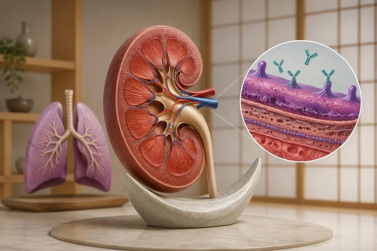
This antibody result can point to a time-sensitive kidney disorder—even when breathing feels normal. Diagnosis depends on the laboratory method, urine findings, kidney function, and sometimes tissue examination.


- Positive anti-GBM antibodies support possible anti-GBM disease but cannot establish a diagnosis from 1 laboratory result alone.
- Kidney-only disease is possible: lung involvement occurs in approximately 40–60% of classic reported cases, not everyone.
- Սուր երիկամային վնասում includes a creatinine rise of at least 0.3 mg/dL, or 26.5 µmol/L, within 48 hours; this threshold is not specific to anti-GBM disease.
- Շտապ օգնության ախտանիշներ include coughing up blood, new breathlessness, or sharply reduced urine output; seek emergency care rather than waiting for a repeat antibody test.
- Մեզի միկրոսկոպիա can identify red-cell casts, which support bleeding from the kidney filters rather than an ordinary bladder infection.
- Antibody cutoffs differ between assays: a result of 21 U/mL has meaning only alongside that laboratory's reference interval.
- Double-positive disease means anti-GBM antibodies and ANCA occur together; it can change relapse surveillance and long-term treatment.
- Ժամանակի նշանակություն matters: specialists may start treatment before confirmation when the clinical picture strongly suggests rapidly progressive disease.
- Transplant timing generally requires anti-GBM antibodies to remain undetectable for at least 6 months, according to KDIGO 2021.
What do positive anti-GBM antibodies actually mean?
Positive anti-GBM antibodies may indicate anti-glomerular basement membrane disease, an autoimmune condition that can rapidly damage kidney filters and sometimes the lungs. They do not, by themselves, confirm Goodpasture syndrome. Contact the ordering clinician today; coughing up blood, new breathlessness, or markedly reduced urine requires emergency assessment.
Anti-GBM describes the antibody's target, not a complete diagnosis: the glomerular basement membrane is part of the kidney's filtration barrier. Most classic disease involves antibodies against the noncollagenous domain of the alpha-3 chain of type IV collagen, a structure also found in lung air sacs; the same immune response can therefore affect 2 organs with very different symptoms.
I'm Thomas Klein, Chief Medical Officer at Kantesti; my first question is whether this result accompanies evidence of organ injury, rather than how alarming the laboratory flag looks. A single mildly positive result with stable creatinine is a different situation from positivity plus a creatinine rise over 48 hours, although both need clinician-led follow-up rather than casual reassurance.
Կանտեստին մի է AI արյան անալիզատոր that can help explain anti-GBM results alongside creatinine, electrolytes, and laboratory reference intervals, but cannot confirm autoimmune kidney disease. Our organization and purpose explain that educational role; our guide to դրական հակամարմնային արդյունքների explains why 1 positive immune marker is not the same as a diagnosis.
Is anti-GBM disease the same as Goodpasture syndrome?
Anti-GBM disease includes kidney-only disease, lung-only disease, and disease affecting both organs. Goodpasture syndrome often refers to the combination of glomerulonephritis and lung bleeding, although clinicians use the name inconsistently. Approximately 40–60% of classic anti-GBM cases have lung involvement, so the terminology should never determine whether kidney assessment is urgent.
The name Goodpasture syndrome can describe a pulmonary–renal presentation rather than prove its cause. ANCA-associated vasculitis and other disorders can also produce kidney inflammation with alveolar bleeding, so a Goodpasture syndrome diagnosis needs etiological clarification; McAdoo and Pusey's 2017 review distinguishes the clinical syndrome from antibody-mediated anti-GBM disease.
Anti-GBM disease is rare, with an estimated incidence around 1 case per million people annually in commonly cited populations, although estimates differ by location and case detection. That rarity matters when interpreting an unexpected screening result: even a good test can generate misleading positives when used in people with little clinical evidence of the disorder.
A low eGFR during this illness may reflect երիկամների սուր վնասում, not an established chronic stage. Chronic kidney disease generally requires abnormalities lasting at least 3 months; our kidney staging guide explains that distinction, but a rapidly changing result needs an acute assessment rather than being filed under a long-term CKD label.
Why can kidney injury occur without lung symptoms?
Kidney-only anti-GBM disease occurs because antibody access and tissue vulnerability differ between kidney filters and lung air sacs. A normal breathing pattern does not protect the kidneys or exclude this diagnosis. Since only approximately 40–60% of classic cases involve the lungs, waiting for a cough can delay treatment.
The glomerular filtration barrier is continuously exposed to circulating plasma under filtration pressure, whereas the lung's alveolar–capillary barrier has different structural protections. Antibody binding also depends on whether relevant collagen epitopes are accessible; this is not simply a situation where antibodies circulate to 1 organ but somehow fail to reach the other.
Smoking and hydrocarbon exposure are associated with pulmonary involvement in anti-GBM disease, and respiratory illness may influence the lung barrier's susceptibility. Those associations do not allow a precise personal risk calculation: a nonsmoker can develop lung involvement, while a smoker may have kidney-only disease, so 0 respiratory symptoms should never be used as a rule-out test.
Consider an illustrative patient with creatinine increasing from 0.9 to 1.8 mg/dL over several days, microscopic urine blood, and no cough at all. That combination needs urgent nephrology assessment because filtration has changed substantially; our eGFR-ի ցածր նախազգուշացման նշանները also explain why significant kidney impairment can initially feel surprisingly quiet.
How should the anti-GBM antibody test number be read?
Այն anti-GBM antibody test must be interpreted using the reporting laboratory's method, units, and cutoff. There is no universal positive concentration that can be transferred between every assay. For example, 21 U/mL is only slightly above a cutoff of 20 U/mL—but that example is not a recommended diagnostic threshold.
Immunoassays may use different antigen preparations and measurement systems, with results reported as U/mL, IU/mL, an index, or a qualitative positive. A result of 30 on an index assay cannot be meaningfully compared with 30 U/mL from another laboratory; the distinction between որակական և քանակական թեստերի վերաբերյալ is particularly useful here.
Low-level positivity deserves more scrutiny when urine findings and kidney function are normal, but a high concentration still cannot measure irreversible damage by itself. I would examine 3 details before interpreting a trend: whether the same assay was used, whether treatment had begun, and whether the laboratory describes the change as analytically meaningful.
Kantesti AI cannot repair an incorrect transcription of a laboratory result, so verify the value, units, reference interval, and collection date against the original report. Our PDF-ի տառադարձման ցուցակը covers those checks; a missing decimal can turn 2.1 into 21 and change the apparent significance of an antibody measurement.
Can a positive anti-GBM result be false or misleading?
A false-positive or clinically misleading anti-GBM result is possible, particularly when positivity is weak and the clinical picture does not fit. A clinician may request confirmation using another method. However, 1 repeat antibody test must not delay assessment when creatinine is rising, urine contains blood, or respiratory symptoms suggest lung involvement.
Pretest probability changes what a positive result means: a person tested for rapidly progressive kidney disease has a different starting likelihood from someone given a broad autoimmune screen without symptoms. Analytical interference, nonspecific reactivity, and differences in antigen recognition can complicate interpretation; repeating the same assay 2 times may reproduce the same problem rather than resolve it.
A reportedly normal urine test should be examined carefully, because a dipstick is not the same as microscopy and a negative result may reflect timing or specimen limitations. Clinicians often pair urine dipstick interpretation with sediment examination and protein measurement; 1 clean-looking urine sample cannot describe everything happening inside the kidney.
Creatinine, urine microscopy, and a urine albumin-to-creatinine or protein-to-creatinine ratio provide complementary information during confirmation. An ACR of at least 3 mg/mmol, approximately 30 mg/g, is abnormal but does not identify anti-GBM disease; our urine creatinine ratio guide explains why dilution-adjusted measurements are more useful than urine concentration alone.
Which urine findings suggest injury to the kidney filters?
Hematuria, proteinuria, dysmorphic red cells, and red-cell casts can support glomerular injury when anti-GBM antibodies are positive. Red-cell casts are especially concerning because they form within kidney tubules. An isolated finding of 3 or more red cells per high-power field warrants clinical interpretation, but does not establish the cause.
Միկրոսկոպիկ հեմատուրիա may appear before urine visibly changes color, so clear urine is not evidence that the kidney filters are unaffected. Conversely, a dipstick positive for blood can reflect hemoglobin or myoglobin without intact red cells; comparing the dipstick with 1 microscopy result helps distinguish these possibilities, particularly after intense exercise or muscle injury.
Protein loss in anti-GBM disease does not have to reach the nephrotic range, conventionally around 3.5 g per 24 hours in adults, to be clinically serious. A patient can have rapidly progressive kidney injury with a much smaller protein measurement; focusing only on whether urine protein is dramatically elevated can miss the more urgent creatinine trajectory.
Sample quality matters because delayed examination can make cellular elements and casts harder to identify, and menstrual contamination can complicate hematuria interpretation. Our միզի նստվածքի ուղեցույցի explains the principal patterns; if the first specimen is inconclusive, clinicians may request a fresh sample rather than assume 0 reported casts means no glomerular disease.
Which kidney function changes make the result urgent?
A rising creatinine level makes positive anti-GBM antibodies more urgent, especially alongside urine blood or protein. Standard acute kidney injury criteria include a rise of at least 0.3 mg/dL, or 26.5 µmol/L, within 48 hours, or at least 1.5 times baseline within 7 days; these criteria do not identify the underlying cause.
An apparently normal creatinine can still represent acute kidney injury if it has risen substantially from a low baseline. An increase from 0.6 to 1.0 mg/dL over 48 hours meets the absolute rise criterion even if 1.0 is inside the laboratory's adult reference interval; the comparison with baseline is therefore more informative than the flag alone.
Estimated GFR equations are less reliable during rapidly changing kidney function because they assume a relatively steady creatinine concentration. Our BUN և կրեատինին համեմատություն explains additional influences such as dehydration and protein intake, but those alternatives do not safely explain away a 2-fold creatinine increase accompanied by positive anti-GBM antibodies and glomerular urine findings.
Kantesti-ն՝ AI արյան անալիզի մեկնաբանություն հարթակ, can organize dated laboratory results so a creatinine change over 48 hours is easier to recognize; treatment decisions still require a clinician. Urine output below 0.5 mL/kg/hour for 6 hours also meets a standard AKI criterion, although patients should seek help for markedly reduced urine without attempting a precise home calculation.
Which symptoms require emergency care rather than a routine referral?
Coughing up blood, new breathlessness, low oxygen, or markedly reduced urine output require emergency assessment when anti-GBM disease is possible. New oxygen saturation at or below 92% at sea level is concerning, but a normal home reading does not exclude lung bleeding. Do not wait for the antibody result to be repeated.
Alveolar hemorrhage can occur without obvious coughing up blood because blood remains within the air sacs rather than reaching the mouth. New breathlessness, an unexplained hemoglobin fall, and new lung imaging abnormalities can therefore be more informative than 0 visible blood; our shortness of breath testing guide explains why respiratory assessment requires more than a single laboratory marker.
Severe kidney injury can produce dangerous potassium accumulation, fluid overload, and acid–base disturbance before an antibody diagnosis is finalized. Potassium at or above 6.5 mmol/L generally requires emergency management; lower levels may also be urgent with AKI or ECG changes, as discussed in our potassium escalation guide.
Chest symptoms, confusion, fainting, or an abrupt fall in urine output should be described clearly to emergency services, including the known positive anti-GBM result. Bring the report and any creatinine measurements from the previous 7 days if readily available, but do not delay departure to gather paperwork; the clinical team can investigate and stabilize complications at the same time.
How do specialists confirm an anti-GBM disease diagnosis?
Anti-GBM disease diagnosis combines serology with evidence of kidney or lung injury, and kidney biopsy often provides decisive information when safe and appropriate. Typical findings include crescentic glomerulonephritis and linear IgG staining along the glomerular basement membrane. These are 2 different observations: tissue injury and the pattern of antibody deposition.
Kidney biopsy can help establish the mechanism of injury and estimate how much potentially recoverable tissue remains, but no single microscopic feature should be read without context. Linear staining can occasionally occur in other settings, and atypical anti-GBM disease may differ from the classic pattern; specialists interpret light microscopy, immunofluorescence, and clinical findings together rather than treating 1 image as definitive.
Այն KDIGO 2021 glomerular diseases guideline recommends starting treatment without delay when anti-GBM disease is strongly suspected, even before confirmation. That does not mean every isolated positive result needs immediate immunosuppression: a patient with rapidly worsening kidney function has a different risk balance from someone with stable creatinine and an equivocal antibody on 2 separate assays.
The broader assessment usually includes ANCA, a full blood count, electrolytes, urine studies, and investigations for alternative immune-mediated kidney disease. ANA, anti-dsDNA, and complement may be useful when lupus is a possibility; our հակադիր-dsDNA մեկնաբանման ուղեցույց explains why a second positive antibody can suggest an alternative or additional process rather than simply confirming the first.
What changes if ANCA is also positive?
Double-positive disease means a patient has both anti-GBM antibodies and ANCA, usually assessed through MPO-ANCA and PR3-ANCA testing. The acute presentation may resemble anti-GBM disease, while later relapse risk can resemble ANCA-associated vasculitis. Those 2 antibody systems therefore affect both immediate treatment and the longer-term follow-up plan.
McAdoo and colleagues' 2017 Kidney International cohort compared 37 double-positive patients with isolated anti-GBM disease and ANCA-associated vasculitis groups. The double-positive group showed features of both conditions, including clinically relevant later relapses; this is why disappearance of the anti-GBM antibody does not automatically justify stopping every form of immune monitoring or maintenance treatment.
An MPO test is not always an MPO-ANCA test: some laboratories offer myeloperoxidase concentration assays for other purposes, which do not answer the same autoimmune question. Confirm that the report identifies antibodies against MPO or PR3; there are 2 major antigen-specific ANCA targets, and a generic enzyme measurement should not be used to classify double-positive disease.
Complement levels can help explore competing diagnoses, but normal C3 and C4 do not exclude anti-GBM disease or ANCA-associated vasculitis. Our low complement interpretation guide explains how reduced complement may point toward another immune process; these 2 results refine the differential diagnosis rather than functioning as a safety check that rules out kidney injury.
How is anti-GBM disease treated, and can kidneys recover?
Classic anti-GBM disease is usually treated with plasma exchange, glucocorticoids, and cyclophosphamide, although specialists adapt treatment to presentation and recovery potential. KDIGO 2021 describes cyclophosphamide for approximately 2–3 months and glucocorticoids for about 6 months. Kidney recovery depends heavily on function and tissue damage when treatment begins.
Plasma exchange removes circulating antibodies, while immunosuppression reduces further antibody production and tissue-directed immune activity. Treatment often continues until anti-GBM antibodies are undetectable, commonly involving daily exchanges over approximately 2–3 weeks, though duration is individualized; antibody disappearance does not instantly repair scarred kidney tissue or guarantee that dialysis can be stopped.
KDIGO allows carefully selected exceptions to intensive treatment when a patient needs dialysis at presentation, has 100% crescents or more than 50% globally sclerotic glomeruli on an adequate biopsy, and has no pulmonary hemorrhage. These criteria require specialist judgment, not self-interpretation: lung involvement, sampling uncertainty, and the amount of viable tissue can change the treatment balance.
Treatment monitoring includes blood counts, kidney function, infection risk, and drug-specific precautions; changes across 2 visits may reflect medication effects rather than renewed autoimmune injury. Kantesti can help arrange those results chronologically, but our դեղորայքային անվտանգության միտումների ուղեցույցը is educational support—not authorization to alter steroids, cyclophosphamide, or a specialist's plasma-exchange schedule.
What follow-up is needed after antibodies become negative?
A negative anti-GBM result after treatment does not mean follow-up can stop, because kidney recovery, medication toxicity, and possible ANCA overlap still need assessment. KDIGO 2021 recommends postponing kidney transplantation until anti-GBM antibodies have remained undetectable for at least 6 months. The monitoring schedule is individualized rather than a universal monthly timetable.
Relapse is uncommon in isolated classic anti-GBM disease, but double-positive disease needs a different long-term strategy because ANCA-associated vasculitis can recur. New urine blood, worsening creatinine, or recurrent breathlessness after 1 apparently successful treatment course deserves assessment; clinicians should not assume every new abnormality is relapse, but should not dismiss it merely because an earlier antibody test was negative.
Այն 6-month antibody-negative interval before transplantation concerns recurrence risk, not simply recovery from the original hospitalization. Transplant teams also consider clinical stability, infection risk, cardiovascular health, and other eligibility factors; a person can be antibody-negative while still requiring dialysis, and that does not mean the original treatment failed to control the immune process.
Diet and supplements cannot remove anti-GBM antibodies or replace immune treatment, and impaired filtration changes the safety of many over-the-counter products. Our երիկամների հավելումների անվտանգության ուղեցույցը explains common concerns; potassium-containing products, herbal mixtures, and high-dose nutrients deserve review when eGFR is reduced, even if only 1 ingredient is advertised as kidney-supporting.
What can AI interpretation help with—and what must stay clinical?
AI interpretation can help organize an anti-GBM report, but cannot diagnose Goodpasture syndrome or decide whether emergency treatment is needed. The most useful handoff includes the antibody method, creatinine trend, urine findings, and symptoms. A change over 48 hours can matter more than the report's color-coded positive flag.
Կանտեստին մի է ԱԻ լաբորատոր թեստի մեկնաբանման ծառայությունում that can explain the relationship between anti-GBM antibodies and accompanying laboratory findings when those results are supplied. Our տեխնոլոգիական բացատրություն describes the interpretation workflow; the original report remains the source for 3 essentials—value, unit, and reference interval—and an AI summary is not independent confirmation.
A technical benchmark is not a clinical diagnosis study, and a tool that explains reports cannot assess lung sounds, examine urine itself, or interpret a kidney biopsy firsthand. Our կլինիկական ստանդարտներ և վավերացում page provides methodological context; even excellent performance on 1 reporting task does not establish safety for every rare autoimmune presentation.
A note from Thomas Klein: the practical question to ask your clinician is, 'Do these antibodies come with evidence of kidney or lung injury, and how quickly should I be assessed?' As of Հոկտեմբեր 6, 2026, this article cites KDIGO 2021 guidance and the named studies below; ask for an explicit plan today rather than leaving a positive result without a follow-up pathway.
Clinical references, related research, and source limitations
The anti-GBM recommendations in this article are supported by 3 directly relevant clinical references, listed separately from our 2 related Zenodo resources. The Zenodo materials concern coagulation and serum proteins; they are supporting laboratory education, not trials, clinical guidelines, or evidence that establishes a Goodpasture syndrome diagnosis.
KDIGO 2021 supplies the treatment and transplantation recommendations; McAdoo and Pusey 2017 explains classic disease mechanisms and presentations; McAdoo and colleagues 2017 addresses double-positive outcomes. These 3 sources answer different questions, and no general protein or coagulation guide can substitute for them; Kantesti's բժշկական խորհրդատվական տեղեկատվություն describes the clinical expertise relevant to interpreting medical content.
aPTT Normal Range: D-Dimer, Protein C Blood Clotting Guide. (n.d.). Zenodo. DOI: 10.5281/zenodo.18262555. This related coagulation interpretation resource explains laboratory concepts relevant to treatment safety, not anti-GBM confirmation; ResearchGate discovery search և Academia.edu discovery search are search links, not verified publication profiles.
Serum Proteins Guide: Globulins, Albumin & A/G Ratio Blood Test. (n.d.). Zenodo. DOI: 10.5281/zenodo.18316300. This related serum protein resource explains why albumin and globulins add context without measuring anti-GBM activity; ResearchGate հրապարակումների որոնում և Academia.edu հրապարակումների որոնում likewise do not establish independent peer review of either of these 2 resources.
Հաճախակի տրվող հարցեր
Положительный результат теста на антитела к базальной мембране клубочков означает ли, что у меня синдром Гудпасчера?
Ընդդեմ GBM հակամարմինների դրական արդյունքը աջակցում է հնարավոր GBM-ի հակա-հիվանդությանը, սակայն ինքնուրույն չի հաստատում Goodpasture-ի համախտանիշը։ Ախտորոշումը պահանջում է երիկամների ֆունկցիայի, միզի հետազոտության և թոքերի հնարավոր ներգրավվածության կլինիկական գնահատում, ընդ որում երիկամների բիոպսիան հաճախ կարևոր ապացույցներ է տալիս։ Թոքերի ներգրավվածությունը դասական հաղորդված դեպքերի մոտ 40% – 60%-ում է, ուստի հնարավոր է միայն երիկամային հիվանդություն։ Հրամանատար բժշկին անհապաղ դիմեք, և շտապ օգնության դիմեք արյուն հազալու, նոր շնչահեղձության կամ միզի զգալի նվազման դեպքում։.
Հակա-ԳԲՄ հիվանդությունն ազդո՞ւմ է երիկամների վրա առանց հազի։
Հակա-ԳԲՄ հիվանդությունը կարող է վնասել երիկամները՝ չառաջացնելով հազ կամ այլ ակնհայտ թոքային ախտանշաններ։ Հակամարմինների հասանելիության և զգայունության մեջ տարբերվող երիկամների զտիչները և թոքերի օդապարկիկները, հետևաբար, 1 օրգանի ներգրավումը չի պահանջում մյուսի ներգրավումը։ 0.3 մգ/դլ կամ 26.5 մկմոլ/լ կրեատինինի բարձրացումը 48 ժամվա ընթացքում համապատասխանում է սուր երիկամային վնասվածքի չափանիշին և պահանջում է շտապ մեկնաբանություն, երբ հակա-ԳԲՄ հակամարմիններն դրական են։ Նորմալ շնչառությունը չպետք է ուշացնի երիկամների գնահատումը։.
Ո՞ր հակա-ԳԲՄ հակամարմնի մակարդակն է համարվում դրական։
Դրական հակա-GBM հակամարմնի կտրվածքը կախված է լաբորատորիայի անալիզից և հաշվետվող միավորներից։ Չկա միասնական համընդհանուր U/mL կամ IU/mL շեմ, որը կիրառվում է բոլոր լաբորատորիաներում։ Օրինակ, 21 U/mL-ը միայն մի փոքր բարձր է 20 U/mL լաբորատոր կտրվածքից, բայց այդ թվերը չպետք է կիրառվեն այլ անալիզի վրա։ Շտապողականությունը կախված է ուղեկցող երիկամների կամ թոքերի վնասվածքից, ոչ միայն հակամարմնի կոնցենտրացիայից։.
Կարո՞ղ է հակա-GBM հակամարմնի դրական թեստը սխալ լինել։
Ուժեղ դրական հակա-GBM հակամարմնային թեստը կարող է մոլորության մատնել վերլուծական միջամտության, ոչ-սպեցիֆիկ ռեակտիվության կամ հիվանդության ցածր կլինիկական հավանականության պատճառով։ Թույլ դրական արդյունքը կայուն երիկամային գործառույթի և նորմալ մեզի արդյունքների դեպքում կարող է հանգեցնել հաստատման՝ օգտագործելով երկրորդ մեթոդ։ Նույն անալիզը 2 անգամ կրկնելը կարող է վերարտադրել նույն վերլուծական խնդիրը, ուստի կլինիցիստները կարող են այլընտրանքային թեստ քննարկել լաբորատորիայի հետ։ Հաստատումը չպետք է ուշացնի շտապ գնահատումը, երբ երիկամային գործառույթը վատթարանում է կամ առկա են թոքային ախտանշաններ։.
Որո՞նք են հակա-GBM ախտանշանները, որոնց դեպքում պետք է դիմեմ շտապօգնության։
Coughing up blood, new or worsening breathlessness, markedly reduced urine output, confusion, or fainting warrant emergency assessment when anti-GBM disease is suspected. New oxygen saturation at or below 92% at sea level is concerning, although a normal home reading does not exclude lung involvement. Potassium at or above 6.5 mmol/L generally requires emergency management, and lower levels can also be dangerous with acute kidney injury or ECG changes. Do not wait for a repeat antibody test before seeking help.
Can anti-GBM disease be treated, and can I have a kidney transplant?
Anti-GBM disease can be treated, usually with plasma exchange, glucocorticoids, and cyclophosphamide, but kidney recovery depends on the severity and duration of injury before treatment. KDIGO 2021 describes cyclophosphamide treatment for approximately 2–3 months and glucocorticoids for about 6 months, with individual adjustments by specialists. People with irreversible kidney failure may be considered for transplantation. KDIGO recommends waiting until anti-GBM antibodies have remained undetectable for at least 6 months before transplantation.
Ստացեք AI-ով աշխատող արյան անալիզի վերլուծություն այսօր
Միացեք ավելի քան 2 միլիոն օգտատերերի ամբողջ աշխարհում, ովքեր վստահում են Kantesti-ին՝ լաբորատոր թեստերի ակնթարթային, ճշգրիտ վերլուծության համար։ Վերբեռնեք ձեր արյան անալիզի արդյունքները և ստացեք 15,000+ բիոմարկերների համապարփակ մեկնաբանություն վայրկյանների ընթացքում։.
📚 Հղված հետազոտական հրապարակումներ
Klein, T., Mitchell, S., & Weber, H. (2026). aPTT-ի նորմալ միջակայքը. D-դիմեր, սպիտակուց C արյան մակարդման ուղեցույց.։ Kantesti AI Medical Research.
Klein, T., Mitchell, S., & Weber, H. (2026). Արյան շիճուկի սպիտակուցների ուղեցույց. գլոբուլինների, ալբումինի և A/G հարաբերակցության արյան ստուգում.։ Kantesti AI Medical Research.
📖 Արտաքին բժշկական հղումներ
Kidney Disease: Improving Global Outcomes Glomerular Diseases Work Group (2021). KDIGO 2021 կլինիկական պրակտիկայի ուղեցույց՝ գլոմերուլային հիվանդությունների կառավարման համար.։ Kidney International։.
McAdoo SP and Pusey CD (2017). Anti-Glomerular Basement Membrane Disease.։ Ամերիկյան Նեֆրոլոգիայի Ընկերության Կլինիկական ամսագիր։.
McAdoo SP et al. (2017). Patients double-seropositive for ANCA and anti-GBM antibodies have varied renal survival, frequency of relapse, and outcomes compared to single-seropositive patients.։ Kidney International։.
⚕️ Բժշկական հրաժարում
Այս հոդվածը միայն կրթական նպատակների համար է և չի հանդիսանում բժշկական խորհրդատվություն։ Ախտորոշման և բուժման որոշումների համար միշտ խորհրդակցեք որակավորված բուժաշխատողի հետ։.
E-E-A-T վստահության ազդանշաններ
Փորձառություն
Բժշկի կողմից ղեկավարվող կլինիկական վերանայում՝ լաբորատոր մեկնաբանության աշխատանքային հոսքերի համար։.
Մասնագիտություն
Լաբորատոր բժշկության կենտրոնացում՝ այն բանի վրա, թե ինչպես են բիոմարկերները դրսևորվում կլինիկական համատեքստում։.
Հեղինակություն
Գրված է դոկտոր Թոմաս Քլայնի կողմից՝ դոկտոր Սառա Միթչելի և պրոֆեսոր դոկտոր Հանս Վեբերի կողմից վերանայմամբ։.
Հուսալիություն
Ապացույցների վրա հիմնված մեկնաբանություն՝ հստակ հետագա քայլերի ուղիներով՝ ահազանգերը նվազեցնելու համար։.