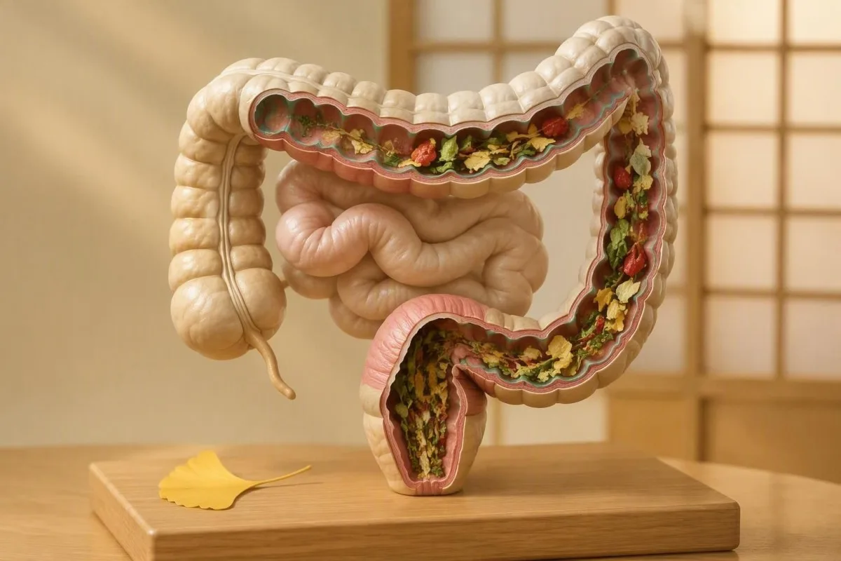
Visible food particles are usually a digestion-speed or food-structure issue, not proof that your body is failing to absorb nutrients. The pattern of the stool, your weight, symptoms, and blood results determine whether it deserves testing.


- Undigested food in stool is commonly harmless when it follows high-fibre meals and occurs without weight loss, chronic diarrhoea, or oily stools.
- Vegetable skins from corn, peppers, tomatoes, leafy greens, and legumes often remain visible because humans do not digest cellulose.
- Rapid transit can leave food particles in stool after gastroenteritis, stress-related diarrhoea, stimulant laxatives, magnesium products, or large caffeine intake.
- Steatorrhoea means bulky, pale, greasy, difficult-to-flush stool and is more concerning than occasional visible food pieces.
- Pérdida de peso involuntaria of 5% or more over 6 to 12 months warrants medical review, particularly with persistent loose stools.
- Faecal calprotectin helps assess intestinal inflammation; values below 50 micrograms/g usually make active inflammatory bowel disease less likely in adults.
- Faecal elastase-1 below 200 micrograms/g suggests possible pancreatic enzyme insufficiency, although watery samples can produce falsely low results.
- Coeliac testing is most reliable while a person is still eating gluten; do not start a gluten-free diet before discussing testing with a clinician.
When visible food in stool is usually normal
Undigested food in stool is usually normal when it appears occasionally after a fibre-rich meal and you otherwise feel well, maintain weight, and have formed stools. The most common explanation is visible plant cell wall material, not failed nutrient absorption.
In my clinical experience, a person who notices corn kernels, tomato skin, spinach fibres, or pepper peel once or twice a week rarely has a serious absorption problem. Plant cell walls contain cellulose, and human digestive enzymes do not break cellulose into absorbable sugar. The softer starch, protein, fat, and many vitamins inside the food may still have been absorbed normally.
The reassuring pattern is remarkably consistent: a recognisable food follows a recognisable meal, stool frequency is roughly your usual baseline, and there is no progressive fatigue or weight loss. Dr. Thomas Klein's practical rule is that the stool should be judged as a pattern over 2 to 4 weeks, not as a single alarming toilet-bowl observation. The Tabla de heces de Bristol can make that pattern easier to describe accurately.
Kantesti es un Analizador de sangre con inteligencia artificial that helps place possible digestive symptoms alongside objective clues such as iron stores, vitamin B12, albumin, liver markers, and inflammation markers. A normal nutritional panel does not rule out every bowel condition, but it makes severe long-standing malabsorption much less likely.
What food particles and vegetable pieces actually mean
Food particles in stool are most often fragments of tough plant material, and their shape can point to the meal rather than to disease. Flat translucent skins and fibrous strands are different from greasy droplets, mucus, or blood.
Corn pericarp, quinoa husks, tomato skins, bell pepper cuticle, mushroom cell walls, and leafy-green fibres are frequent examples. Their bright colour may look startling because pigments can survive transit; beetroot can redden stool, while spinach may leave dark green flecks. This is especially common after meals containing 25 to 35 g of fibre, close to many adult dietary targets.
A useful distinction is whether the material is recognisable. A few identifiable pieces among otherwise ordinary stool generally point toward food structure or fast transit; a diffuse oily sheen, pale clay-like stool, or stool that floats repeatedly because of trapped fat requires a different line of inquiry. Persistent pale stool deserves attention because it can reflect reduced bile reaching the intestine; see our guide to pale stool patterns.
Do not try to diagnose yourself by photographing every bowel movement. Instead, write down the preceding 24 hours of food, supplements, alcohol, illness, and stool frequency for 7 days. That small diary often exposes a very ordinary explanation—particularly a new high-fibre breakfast, magnesium supplement, or diarrhoeal illness.
Why high-fibre foods survive digestion
High-fibre foods can produce visible vegetable pieces because human enzymes cannot digest cellulose, lignin, and some resistant starch structures. Seeing the outer layer of a food does not mean the calorie or micronutrient content passed through untouched.
Chewing and cooking change how much plant matter remains visible. A raw salad with kale, carrots, pepper skins, seeds, and legumes creates more recognisable residue than the same ingredients cooked into a soup. Even so, colonic bacteria ferment part of the fibre into short-chain fatty acids, which support the colonic lining and are absorbed as energy.
Rapidly raising fibre from 10 g to 30 g daily can also cause gas, urgency, and looser stools for 1 to 2 weeks. Most patients find a slower increase—about 5 g every 3 to 4 days—with adequate fluids is more comfortable. A sudden fibre increase may make food particles more obvious without causing any nutritional deficit.
If a high-fibre diet causes daily pain, nocturnal diarrhoea, or worsening bloating, do not simply force more fibre. Fermentable carbohydrates can aggravate symptoms in some people with irritable bowel syndrome, while coeliac disease and inflammatory conditions need different management. Our article on foods that shift stool tests explains why diet changes should precede stool testing by a sensible interval.
How fast intestinal transit leaves food visible
Fast intestinal transit can leave vegetable pieces in stool because food spends less time being mechanically broken down and exposed to digestive enzymes. Acute diarrhoea is the classic setting, but medication, stress, and diet can do the same thing.
Stool normally takes roughly 24 to 72 hours to travel through the colon, although healthy variation is broad. When transit speeds up, more water remains in stool and food is less fragmented. Viral gastroenteritis, food poisoning, a large coffee intake, endurance running, anxiety, and menstruation-related bowel changes can all temporarily shorten transit.
Magnesium citrate and magnesium oxide commonly loosen stool because poorly absorbed magnesium draws water into the bowel; doses above 350 mg daily from supplements are more likely to cause gastrointestinal effects. Metformin, antibiotics, GLP-1 medicines, laxatives, and some sugar alcohols can also change stool form. Do not stop a prescribed medicine without discussing it with the prescriber.
Chronic diarrhoea means loose or watery stool for 4 weeks or longer, not merely three bowel movements after a spicy meal. The British Society of Gastroenterology guideline recommends a structured history, medication review, coeliac serology, and targeted stool testing when diarrhoea persists (Arasaradnam et al., 2018). For related warning patterns, read our guía de análisis de sangre para diarrea.
Chewing, meal speed and the overlooked mechanical cause
Poor chewing can make food look less digested even when pancreatic enzymes and intestinal absorption are normal. The mouth is the first grinding stage of digestion, and rushed eating changes what reaches the colon.
I see this most often in people who eat lunch at a desk, have dental discomfort, wear poorly fitting dental appliances, or swallow quickly after a long fast. Large pieces of mushroom, carrot, nuts, leafy greens, and legumes are mechanically harder for the stomach and bowel to reduce. The practical experiment is simple: chew deliberately for 7 days before changing supplements or buying tests.
A 52-year-old runner I reviewed had visible kale and lentil fragments after race weekends, but no weight loss and a stable ferritin of 64 ng/mL. His pattern improved when he ate a smaller pre-run fibre load, drank less coffee immediately before exercise, and slowed his evening meal. That is not a universal prescription, but it shows why context beats panic.
Difficulty swallowing, choking, pain with chewing, or an unexpected change in dentition deserves dental or medical assessment rather than a digestive cleanse. If you have coexisting tiredness, paleness, or poor appetite, the guía de síntomas de ferritina baja may help you recognise when a blood count and iron studies are reasonable.
How malabsorption differs from harmless visible fibre
Malabsorption usually causes a repeated cluster of symptoms—weight loss, chronic loose stool, excess gas, nutrient deficiency, or greasy bulky stool—not isolated vegetable pieces. The combination matters far more than the visual appearance of one bowel movement.
Steatorrhoea describes excess fat in stool and often looks pale, bulky, oily, foul-smelling, or difficult to flush. It may float, although floating alone is not diagnostic because gas can do that too. A repeated greasy film plus weight loss is a much stronger warning sign than seeing corn or seeds.
Malabsorption symptoms can include iron-deficiency anaemia, recurrent mouth ulcers, low folate, vitamin B12 deficiency, low albumin, bruising from vitamin K deficiency, or bone pain from low vitamin D. No single blood test proves malabsorption, and normal results do not exclude early coeliac disease. Kantesti's plataforma de interpretación de biomarcadores con IA reviews iron indices, full blood count, albumin, calcium, liver tests, and nutrient markers as a pattern rather than treating one low value as a diagnosis.
The reason clinicians worry about loose oily stool combined with a falling weight is that together they suggest lost calories and fat-soluble nutrients, whereas food particles alone usually do not. If stool appears greasy or unusually light for more than 2 weeks, our faecal fat test overview explains what collection-based testing can and cannot establish.
Warning signs that need prompt medical review
Visible food needs medical review when it occurs with blood in stool, black tarry stool, fever, persistent pain, dehydration, or unintentional weight loss. These features raise concern for inflammation, infection, bleeding, obstruction, or clinically meaningful malabsorption.
Seek urgent care today for black tar-like stool, maroon or large-volume red stool, severe constant abdominal pain, a rigid abdomen, fainting, confusion, or inability to keep fluids down. Black stool can be caused by iron or bismuth, but true melena is sticky, offensive, and may signal upper gastrointestinal bleeding. A positive stool blood test needs clinician-directed follow-up rather than repeated home tests.
Arrange a routine appointment within days to weeks if diarrhoea lasts beyond 4 weeks, stools wake you at night, weight drops by 5% or more in 6 to 12 months, or you develop persistent fatigue. Fever above 38.0°C with diarrhoea, recent international travel, recent antibiotics, or immune suppression lowers the threshold for stool infection testing.
Age changes the threshold too. New bowel habit change after age 50, or at any age with a first-degree relative with colorectal cancer or inflammatory bowel disease, deserves a proper review. Clinicians may use a faecal immunochemical test for occult bleeding, but it does not diagnose coeliac disease, pancreatic insufficiency, or all bowel conditions.
Coeliac disease clues behind persistent food particles
Coeliac disease should be considered when persistent loose stools or food particles accompany iron deficiency, weight loss, recurrent mouth ulcers, low bone density, or a family history of coeliac disease. Many adults have non-classic symptoms and do not look underweight.
The first-line blood test is usually tissue transglutaminase IgA together with total IgA. A negative tissue transglutaminase IgA result is less reliable in IgA deficiency, which is why total IgA matters; IgG-based tests may then be used. The American College of Gastroenterology advises testing while the patient is consuming gluten, not after a self-started gluten-free diet (Rubio-Tapia et al., 2023).
Low ferritin can precede anaemia in coeliac disease. Ferritin below 15 ng/mL strongly supports iron deficiency in many laboratories, although inflammation can make ferritin appear falsely reassuring; transferrin saturation below 20% adds useful context. Our guía de desafío de gluten discusses why stopping gluten before testing can create a false-negative result.
Dr. Thomas Klein has seen patients delay diagnosis because they assumed bloating and vegetable pieces meant they simply needed probiotics. Probiotics may change symptoms in selected cases, but they do not diagnose or treat immune-mediated gluten injury. Kantesti is an Herramienta de análisis de pruebas de sangre con IA that can identify a blood-test pattern worth discussing with a clinician, not replace confirmatory coeliac assessment.
When pancreatic or bile problems are more likely
Pancreatic enzyme deficiency and reduced bile delivery are more likely when stool is repeatedly greasy, pale, bulky, and associated with weight loss than when it simply contains vegetable skins. Both conditions affect fat digestion, so the stool changes are usually conspicuous.
The pancreas supplies lipase, proteases, and amylase to the small intestine. Faecal elastase-1 below 200 micrograms/g may suggest pancreatic exocrine insufficiency, while values below 100 micrograms/g are more strongly suggestive; a watery stool sample can dilute elastase and give a falsely low result. This test should be interpreted alongside symptoms, nutrition, imaging, and history of pancreatitis, pancreatic surgery, cystic fibrosis, or heavy alcohol exposure.
Bile obstruction can make stool pale or putty-coloured and may occur with dark urine, yellowing of the eyes, itching, or right-upper abdominal pain. Blood tests may show raised alkaline phosphatase, gamma-glutamyl transferase, and direct bilirubin, but normal liver tests do not explain every pale stool episode. Review our patrones de análisis de sangre de colestasis for the combinations clinicians use.
Do not take over-the-counter digestive enzymes indefinitely to mask these symptoms. Enzymes can be appropriate after a diagnosis, but they may delay recognition of a bile duct or pancreatic condition. A stool elastase result is most helpful when the sample is formed or semi-formed and the symptom history is clear.
Which stool tests are appropriate and why
Stool testing is appropriate for persistent diarrhoea, greasy stools, blood, fever, travel-related symptoms, or suspected bowel inflammation—not for one isolated piece of corn. The best test depends on the clinical question.
Faecal calprotectin measures neutrophil-related intestinal inflammation. In many adult laboratories, a result below 50 micrograms/g makes active inflammatory bowel disease less likely, while 50 to 150 micrograms/g often leads to repeat testing after infection, NSAID use, or a flare settles. Values above 250 micrograms/g more strongly support prompt clinical investigation, although cut-offs vary by assay and age.
Stool culture or molecular pathogen testing is most useful with acute diarrhoea plus fever, blood, severe illness, recent travel, outbreaks, or immune compromise. Ova-and-parasite testing should be targeted to travel, exposure, prolonged diarrhoea, or specific risks rather than ordered automatically. Our stool culture results guide explains why a report can show normal flora without answering every symptom.
For pancreatic questions, request faecal elastase; for fat loss, clinicians may use a qualitative stain or quantitative faecal fat collection; for inflammatory questions, calprotectin or lactoferrin is usually more useful. The calprotectin versus lactoferrin comparison helps patients understand why these tests are not interchangeable.
Blood tests that can reveal long-term absorption problems
Blood tests can reveal the consequences of chronic malabsorption, especially iron deficiency, anaemia, low albumin, vitamin deficiencies, and electrolyte disturbance. They do not identify food particles directly, but they can show whether digestion problems are affecting the rest of the body.
A sensible initial panel often includes full blood count, ferritin with transferrin saturation, folate, vitamin B12, albumin, liver tests, renal function, calcium, and coeliac serology when symptoms fit. Albumin below 35 g/L may reflect poor intake, inflammation, liver disease, kidney loss, or intestinal protein loss; it is not a stand-alone malabsorption diagnosis. Persistent low magnesium or potassium in someone with diarrhoea can signal clinically significant fluid losses.
De Kantesti servicio de interpretación de pruebas de laboratorio de IA can organise these results against the laboratory's own ranges and show trends across dates. In our analysis of more than 2 million interpreted tests, the most useful signal is often a direction of travel—ferritin falling from 42 to 18 ng/mL, for example—rather than one borderline result.
Vitamin B12 below 200 pg/mL, or approximately 148 pmol/L, is commonly considered low, though laboratory thresholds differ and methylmalonic acid can clarify equivocal cases. Folate deficiency, iron deficiency, and low vitamin D together raise the index of suspicion for small-bowel disease, but diet restriction alone can produce the same combination. See our comparación de B12 y folato before treating a low value in isolation.
Special considerations for children and older adults
Children with visible food in stool are often experiencing normal dietary fibre passage or a short-lived viral illness, but poor growth and persistent diarrhoea need paediatric assessment. In older adults, a new bowel change deserves a lower threshold for review.
For children, contact a clinician promptly if there are fewer wet nappies or urinations, dry mouth, unusual sleepiness, fever, blood in stool, repeated vomiting, or slowed growth. Babies and toddlers can pass partially digested peas, carrots, and corn after diet changes because their chewing and digestive capacity are still developing. Never use adult anti-diarrhoeal medicines in a child without professional guidance.
In older adults, poor dentition, reduced appetite, polypharmacy, diabetes medicines, antibiotics, and reduced mobility can all alter stool appearance. New constipation alternating with diarrhoea, iron-deficiency anaemia, or a positive stool blood test should not be blamed on vegetables. Our article about senior medication effects may help prepare a medication list for the appointment.
Weight should be measured rather than guessed. A 3 kg loss matters differently in a 45 kg person than in a 100 kg person, which is why clinicians use percentage loss; 5% over 6 to 12 months is a common trigger for assessment. For paediatric laboratory questions, interpretación segura de IA para niños explains the limits of automated result support.
A practical 7-day plan before your appointment
A 7-day food, stool, and symptom record often separates harmless food residue from a persistent clinical pattern before any test is ordered. Record details that change medical decisions, not every minor variation.
Record meal timing, high-fibre foods, alcohol, caffeine, supplements, medicines, stool frequency, Bristol type, pain, urgency, and whether stool was oily or difficult to flush. Weigh yourself once at the start and once at day 7 under similar conditions. Avoid abruptly eliminating gluten if coeliac testing may be needed, and avoid starting enzyme products just before a clinical review.
Hydration is particularly important during diarrhoea. Adults with uncomplicated fluid loss can usually use oral rehydration solution or frequent small drinks; dizziness on standing, minimal urination for 8 to 12 hours, or inability to retain fluids needs same-day advice. If fasting has coincided with symptoms, our fasting and stool-change guide covers common non-dangerous mechanisms.
Bring a photograph only if it genuinely shows blood, black stool, pale stool, or repeated oily residue; most clinicians do not need routine images of vegetable pieces. Bring the diary and a medication list instead. Those two items often shorten the path to the right test more than an unstructured description of “undigested food.”
Misconceptions about food particles, detoxes and enzymes
Visible food particles do not prove a “leaky gut,” toxin buildup, parasite infection, or lack of stomach acid. These claims sound plausible because the symptom is visual, but they are not reliable diagnostic shortcuts.
Commercial detoxes and colon cleanses can actually create the diarrhoea that makes food more visible. Stimulant laxatives may cause urgency, cramping, electrolyte shifts, and dependence when used regularly. Magnesium-containing products can be useful for constipation under guidance, but a new loose-stool pattern after starting them is often pharmacological rather than mysterious.
Parasites are possible after relevant travel, contaminated water exposure, certain occupational exposures, or prolonged diarrhoea, but food pieces are not a parasite test. Testing is more accurate than empiric anti-parasitic treatment because different organisms need different medicines. Our ova and parasite testing guide explains when sample collection is actually worthwhile.
Digestive enzymes can be medically appropriate for confirmed pancreatic exocrine insufficiency, but dosing depends on fat intake and diagnosis. “Low stomach acid” supplements may be unsafe for people with ulcers, gastritis, reflux, or certain medicines. Evidence-based review means identifying the mechanism first, then choosing treatment—not trying every bottle on the shelf.
When to book a clinician review and what to ask
Book a clinician review if food particles persist for more than 2 to 4 weeks with diarrhoea, pain, weight loss, greasy stools, or abnormal blood results. A focused history and a few targeted tests are usually more useful than broad commercial panels.
Ask: “Does my stool pattern suggest inflammation, infection, fat malabsorption, coeliac disease, or rapid transit?” Then ask which one or two tests would change management. This phrasing helps avoid scattershot testing. If you have existing results, include the laboratory name, collection date, reference ranges, and whether you were ill or taking supplements at the time.
Kantesti can help you organise a blood-test PDF or photo for discussion, flagging linked abnormalities and longitudinal changes in about 60 seconds; it cannot examine you, inspect a stool sample, or replace a clinician's diagnosis. Our approach is reviewed against clinical standards described in our resumen de validación médica, with physician input from the Consejo Asesor Médico.
As of September 20, 2026, the sensible bottom line remains reassuring but specific: occasional vegetable pieces in otherwise normal stool are common; persistent greasy diarrhoea, blood, weight loss, fever, or nutrient-deficiency clues are not. The guía de biomarcadores de Kantesti can help you prepare informed questions from laboratory results, while your own clinician determines the diagnosis and next step.
Preguntas frecuentes
¿Es normal la presencia de comida no digerida en las heces?
La comida sin digerir en las heces es a menudo normal cuando el material es fibra vegetal reconocible, como maíz, piel de tomate, espinacas, pimientos o legumbres, y no hay pérdida de peso ni diarrea persistente. Los humanos no pueden digerir la celulosa, por lo que las estructuras externas de las plantas pueden permanecer visibles incluso cuando los nutrientes del alimento se absorbieron. La revisión médica es apropiada si este patrón dura más de 2 a 4 semanas con heces grasosas, dolor, fiebre, sangre o una pérdida de peso de 5% o más en 6 a 12 meses.
¿Por qué veo trozos de verduras en mis heces?
Los trozos de verdura en las heces suelen reflejar pieles ricas en celulosa, masticación inadecuada o tránsito intestinal rápido en lugar de malabsorción de nutrientes. La col rizada cruda, el maíz, los champiñones, las pieles de pimiento, las pieles de tomate y las cáscaras de quinoa son ejemplos comunes porque su material estructural resiste la digestión humana. Un aumento repentino de aproximadamente 10 g a 30 g de fibra al día puede hacer que estos fragmentos sean más notables durante 1 a 2 semanas mientras el intestino se ajusta.
¿Significa la comida en las heces que tengo malabsorción?
La comida en las heces por sí sola no significa malaabsorción. La malaabsorción causa más a menudo una combinación repetida de heces sueltas o grasas voluminosas, pérdida de peso involuntaria, fatiga, deficiencia de hierro, niveles bajos de vitaminas o albúmina baja. La elastasa fecal-1 por debajo de 200 microgramos/g puede sugerir insuficiencia de enzimas pancreáticas, mientras que las pruebas de sangre celíacas se utilizan cuando se sospecha enfermedad del intestino delgado relacionada con el gluten.
¿Cómo se ven las heces grasosas?
Heces grasas, también llamadas esteatorrea, son típicamente pálidas, voluminosas, grasosas, con un olor inusualmente desagradable y difíciles de tirar en el inodoro en lugar de simplemente contener verduras visibles. Las heces pueden flotar, pero flotar por sí solo no es diagnóstico porque el gas intestinal también puede hacer que las heces floten. Heces aceitosas repetidas con pérdida de peso deben ser evaluadas clínicamente, a menudo incluyendo elastasa fecal, pruebas de hígado, marcadores de nutrición y, a veces, pruebas cuantitativas de grasa fecal.
¿Puede la ansiedad causar alimentos sin digerir en las heces?
La ansiedad puede contribuir a la presencia de alimentos sin digerir en las heces al acelerar el tránsito intestinal, especialmente cuando desencadena diarrea, movimientos intestinales frecuentes, consumo de cafeína o comer apresuradamente. Un tránsito más rápido deja menos tiempo para que los alimentos se descompongan en fragmentos más pequeños, por lo que las pieles y las fibras de las verduras pueden permanecer reconocibles. La ansiedad no explica la sangre roja, las heces negras alquitranadas, la fiebre superior a 38,0 °C, la diarrea nocturna persistente o la pérdida de peso involuntaria.
¿Qué pruebas detectan la malabsorción?
Tests for malabsorption are selected by the suspected cause and may include a full blood count, ferritin with transferrin saturation, vitamin B12, folate, albumin, calcium, coeliac serology, faecal elastase, and faecal calprotectin. Faecal calprotectin below 50 micrograms/g often makes active inflammatory bowel disease less likely in adults, while faecal elastase below 200 micrograms/g can suggest pancreatic exocrine insufficiency. A clinician should interpret results with stool appearance, medicines, diet, weight trend, and medical history.
Obtén hoy un análisis de sangre con IA
Únete a más de 2 millones de usuarios en todo el mundo que confían en Kantesti para el análisis instantáneo y preciso de pruebas de laboratorio. Sube tus resultados de análisis de sangre y recibe una interpretación completa de los biomarcadores de 15,000+ en segundos.
📚 Publicaciones de investigación citadas
Klein, T., Mitchell, S., & Weber, H. (2026). Diarrea después del ayuno, manchas negras en las heces y guía gastrointestinal 2026. Investigación médica con IA de Kantesti.
Klein, T., Mitchell, S., & Weber, H. (2026). Guía de salud femenina: ovulación, menopausia y síntomas hormonales. Investigación médica con IA de Kantesti.
📖 Referencias médicas externas
⚕️ Descargo de responsabilidad médica
Este artículo es solo con fines educativos y no constituye asesoramiento médico. Consulta siempre a un profesional sanitario cualificado para decisiones de diagnóstico y tratamiento.
Señales de confianza E-E-A-T
Experiencia
Revisión clínica dirigida por un médico de los flujos de interpretación de análisis.
Pericia
Enfoque en medicina de laboratorio sobre cómo se comportan los biomarcadores en el contexto clínico.
Autoridad
Escrito por el Dr. Thomas Klein, con revisión de la Dra. Sarah Mitchell y el Prof. Dr. Hans Weber.
Integridad
Interpretación basada en la evidencia con vías de seguimiento claras para reducir la alarma.