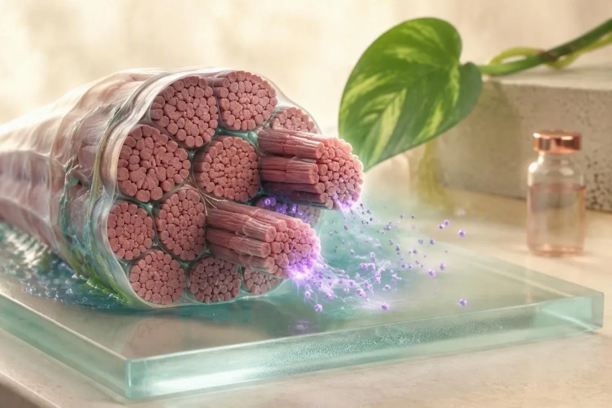
Aldolase is a muscle-associated enzyme, but an abnormal result is a pattern to investigate—not a diagnosis. The accompanying CK, AST, symptoms, training history and sample quality usually matter more than the aldolase number alone.


- Referenca intervalo for serum aldolase is commonly about 1.0–7.5 U/L in adults, but the reporting laboratory’s interval is the only range that applies to your result.
- Efiko de ekzercado can raise aldolase, CK and AST for 24–72 hours after unfamiliar resistance work, a marathon, or hard intervals.
- CK comparison matters: high aldolase with high CK supports active muscle-cell injury; high aldolase with normal CK can still occur in selected inflammatory myopathies.
- Hemolysis flag means damaged red cellular elements may have altered the specimen, so a fresh sample is often more informative than an expanded work-up.
- Urĝa ŝablono is severe muscle pain or weakness with dark urine, low urine output, fever, or CK above 5,000 U/L—seek same-day medical assessment.
- Follow-up panel usually includes CK, AST, ALT, LDH, creatinine, electrolytes, urinalysis and sometimes haptoglobin, reticulocytes, ESR/CRP and myositis antibodies.
- Myositis clue is objective, progressive proximal weakness lasting weeks, especially with rash, swallowing trouble, breathlessness or interstitial lung symptoms.
- Retest preparation is often 5–7 days without strenuous exercise, good hydration, and a medication/supplement review unless a clinician advises otherwise.
What a high aldolase result usually means
A high aldolase level usually reflects release of this glycolytic enzyme from stressed or injured skeletal muscle, although liver tissue and red cellular elements also contain aldolase. In adults, many laboratories use a serum reference interval near 1.0–7.5 U/L; a result of 9 U/L deserves context, while 40 U/L alongside weakness deserves prompt evaluation.
Aldolase participates in glucose breakdown inside cells. Its older reputation as a “muscle enzyme” is fair, but incomplete: it does not identify the cause, the exact muscle, or the severity of illness by itself. I have seen a value of 11 U/L normalize after a patient paused new boot-camp sessions; I have also seen modest persistent elevations reveal inflammatory muscle disease.
The most useful first comparison is kreatin-kinazo (CK). CK is more concentrated in skeletal muscle and often rises more dramatically in rhabdomyolysis, whereas aldolase can be disproportionately elevated in some regenerating muscle-fiber disorders. Our CK interpretation guide explains why one enzyme cannot stand in for the whole clinical picture.
Kantesti estas AI-sangotesta analizilo that reads aldolase alongside CK, AST, ALT, LDH, kidney markers and prior results rather than treating a single laboratory flag as a diagnosis. That comparison is especially useful when the same laboratory has tested you more than once.
Why muscle, liver and red cells can raise aldolase
Aldolase rises from skeletal muscle most often in this setting, but liver-cell injury and red-cell breakdown can contribute. A high result with normal bilirubin, ALT and CK is therefore not automatically reassuring or alarming; it is an invitation to check the pattern.
The assay measures enzyme activity in serum, not a muscle-specific isoenzyme in routine practice. Skeletal muscle injury commonly produces a cluster of high CK, aldolase, AST, LDH and sometimes myoglobin; ALT can rise too, which is why isolated “liver enzyme” interpretation can go wrong. See our practical guide to AST patterns when AST is part of the result.
Red cellular elements contain aldolase, so in vivo hemolysis may increase it alongside indirect bilirubin, LDH and reticulocytes, with reduced haptoglobin. In vitro hemolysis during collection is different: it can interfere with some assays and should prompt the laboratory’s own sample-quality review rather than assumptions about a hemolytic illness.
Dr. Thomas Klein’s clinical rule is simple: if a value is biologically surprising, verify the specimen before pursuing rare disease. A visibly hemolyzed sample, difficult collection, prolonged tourniquet time, or delayed processing changes the confidence we can place in a borderline result.
A liver pattern is not a muscle diagnosis
ALT and AST above the reference interval with normal CK and normal aldolase more often point clinicians toward hepatic rather than muscle causes. A concurrent GGT, alkaline phosphatase and bilirubin pattern helps separate these possibilities; our superrigardo de hepata panelo lays out that sequence.
How exercise causes a temporary aldolase rise
Hard or unfamiliar exercise can temporarily increase aldolase because contracting muscle fibers develop microscopic membrane stress and release intracellular enzymes. The rise is most plausible when training occurred within the previous 24–72 horoj, symptoms are improving, kidney function is normal and the result falls on repeat testing.
Eccentric loading—downhill running, heavy negatives, high-volume squats, or a first CrossFit session after a break—often causes more enzyme leakage than steady walking or easy cycling. CK frequently peaks later than soreness, sometimes 24–72 hours after exertion, and can remain high for several days. A gvidilo pri rabdomiolizo de CrossFit covers the danger signs that should not be managed as routine recovery.
A 52-year-old recreational runner with AST 89 U/L, CK 1,240 U/L and mildly raised aldolase after a hilly race is a familiar pattern; I would ask about urine color, hydration, medicines and prior CK before calling it liver disease. For an elective repeat, many clinicians advise 5–7 tagoj without strenuous activity, though elite training schedules and symptoms can make that impractical.
Kantesti AI estas AI sangotesta interpretada platformo that can place a training-day result beside your prior baseline and flag whether CK, creatinine and potassium moved together. The trend matters: an enzyme that declines after rest behaves differently from one that rises over 2–4 weeks.
When exercise is not a sufficient explanation
Exercise should not be used to explain progressive weakness, swallowing difficulty, tea-colored urine, reduced urine output or a CK above 5,000 U/L. Those features require same-day assessment for substantial muscle injury and kidney complications.
When high aldolase suggests significant muscle injury
Aldolase supports significant muscle injury when it rises with CK, muscle pain or weakness, and urine or kidney abnormalities. Rhabdomyolysis is usually defined clinically by CK at least five times the upper reference limit, not by aldolase alone.
The risk in severe muscle breakdown is not the aldolase itself; it is myoglobin-related kidney stress, electrolyte disturbance and, occasionally, cardiac rhythm risk from potassium abnormalities. A CK of 8,000 U/L with dark urine and creatinine rising from 0.8 to 1.4 mg/dL needs urgent clinical management even if aldolase is only moderately high.
I ask patients about crush injury, heat illness, seizures, prolonged immobility, stimulant use, alcohol, viral symptoms and new medication combinations. Statins are common enough to review, but stopping a prescribed medicine without speaking to the prescriber is not sensible unless urgent clinicians instruct it. Our article on danĝeraj CK-altiĝoj details the emergency pattern.
Urinalysis can show heme positivity with few red cells when myoglobin is present, although it is not perfectly sensitive. Creatinine, potassium, phosphate, calcium and bicarbonate should be checked promptly in suspected rhabdomyolysis; potassium above 6.0 mmol/L is potentially dangerous and needs immediate evaluation.
Pain is less reliable than weakness
Soreness after exertion can be intense without dangerous injury, while some inflammatory myopathies cause little pain despite substantial weakness. Objective function—standing from a chair, climbing stairs, lifting arms overhead—often provides better triage information than a 0–10 pain score.
When elevated aldolase raises concern for inflammatory myopathy
Elevated aldolase raises concern for inflammatory myopathy when it persists and accompanies objective proximal weakness, characteristic rash, dysphagia, lung symptoms or systemic inflammation. Normal CK does not fully exclude myositis, particularly in some dermatomyositis-spectrum and perimysial disorders.
The 2017 EULAR/ACR classification criteria combine age, weakness pattern, skin findings, laboratory enzymes, biopsy features and antibodies; they do not use aldolase as a standalone diagnostic test (Lundberg et al., 2017). Weak hip flexors and shoulder abductors that worsen over weeks are more concerning than isolated calf cramps after a workout.
Aldolase may be elevated when CK is normal in some patients with immune-mediated muscle disease, possibly because regenerating fibers express aldolase prominently. This is a useful clue, but the evidence base is smaller than many online summaries suggest. A miositaj antikorpa superrigardo can help readers understand what antibody panels can and cannot establish.
Dr. Thomas Klein has seen delayed diagnoses when a “normal CK” reassured everyone despite clear functional decline. If a person cannot rise from a low chair without pushing off, develops new choking, or notices breathlessness with a dry cough, referral to rheumatology or neuromuscular medicine should not wait for repeated enzyme panels.
Why antibody tests are not screening tests
Myositis-specific antibodies can refine a suspected syndrome, but a negative panel does not rule it out and a low-level positive result without compatible symptoms can mislead. ESR and CRP may be normal in active myositis, so normal inflammatory markers do not settle the question.
Medicines and metabolic conditions that can mimic muscle disease
Medicines, thyroid disease, electrolyte disorders and vitamin deficiencies can cause muscle symptoms and enzyme abnormalities that resemble inflammatory myopathy. The practical next step is a careful timeline: symptoms, dose changes and training changes over the preceding 2–12 semajnoj.
Statins, fibrates, colchicine, antiretrovirals, antipsychotics, corticosteroids and some supplements deserve review when muscle enzymes are abnormal. The concerning combination is a medication exposure plus progressive weakness and CK elevation; a statin-associated autoimmune process is uncommon but can persist after stopping the drug and usually requires specialist testing.
Hypothyroidism can cause myalgias, cramps and CK elevation, while severe hyperthyroidism can cause weakness through a different mechanism. Low phosphate, potassium, vitamin D or magnesium may worsen muscle function even when they do not fully explain aldolase elevation. Our gvidilo pri malaltaj fosfataj simptomoj explains why severe depletion can be medically urgent.
Kantesti estas AI-movita ilo por analizo de sangotestoj that organizes medication timing with thyroid, electrolyte and muscle-marker trends for discussion with a clinician. It does not diagnose medication toxicity, but it can help prevent the common error of reviewing CK without TSH, potassium, phosphate or creatinine.
Supplements are not automatically harmless
Creatine itself does not usually raise aldolase directly, but intense training, dehydration, stimulant-containing pre-workout products or contaminated supplements can complicate the picture. Bring containers or clear photographs to the appointment; ingredient lists change more often than patients expect.
How hemolysis and collection problems affect aldolase results
Hemolysis can make a high aldolase result less reliable because red cellular elements release intracellular contents and free hemoglobin may interfere with photometric assays. When the laboratory reports a hemolysis index or rejects the sample, a clean redraw is generally the correct first test.
Not all hemolysis is visible to the eye, and laboratories use analyzer-specific thresholds to decide whether to report, qualify or suppress a result. A difficult collection, transport vibration, extreme temperature or delayed separation can create in vitro hemolysis. The appropriate question is not “is my blood breaking down?” but “did the lab flag the specimen as compromised?”
True hemolysis inside the body produces a different cluster: high LDH and indirect bilirubin, low haptoglobin, reticulocytosis and sometimes anemia. A low haptoglobin review is useful because liver disease and inherited low values can also lower haptoglobin.
For a repeat sample, hydrate normally, avoid a clenched-fist pump during collection, and tell the phlebotomist if prior draws were difficult. Our hemolyzed sample redraw guide explains why a repeat result should be compared with the original rather than viewed as a separate mystery.
A normal repeat changes the probability
If aldolase normalizes on a non-hemolyzed, rested sample and CK, AST, creatinine and urinalysis are normal, serious ongoing muscle disease becomes much less likely. It does not erase symptoms, but it often avoids unnecessary antibody panels or imaging.
The follow-up blood tests clinicians order after aldolase
Clinicians usually follow a high aldolase level with CK, AST, ALT, LDH, creatinine, electrolytes and urinalysis because the combination separates muscle injury, liver patterns, hemolysis and kidney risk. A single repeat aldolase without these companion tests often produces more uncertainty than clarity.
CK and aldolase both high favors active skeletal muscle injury; AST pli alta ol ALT may follow muscle exercise but is not specific. GGT and alkaline phosphatase are helpful when a liver source is in question because skeletal muscle does not meaningfully raise GGT. Our GGT reference guide adds useful context for that distinction.
LDH is sensitive to tissue turnover but too nonspecific to localize injury. Creatinine and urinalysis are safety tests, not merely add-ons: rising creatinine, protein, heme positivity or low urine output shifts the urgency substantially. In a stable outpatient with no red flags, repeat labs after rest are usually more revealing than same-day imaging.
Kantesti AI interprets aldolase by checking discordance—such as high aldolase with normal CK but rising AST, or high CK with normal creatinine—against prior values. Readers can explore how pattern logic is reviewed in our medicina validiga kadro.
When EMG, MRI and muscle biopsy become useful
EMG, muscle MRI and biopsy are useful when weakness persists, the enzyme pattern remains abnormal, or antibody results do not explain the clinical picture. They are not routine tests for one mild aldolase elevation after exercise.
Muscle MRI can identify edema-like signal, fatty replacement and the best muscle for targeted tissue examination; it can also reveal an asymmetrical pattern that changes the differential. EMG can support a myopathic process but is neither perfectly sensitive nor specific, especially early in disease.
Biopsy is considered when the result will change treatment or clarify a difficult diagnosis. A sample from severely replaced muscle can be unhelpful, which is why MRI-guided site selection is more than a technical detail. The EULAR/ACR framework recognizes biopsy as one component of classification, not an automatic requirement for every patient (Lundberg et al., 2017).
If unexplained weakness accompanies a persistent enzyme rise for more than 4–6 semajnoj, specialist referral is reasonable even with a normal CK. For broader preparation, our gvidilo pri dua opinio pri sangotestoj suggests what history, medication lists and prior laboratory PDFs to bring.
MRI is not a replacement for examination
An abnormal MRI signal can occur after vigorous exercise, trauma or immune-mediated disease. A clinician still needs strength testing, reflexes, sensation and a history of onset to determine whether imaging actually fits the patient’s condition.
How to prepare for a repeat aldolase blood test
For a non-urgent, unexplained mild high aldolase level, a useful repeat is often collected after 5–7 days without strenuous exercise, normal hydration and a documented medication/supplement list. Do not delay testing if you have dark urine, severe pain, new weakness or reduced urine output.
Keep normal daily movement; the goal is to avoid unaccustomed heavy lifting, long races or intense intervals, not bed rest. Avoid alcohol binges and dehydration for 24–48 hours where possible, and record any intramuscular injections, falls, fever or viral illness. A gvidilo pri preparado por bazlinia sangokontrolo explains why stable pre-test conditions improve comparisons.
Fasting is not universally required for aldolase, but follow the laboratory’s instructions if other tests are ordered. I prefer the repeat draw at a similar time of day and, when possible, at the same laboratory because assay methods and reference intervals vary. Some European laboratories use different upper limits than US laboratories, which can make raw numbers look falsely discordant.
As of September 13, 2026, no major guideline sets a universal aldolase retest cutoff or timing rule. The plan should be individualized around symptoms, CK level, kidney function and the probability that exertion or sample quality explains the result.
Record the details that change interpretation
Write down the last hard workout, distance or weight session, recent vaccination or injection, alcohol intake, viral symptoms and new medicines. Those details often explain a 20–50% shift in muscle enzymes more effectively than another broad screening panel.
Symptoms that need urgent assessment with high aldolase
A high aldolase level needs urgent assessment when it occurs with dark cola-colored urine, markedly reduced urine, severe generalized muscle pain, rapidly worsening weakness, confusion, chest symptoms or fever after heat exposure. These symptoms can signal significant muscle breakdown or an electrolyte complication even before every result is available.
Seek same-day emergency care if you cannot keep fluids down, cannot stand from a chair because of new weakness, or have palpitations with suspected muscle injury. CK above 5,000 U/L, kalio super 6.0 mmol/L, or a rising creatinine level are laboratory features that increase urgency, but symptoms should not wait for a portal result.
New trouble swallowing, shortness of breath, weak cough or rapidly progressive shoulder and hip weakness also deserve urgent medical contact. Immune-mediated myopathy can involve swallowing or breathing muscles, although this is far less common than exercise-related enzyme elevation. Our senspira testogvidilo explains which associated findings clinicians assess.
Most mildly elevated aldolase results are not emergencies. The reason clinicians worry about the combinations above is that muscle enzyme release plus kidney, urine or neurologic symptoms suggests active injury with consequences beyond an abnormal number.
Do not force fluids if you are medically restricted
People with heart failure, advanced kidney disease or fluid restrictions should not try to “flush out” an enzyme elevation with large volumes of water. Ask the treating clinician for individualized advice, especially if swelling or breathlessness is present.
Common mistakes when interpreting aldolase results
The commonest mistake is assuming that aldolase proves a specific muscle disease. Aldolase is a supporting marker, and its diagnostic value rises only when the sample, symptoms, examination and companion tests tell the same story.
“My AST is high, so my liver is damaged” is another frequent error. Muscle contains AST, and strenuous exercise may raise AST more than ALT; GGT, bilirubin, CK and history help decide whether liver evaluation is needed. Our ALT klarigo clarifies what ALT adds to that distinction.
“Normal CK rules out myositis” is too absolute. Most active necrotizing muscle injury raises CK, but selected inflammatory myopathies can show normal CK with high aldolase and clear weakness. Conversely, a high CK can occur in healthy, muscular people after training without immune disease.
“I should stop every medicine immediately” is also unsafe. A medication may be relevant, but abrupt changes to statins, corticosteroids, antiseizure drugs or psychiatric medicines can create their own harm; contact the prescriber unless emergency clinicians advise otherwise.
Reference ranges are statistical, not personal baselines
A laboratory interval usually captures the middle 95% of a reference population, so about 1 in 20 healthy results may lie outside it. A stable personal aldolase of 8 U/L may be less concerning than a new rise from 3 to 7 U/L in someone developing weakness.
Why aldolase trends are more useful than one result
A falling aldolase trend after rest supports a transient trigger, while a persistent or rising result over several weeks increases the case for directed evaluation. Trends are meaningful only when the same assay, comparable activity level and clinical context are considered.
Biological variation is real. A small difference near the upper limit may reflect activity, hydration, sampling conditions or assay variation rather than new tissue injury, whereas a doubling from 8 to 16 U/L with worsening weakness is more persuasive. Our gvidilo pri diferencoj en sangotestoj explains why not every flagged change is clinically meaningful.
Kantesti, kiu estas AI-labora testa interpretservo, can organize prior aldolase, CK, AST, creatinine and symptom notes into a time-based view; it should support—not replace—clinical assessment. We deliberately flag incompatible patterns for review, because a neat trend graph cannot detect weakness on examination or a compromised specimen.
For long-term monitoring, keep the original PDFs and note the laboratory, units, workout timing and medicine changes. A one-line note saying “heavy deadlifts 36 hours before draw” can be more clinically useful than a page of generic wellness data.
A stable mild elevation may still need one careful review
Persistent values just above the laboratory interval are often benign, but a clinician should still verify CK, liver markers, thyroid status and symptoms at least once. This is particularly true if there is a family history of neuromuscular disease or autoimmune illness.
A practical follow-up workflow for a high aldolase level
A practical high aldolase workflow is: verify the reference range and sample quality, check symptoms and recent exercise, order CK plus renal, liver and urine markers, then repeat or refer according to the pattern. This sequence avoids both missed muscle disease and unnecessary alarm after a hard workout.
First, confirm whether the result was serum or plasma, whether the laboratory flagged hemolysis, and how far it exceeds that laboratory’s upper limit. Second, assess red flags and obtain CK, creatinine, electrolytes and urinalysis when muscle injury is possible. Our biomarkila gvidilo offers plain-language context for many of these companion markers.
Third, if exercise, injection, viral illness or a poor sample is likely and safety tests are normal, repeat under standardized conditions. Fourth, if weakness is objective or results persist, add TSH, ESR/CRP, targeted autoantibodies and specialist assessment; MRI, EMG or tissue examination are selected rather than automatic tests.
La klinika enhavo de Kantesti AI estas reviziata kun kontribuo de niaj Medicina Konsila Komisiono, and our AI is designed to surface follow-up questions rather than issue a diagnosis. In my experience, that is the safer use of laboratory interpretation: turn an unexpected number into a better conversation with the clinician who can examine you.
Demandoj por preni al via rendevuo
Ask: “Was the sample hemolyzed?” “How does my CK compare?” “Could my recent exercise or medicine timing explain this?” and “What symptom or number would make you refer me?” Those four questions usually produce a clearer plan than asking whether aldolase is simply high or low.
Oftaj Demandoj
Kion signifas alta nivelo de aldolazo?
Alta aldolaza nivelo plejofte reflektas skeletmuskolan streĉon aŭ vundon, sed ĝi ankaŭ povas rilati al hepata ĉela vundo, vera hemolizo, aŭ kompromitita laboratoria specimeno. Multaj laboratorioj uzas plenkreskan serumintervalon proksime de 1.0–7.5 U/L, kvankam la preciza intervalo varias laŭ metodo. Milde alta rezulto post malfacila ekzercado povas establiĝi ene de tagoj, dum persista altigo kun progresiva malforto bezonas medicinan revizion. CK, AST, ALT, LDH, kreatinino kaj urinanalizo determinas kion la rezulto signifas pli precize ol aldolazo sola.
Ĉu ekzerco povas kaŭzi altan aldolazon?
Jes, nekonata aŭ peniga ekzercado povas altigi aldolazon dum proksimume 24–72 horoj, precipe post ekscentra rezista laboro, montara kurado aŭ maratono. CK povas pinti pli malfrue ol muskola doloro kaj povas resti levita dum pluraj tagoj. Por planita ripettesto, klinikistoj ofte sugestas eviti penigan ekzercon dum 5–7 tagoj, se simptomoj kaj sekurecaj markiloj estas trankviligaj. Malhela urino, reduktita urina produktado, severa doloro aŭ CK super 5.000 U/L ne estas rutinaj ekzerco-trovaĵoj kaj postulas urĝan taksadon.
Ĉu aldolazo povas esti alta kun normala CK?
Jes, aldolazo povas esti alta kun normala CK en elektitaj inflammaj miopatioj, inkluzive de kelkaj dermatomiosito-spektraj aŭ perimisaj malordoj. Tiu padrono estas nekutima kaj ne estas diagnoza memstare; objektiva proksimala malforteco, haŭtaj ŝanĝoj, glutaj simptomoj kaj pulmaj simptomoj gravas multe. La EULAR/ACR miositaj kriterioj de 2017 uzas plurajn klinikajn kaj laboratoriajn trajtojn prefere ol iu ajn ununura enzima rezulto. Normala CK ne devus forsendi persistan funkcian malfortecon daŭrantan pli ol 4–6 semajnoj.
Ĉu hemolizo kaŭzas altan aldolazan rezulton?
Hemolysis can affect aldolase interpretation because red cellular elements contain aldolase and free hemoglobin can interfere with some laboratory methods. In vitro hemolysis from collection or transport is usually identified by a laboratory hemolysis index and often warrants a fresh sample. True in-body hemolysis is more likely to produce high LDH and indirect bilirubin, low haptoglobin, reticulocytosis and sometimes anemia. A repeat non-hemolyzed sample is often more informative than immediately ordering rare-disease tests.
What tests should follow a high aldolase level?
The usual follow-up tests are CK, AST, ALT, LDH, creatinine, potassium, phosphate, calcium, bicarbonate and urinalysis. If hemolysis is possible, clinicians may add bilirubin fractions, haptoglobin, reticulocyte count and a peripheral smear. Persistent weakness may justify TSH, ESR/CRP, targeted myositis antibodies and specialist-directed MRI, EMG or tissue examination. The urgency rises when CK exceeds five times the upper reference limit or creatinine, potassium or urine findings are abnormal.
Is high aldolase dangerous?
A high aldolase level is not dangerous by itself; it is a signal that may reflect muscle-cell stress, liver involvement or sample interference. It becomes concerning when it accompanies dark urine, reduced urine output, severe weakness, severe muscle pain, rising creatinine, potassium above 6.0 mmol/L or CK above 5,000 U/L. Mild isolated elevations often prove transient after exercise or a problematic sample. The safest interpretation combines the number with symptoms, the specimen comment and companion tests.
Akiru hodiaŭ AI-movitan analizon de sangoanalizo
Aliĝu al pli ol 2 milionoj da uzantoj tutmonde, kiuj fidas je Kantesti por tuja, preciza analizo de laboratoriaj testoj. Alŝutu viajn rezultojn de sangoanalizo kaj ricevu ampleksan interpretadon de 15,000+-biomarkiloj en sekundoj.
📚 Referencitaj esplorpublikaĵoj
Klein, T., Mitchell, S., & Weber, H. (2026). Kantesti LTD. (2026). Antaŭregistrita, Rubrik-bazita Aŭtomatigita Teknika Komparnormo de la Kantesti Blood-Test Interpretation Engine sur 100,000 Sintezaj Testkazoj. Figshare.. Kantesti AI Medicina Esploro.
Klein, T., Mitchell, S., & Weber, H. (2026). Kantesti LTD. (2026). Klinika Valideca Kadro v2.0 (Paĝo pri Medicina Valido). Zenodo.. Kantesti AI Medicina Esploro.
📖 Eksteraj medicinaj referencoj
⚕️ Medicina Deklaro
Ĉi tiu artikolo estas nur por edukaj celoj kaj ne konsistigas medicinan konsilon. Ĉiam konsultu kvalifikitan sanprovizanton por diagnozaj kaj traktadaj decidoj.
Signaloj de fido E-E-A-T
Sperto
Kuracista gvidata klinika revizio de laboratoriaj interpretaj laborfluoj.
Kompetenteco
Laboratoria medicino fokusiĝas pri kiel biomarkiloj kondutas en klinika kunteksto.
Aŭtoritateco
Skribita de d-ro Thomas Klein kun revizio de d-ro Sarah Mitchell kaj prof. d-ro Hans Weber.
Fidindeco
Evident-bazita interpretado kun klaraj sekvaj vojoj por redukti alarmon.