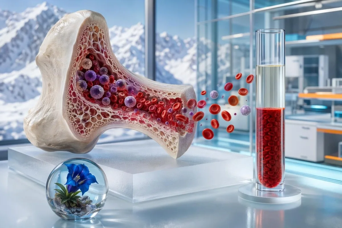
انخفاض الهيماتوكريت ماشي غير تشخيص واحد. النمط اللي كيدور حولو — الهيموغلوبين، عدد RBC، MCV، الريتيكولوسايت، الفيريتين، مؤشرات الكِلى وعلامات الترطيب — كيعطيك واش كاين نقص حقيقي فالكريات الحمراء ولا غير كاين تخفيف ديال الدم.


- انخفاض الهيماتوكريت كيعني أن كريات الدم الحمراء كتشغل نسبة أصغر من حجم الدم؛ الحدود ديال البالغين تقريباً 40-52% للرجال و36-46% للنساء اللي ماشي حاملين.
- فقر دم حقيقي غالبا كيكون ملي الهيماتوكريت والهيموغلوبين وعدد كريات الدم الحمراء كاملين منخفضين مع بعضهم، خصوصا إلا كانت كاينة أعراض.
- هيماتوكريت منخفض بسبب التخفيف ممكن يوقع بعد سوائل IV، شرب سوائل بزاف، الحمل ولا فرط الترطيب؛ الألبومين والصوديوم كذلك ممكن يهبطو.
- نزيف حديث ماشي بالضرورة كينقص الهيماتوكريت مباشرة؛ النقص كيبان أكثر غالبا بعد 6-24 ساعة حيث كيديرو تحولات فالسوايل.
- نقص الحديد غالبا كيبان MCV تحت 80 fL، فيريتين تحت 30 ng/mL، ولا تشبع الترانسفيرين تحت 20%.
- نقص B12 أو حمض الفوليك غالبا كيدفع MCV لفوق فوق 100 fL، مع B12 تحت 200 pg/mL ولا MMA فوق 0.40 µmol/L وهاد الشي كيدعم النقص.
- الخلايا الشبكية كيساعد يفصل بين نقص الإنتاج وفقدان الدم؛ مؤشر إنتاج الريتيكولوسايت تحت 2 كيشير لاستجابة ضعيفة من النخاع.
- الحمل كينقص الهيماتوكريت بسبب توسع البلازما؛ الهيموغلوبين تحت 11.0 g/dL فـ 1st ولا 3rd trimester غالبا خاصو مراجعة.
- أعراض عاجلة يشمل ألم الصدر، الإغماء، ضيق التنفس أثناء الراحة، براز أسود، أو نزيف مستمر وغزير، أو هيموغلوبين قريب من 7-8 g/dL.
شنو كيعني فعلا نتيجة انخفاض الهيماتوكريت
انخفاض الهيماتوكريت غالباً يعني واحداً من ثلاثة أشياء: عدد قليل جداً من كريات الدم الحمراء، فقدان حديث لكريات الدم الحمراء، أو زيادة في البلازما تُخفّف العينة. عملياً، لا أفسّر الهيماتوكريت وحده؛ أقارنه مع الهيموغلوبين، وعدد RBC، وMCV، وRDW، والريتيكولوسايت، والقصة حول سحب العينة. قيمة منخفضة بعد 2 لترات من سوائل IV تختلف عن قيمة منخفضة مع فيريتين 9 ng/mL. هذا الفرق يغيّر الفحص التالي، ومدى الاستعجال، والعلاج.
الهيماتوكريت هو نسبة حجم الدم الكامل التي تشغلها العناصر الخلوية الحمراء. هيماتوكريت 33% يعني أن حوالي ثلث حجم العينة عبارة عن عناصر خلوية وحوالي ثلثين عبارة عن بلازما، لذلك فإن التحولات في أي جهة من هذه المعادلة يمكن أن تغيّر النتيجة.
Kantesti هو منصة لتفسير تحليل الدم بالذكاء الاصطناعي تقرأ الهيماتوكريت إلى جانب باقي عناصر CBC بدل أن تعامل النسبة كتشخيص قائم بذاته. فريقنا السريري، كما هو موصوف في معلومات عنا, ، يرى نفس الإشكال العملي كل أسبوع: هيماتوكريت منخفض مُعلَّم قد يعني نقص الحديد، أو تخفيف السوائل الحاد، أو فسيولوجيا الحمل، أو نقص إنتاج مرتبط بالكلى، أو مشكلة في توقيت التحليل.
إرشادات منظمة الصحة العالمية لعام 2011 بشأن فقر الدم تُعرّف فقر الدم بالهيموغلوبين وليس بالهيماتوكريت: هيموغلوبين أقل من 13.0 g/dL عند الرجال، وأقل من 12.0 g/dL عند النساء غير الحوامل، وأقل من 11.0 g/dL أثناء الحمل يحقق معايير فقر الدم (WHO، 2011). إذا كنت تحاول فكّ بقية عناصر اللوحة، دليلنا إلى مكونات تحليل الدم الشامل يشرح أين يقع الهيماتوكريت داخل تحليل الدم الشامل.
الحدود الطبيعية ديال الهيماتوكريت كتختلف حسب الجنس، العمر والحمل
هيماتوكريت البالغين غالباً حوالي 40-52% عند الرجال و36-46% عند النساء غير الحوامل، رغم أن كل مختبر يحدد مجاله الخاص. نتيجة أقل بنقطة إلى نقطتين مئويتين فقط من المجال تكون عادة أقل إثارة للقلق من انخفاض مفاجئ بـ 6-10 نقاط عن خط أساسك المعتاد.
بعض المختبرات الأوروبية تستخدم مجالات أضيق قليلاً، وقد تُبلّغ مناطق المرتفعات عن هيماتوكريت أعلى متوقعاً لأن الضغط المزمن الأقل للأكسجين يحفّز إنتاج كريات الدم الحمراء. من تجربتي السريرية، غالباً ما تكون المقارنة الأكثر فائدة هي نتيجتك السابقة أنت، وليس المجال المطبوع بجانب رقم اليوم.
تختلف مجالات المختبر لأن الهيماتوكريت يتأثر بهرمونات الجنس، والارتفاع، والتدخين، والحمل، والترطيب، وطريقة جهاز التحليل. مقالنا حول نطاقات التحاليل الخاصة بالجنس يشرح لماذا قد تكون قيمة منخفضة بالنسبة لبالغ واحد طبيعية بالنسبة لآخر.
قاعدة عملية أستخدمها: الهيماتوكريت يساوي تقريباً ثلاثة أضعاف الهيموغلوبين عندما يتم الإبلاغ عنهما بوحدات تقليدية. إذا كان الهيموغلوبين 10.5 g/dL، فإن هيماتوكريت قريب من 31-32% يكون متسقاً داخلياً؛ وإذا كان الزوج غير متطابق بشكل كبير، أتحقق من الوحدات، وإشارات جهاز التحليل، وملاحظات العينة قبل افتراض وجود مرض.
مؤشرات فـ CBC كتفصل بين فقر الدم والتخفيف
فقر الدم الحقيقي يكون أكثر احتمالاً عندما يكون الهيماتوكريت والهيموغلوبين وعدد كريات الدم الحمراء منخفضين معاً. يكون التخفيف أكثر احتمالاً عندما ينخفض الهيماتوكريت بعد السوائل ويكون النمط مصحوباً بصوديوم منخفض-طبيعي، أو ألبومين منخفض، أو انخفاض مفاجئ في نفس اليوم دون تطابق مع الأعراض.
انخفاض عدد كريات الدم الحمراء مع انخفاض الهيموغلوبين عادة يشير إلى انخفاض حقيقي في القدرة على حمل الأكسجين. للحصول على شرح أعمق حول لماذا قد يختلف عدد RBC والهيموغلوبين أحياناً، انظر دليلنا إلى RBC مقابل الهيموغلوبين.
MCV و RDW كيدّوزو لطبقة اللي من بعد. MCV تحت 80 fL كيدلّ على صِغَر حجم الكريات (microcytosis)، و MCV فوق 100 fL كيدلّ على كِبَر حجم الكريات (macrocytosis)، و RDW فوق حوالي 14.5% كيعني تباين كبير فحجم الخلايا بما يكفي باش يدعم نقص كيتطور ولا أسباب مختلطة؛ ديالنا دليل RDW كيدخل فالنمط بالتفصيل أكثر.
Kantesti AI كفسّر الهيماتوكريت عبر التحقق واش العلاقة بين الهيموغلوبين والهيماتوكريت منطقية بيولوجياً، ومن بعد كيدور على إشارات متناقضة بحال تكتّل الصفائح، عمر العينة ولا دلتا مفاجئة مقارنة مع آخر CBC. هاد النوع ديال فحص النمط غالباً هو اللي كيمانع نتيجة تخفيفية خفيفة (dilutional) باش ما تتوسمّش خطأ كأنها فقر دم مزمن.
النزيف الأخير ممكن يختبي فالهيماتوكريت الأول
النزيف الحاد ماشي بالضرورة كينقص الهيماتوكريت مباشرة، حيث كريات الدم والبلازما كيتفقدو مع بعض فالبداية. الهيماتوكريت غالباً كينقص بشكل أوضح بعد 6-24 ساعة حيث السائل كيدخل للدم ولا كيتعطى سوائل وريدية (IV fluids).
شفت مرضى عندهم نزيف أنفي قوي وواضح ولا فقدان هضمي كيجيو بهيماتوكريت عادي بشكل مدهش، ومن بعد كيهبط من 39% حتى 31% فالصباح اللي من بعد. الرقم الأول ماكانش مزوّر؛ غير كان متسحب قبل ما إعادة توزيع البلازما تكشف النقص فعدد كريات الدم.
توقيت الدورة كاهم كذلك. مريضة عندها دورة قوية ممكن تبان عندها الهيموغلوبين 11.2 g/dL والهيماتوكريت 34% خلال الأسبوع اللي من بعد النزيف، فحيت الفيريتين كينقص من شهور؛ ديالنا على تحولات الهيموغلوبين المرتبطة بالدورة كشرح هاد الجدول الزمني.
النزيف الأخير كيكون أكثر مقلق ملي الهيماتوكريت كيهبط بأكثر من 3-5 نقاط مئوية من خط الأساس، النبض فوق 100 ضربة فالدقيقة، البراز كيبان أسود، البول كيبان أحمر بشكل واضح ولا كاين إغماء. إلا كانت القصة كتجي بحال كاين فقدان مستمر، انتظار شهر باش تعاود CBC غالباً ماشي هو الخطة الصحيحة.
نقص الحديد هو النمط الأكثر شيوعا ديال الهيماتوكريت المنخفض اللي كيدورو عليه الأطباء
نقص الحديد غالباً كيسبب هيماتوكريت واطي مع هيموغلوبين واطي، MCV واطي و RDW عالي. الفيريتين تحت 15 ng/mL كيتعتبر محدِّد بزاف لنقص مخزون الحديد، و الفيريتين تحت 30 ng/mL كيدل بقوة فحالات بزاف من البالغين.
إرشادات الجمعية البريطانية لأمراض الجهاز الهضمي (British Society of Gastroenterology) كتقول إن الفيريتين هو المؤشر الوحيد الأكثر فائدة لفقر الدم بسبب نقص الحديد، ولكن ممكن يبان طبيعي بشكل كاذب ملي كاين التهاب (Snook et al., 2021). فحالات الالتهاب، تشبع الترانسفيرين تحت 20% مع فيريتين تحت 100 ng/mL مازال ممكن يوافق إنتاج كريات دم محكومة بنقص الحديد.
الحديد فالمصل كيتأرجح خلال النهار وبعد الوجبات، لذلك نادراً ما كنستعمله بوحدو. إلا كان تقريرك كيبين الحديد فالمصل واطي، TIBC عالي وتشبع الترانسفيرين واطي، ديالنا حديد فالمصل منخفض و دليل دراسات الحديد يقدر يساعدك تفسّر هاد النمط.
فالمستعجلات/العيادة، كنطرح 3 أسئلة ماشي مزيانة ولكن مهمة ملي الهيماتوكريت واطي والحديد باين واطي: دورات قوية، التبرع بالدم، وفقدان هضمي. عند البالغين اللي فوق حوالي 50 سنة، فقر الدم الجديد بسبب نقص الحديد كيتطلب بحث يقوده طبيب/ة عن أسباب فالجهاز الهضمي حتى إلا كان النظام الغذائي كيبان معقول.
MCV مرتفع مع هيماتوكريت منخفض كيشير لمشاكل ديال B12 ولا الفولات
هيماتوكريت واطي مع MCV فوق 100 fL غالباً كيدلّ على نقص B12، نقص الفولات، تأثير الكحول، مرض الكبد، قصور الغدة الدرقية، ولا تداخل من دواء. B12 تحت 200 pg/mL غالباً كيدعم النقص، بينما 200-300 pg/mL كيبقى منطقة حدّية حيث MMA يقدر يوضح.
حمض الميثيل مالونيك (Methylmalonic acid) فوق حوالي 0.40 µmol/L كيدعم نقص B12 الوظيفي، خصوصاً ملي الأعراض كتضمن تنميل، مشاكل فالتوازن، حريق فالرجلين، ولا تغيّر فالميموري/الذاكرة. الهوموسيستين يقدر يرتفع فكل من نقص B12 ونقص الفولات، لذلك ماشي محدِّد بوحدو.
مريض ممكن يكون عندو أعراض نقص الهيموغلوبين قبل ما MCV تولّي عالية بزاف. شفت نقص B12 مع MCV ديال غير 94 fL ملي كان كاين كذلك نقص الحديد، حيث microcytosis وmacrocytosis كيلغيو بعضهم جزئياً فالمتوسط.
إلا كان B12 ديالك حدّي/بين بين، قارن الوحدات بعناية حيث pg/mL و pmol/L ماشي قابلين للتبديل. ديالنا دليل نطاق فيتامين ب12 كشرح هاد الحدود وليش MMA مرات كيهم أكثر من رقم B12 اللي باين فالعنوان.
مرض الكِلى والالتهاب كينقصو إنتاج كريات الدم الحمراء
هيماتوكريت واطي بسبب نقص الإنتاج غالباً كيبان مع استجابة reticulocyte واطية، MCV طبيعي ولا صغير، وعلامات التهاب مزمن أو ديال الكِلى. فقر الدم المرتبط بالـ CKD كيبدا يكون أكثر شيوعاً ملي eGFR كينقص تحت 60 mL/min/1.73 m² وكيكون شائع بزاف خصوصاً تحت 30.
الكلي تنتج إريثروبويتين، الإشارة الهرمونية التي تخبر نخاع العظم بإنتاج خلايا الدم الحمراء. توصي إرشادات KDIGO لالتهاب فقر الدم بتقييم حالة الحديد، B12، الفولات والالتهاب قبل عزو فقر الدم إلى مرض الكلي المزمن وحده (KDIGO، 2012).
الالتهاب يغيّر طريقة تدبير الحديد عبر رفع الهيبسيدين، مما يحبس الحديد في مواقع التخزين حتى عندما لا يكون الحديد الكلي في الجسم غائبًا فعليًا. لهذا قد تكون الفيريتين 150 نغ/مل والتشبع بالترانسفيرين 14% في فقر الدم بسبب الالتهاب — كمية كافية من الحديد المخزّن، لكن توصيله ضعيف.
Kantesti هي أداة لتحليل فحوصات الدم مدعومة بالذكاء الاصطناعي، تقرأ الهيماتوكريت إلى جانب eGFR، الكرياتينين، CRP، الألبومين ومؤشرات الحديد. إذا كانت أرقام الكِلى جزءًا من نمطك، فإن دليل مراحل CKD يقدّم سياق eGFR و ACR للبول الذي يحتاجه المرضى غالبًا.
الريتيكولوسايت كيبينو واش النخاع كيردّ
الشبكيات (Reticulocytes) هي كريات دم حمراء شابة، وتخبرنا إن كان نخاع العظم يحاول التعويض. في فقر الدم، إذا كان مؤشر إنتاج الشبكيات أقل من 2 فهذا يشير إلى نقص الإنتاج، بينما مؤشر يفوق 3 يشير إلى فقد حديث أو انحلال دم إذا كان النخاع سليمًا.
يُشتبه في الانحلال الدموي عندما يقترن انخفاض الهيماتوكريت بارتفاع الشبكيات، وارتفاع LDH، وارتفاع البيليروبين غير المباشر، وانخفاض الهابتوغلوبين. انخفاض الهابتوغلوبين إلى أقل من حوالي 25 مغ/دل يدعم تكسّر كريات الدم الحمراء، رغم أن أمراض الكبد قد تُخفض الهابتوغلوبين أيضًا وتُعقّد القراءة.
اختبار الأجسام المضادة المباشرة، ويُسمّى أحيانًا DAT أو اختبار كومبس، يساعد على كشف الانحلال الدموي المناعي. غالبًا ما يلاحظ المرضى اصفرارًا، بولًا داكنًا، انزعاجًا في الظهر أو تعبًا مفاجئًا، لكن الانحلال الدموي الخفيف قد يكون هادئًا بشكل مدهش.
عندما أراجع هذا النمط، أبحث عن المجموعة الكاملة بدل قيمة واحدة لافتة. دليلنا إلى نتائج الهابتوغلوبين ومراجعتنا الدموية إلى مؤشرات الرتِيكولوسايت تشرح لماذا يكون الجمع أكثر موثوقية من أي مؤشر منفرد.
فرط الترطيب وسوائل IV ممكن يخففو الهيماتوكريت
يحدث انخفاض الهيماتوكريت بسبب التخفيف عندما يتوسع حجم البلازما أسرع من تغيّر كتلة كريات الدم الحمراء. في سجلات المستشفى، يمكن لـ 1-2 لتر من سوائل البلّوريات (crystalloid) أن تُخفض الهيماتوكريت بنحو 2-5 نقاط مئوية دون أي فقد جديد لكريات الدم الحمراء.
الدلائل ليست خفية إذا عرفت أين تبحث: ينخفض الهيماتوكريت بسرعة، قد ينخفض الألبومين، قد يميل الصوديوم للانخفاض، وقد يكون المريض قد تلقّى سوائل، أو رعاية حمل، أو إعادة ترطيب بعد حدث تحمّل، أو علاجًا للقيء. يبدو الـ CBC أسوأ، لكن توصيل الأكسجين قد لا يكون تغيّر بقدر ما توحي به النسبة.
الرياضيون ذوو التحمل حالة خاصة. بعد فترات تدريب طويلة، قد يؤدي توسع حجم البلازما إلى هيماتوكريت منخفض-طبيعي يبدو كأنه فقر دم على الورق، بينما يبقى الأداء مستقرًا ويكون الفيريتين مقبولًا؛ مؤشرات الترطيب مثل تركيز البول تضيف سياقًا.
إذا كان ميزان السوائل جزءًا من سؤالك، قارن الـ CBC مع الصوديوم، الألبومين، BUN/اليوريا وتركيز البول. دليلنا إلى أسمولالية البول مفيد عندما يكون السؤال جفافًا، فرط ترطيب أو صورة مختلطة للسوائل.
الحمل كينقص الهيماتوكريت بسبب التوسع الطبيعي ديال البلازما
غالبًا ما يُنقص الحمل الهيماتوكريت لأن حجم البلازما يرتفع بحوالي 40-50%، بينما ترتفع كتلة كريات الدم الحمراء بنحو 20-30%. يُتوقع هذا التخفيف الفسيولوجي، لكن الهيموغلوبين أقل من 11.0 غ/دل في الثلث الأول أو الثالث ما يزال يستحق المراجعة.
الثلث الثاني هو حيث تبدو القيم غالبًا في أدنى مستوياتها. يستعمل كثير من الأطباء الهيموغلوبين أقل من 10.5 غ/دل في الثلث الثاني كعتبة لفقر الدم، رغم أن البروتوكولات التوليدية المحلية تختلف، وغالبًا ما يُفحص الفيريتين مبكرًا الآن.
هيماتوكريت ما بعد الولادة أكثر تعقيدًا لأن الفقد المرتبط بالولادة، والـ IV fluids، والالتهاب تتداخل كلها. قد تكون لدى المرأة هيماتوكريت 29% صباح اليوم التالي للولادة، ثم 32% بعد أسبوع دون أي إعطاء للحديد فقط لأن ميزان السوائل عاد إلى طبيعته.
اعتبارًا من 15 يوليوز 2026، نهجي العملي هو التعامل مع هيماتوكريت الحمل كاتجاه مع الأعراض، لا كإنذار واحد أحمر. دليلنا إلى تحاليل الدم للحمل يغطي أنماط التحذير في نفس اليوم التي لا ينبغي انتظارها إلى حين المتابعة الروتينية.
أعراض الهيماتوكريت المنخفض كتعلق بالسرعة ديال حدوثه، ماشي غير بالرقم
أعراض انخفاض الهيماتوكريت غالبًا تنجم عن نقص توصيل الأكسجين وتشمل التعب، ضيق النفس، الدوخة، الخفقان، الصداع وشحوب الجلد. قد يبدو الانخفاض البطيء إلى هيموغلوبين 9.5 غ/دل خفيفًا، بينما قد يبدو الانخفاض السريع إلى 10.5 غ/دل مخيفًا.
أعراض نقص الهيموغلوبين كتولي أكثر احتمال ملي كينقص الهيموغلوبين لّـي تحت 10 غ/ديسيلتر، خصوصا أثناء الرياضة ولا طلوع السلالم. تحت 8 غ/ديسيلتر، بزاف ديال البالغين كيحسو بضيق فالتنفس، الضعف ولا الدوخة الخفيفة، والناس اللي عندهم مرض فالقلب ممكن يبان ليهم ضغط فالصدر فمستويات أعلى.
الرايات الحمراء اللي ما نْحبّش اللي كتْتْجاهل هي: الإغماء، ضيق فالتنفس حتى ورا الراحة، ألم فالصدر، تشوش، براز كحل، نزيف قوي مستمر، نبض فوق 120 ضربة فالدقيقة ولا تشبع الأكسجين تحت المستوى المعتاد ديال المريض. هادشي كيعني علامات لمراجعة طبية فنهار نفسو، ماشي علامات لتعديل نمط العيش.
الأعراض كتتداخل مع مشاكل الغلوكوز، الغدة الدرقية، الملح، وإيقاع القلب. إلا كان الشكوى هي الدوخة ماشي الراية ديال التحليل، دليلنا ديال تحاليل الدم ديال الدوخة كْيشرح علاش CBC غير جزء واحد من التقييم.
إعادة التحليل للهيماتوكريت المنخفض خاصها تطابق السبب اللي كيتشتبه فيه
نقص خفيف فالهيماتوكريت غالبا تقدر تعاود التحقق منه فـ 1-4 أسابيع إلا كنتي حاسّ براسك مزيان وباقي CBC ثابت. إلا كان كاين هبوط سريع، أعراض، رايات حمراء ديال الحمل، نزيف مشتبه، ولا الهيموغلوبين قريب من 7-8 غ/ديسيلتر، خاصك نصيحة طبية فنهار نفسو.
من بعد شرب سوائل بكثرة، سوائل وريدية (IV) ولا مرض حاد صعيب، إعادة CBC بعد 48-72 ساعة تقدر تبين واش النقص فالهيماتوكريت كان مؤقت بسبب التخفيف. من بعد ما كتبدأ الحديد الفموي، عادة كنْتوقع ارتفاع فالهيموغلوبين بحوالي 1-2 غ/ديسيلتر خلال 3-4 أسابيع إلا كان الامتصاص والتشخيص صحيحين.
الهيماتوكريت غير الطبيعي بوحدة واحدة خاصو يتقارن مع النتيجة اللي قبلها، ملاحظات العينة والوحدات. دليلنا ديال إعادة فحص التحاليل غير الطبيعية كْيشرح علاش بعض النتائج خاصها تأكيد سريع، فحيت شي نتائج تقدر تتْتْبع بأمان مع الوقت.
فحوصات الفرق (Delta checks) مهمة. هبوط من هيماتوكريت 42% حتى 36% فشي أسبوع يستاهل انتباه أكثر من شخص عاش عند 35-36% لسنوات؛ مقالنا ديال تغيّرات مفاجئة في التحاليل كيبين كيفاش المختبرات والأطباء كْيفكرو فالتغييرات اللي ما كانتش متوقعة.
كيفاش Kantesti كيقرا أنماط الهيماتوكريت المنخفض
Kantesti كْيقرأ الهيماتوكريت ناقص من خلال جمع مؤشرات CBC، مؤشرات الحديد، وظائف الكلى، مؤشرات الالتهاب، دلائل الترطيب، الجنس، حالة الحمل، والاتجاهات اللي كانت من قبل. الهدف ماشي هو التشخيص من رقم واحد؛ الهدف هو تحديد أكثر نمط محتمل، وأأمن خطوة من بعد.
Kantesti هي منصة ديال تفسير مؤشرات حيوية (biomarker) بالذكاء الاصطناعي كتقدر تعالج PDFs ديال تحاليل الدم اللي كتْرفع ولا صور فحوالي 60 ثانية، بما فيها CBC حيث الهيماتوكريت هو اللي طلع كراية. الشبكة العصبية ديالنا كتفحص أكثر من 15,000 مؤشر حيوي، ولكن القيمة السريرية كتجي من التوليفات — مثلا، هيماتوكريت 33%، فيريتين 7 ng/mL و MCV 76 fL ماشي نفس القصة ديال هيماتوكريت 33%، ألبومين 30 g/L و صوديوم 130 mmol/L.
أطبائنا كْيْراجعو المنطق السريري اللي مستعمل فالنظام، والمنهجية موصوفة فـ دليل التكنولوجيا. بالنسبة للهيماتوكريت، النموذج كْيْوزن الشدة، اتجاه التغيير، التناسق الداخلي ديال CBC، تحويلات الوحدات، وواش مؤشرات أخرى كْتدعم نقص الحديد، الماكروسيطوز (macrocytosis)، نقص الإنتاج المرتبط بالكلى، ولا التخفيف.
الدقة فالشغل ماشي غير ادعاء مرة وحدة. عملية التحقق ديال Kantesti، بما فيها الفحص اعتمادا على Rubric (قواعد تقييم) والإشراف الطبي، مْلخّصة فـ التحقق الطبي ; ومن وجهة نظري كمّا Thomas Klein, MD، هادي هي اللي فيها الذكاء الاصطناعي كسب الثقة ولا كْيخسرها.
ملاحظات ديال البحث ومتى خاصك تجيب النتائج لطبيب
جِيب نتيجة هبوط فالهيماتوكريت لطبيب/ة بسرعة إلا كانت القيمة جديدة، كتهبط، فيها أعراض، مرتبطة بالحمل، مرتبطة بنزيف مشتبه، ولا كانت مْقترنة مع خلايا بيضاء ولا صفائح غير طبيعية. هبوط خفيف ومْستقر فالهيماتوكريت مازال خاصو تفسير، ولكن الإطار الزمني كيكون مختلف.
Thomas Klein, MD، وفريق Kantesti الطبي كينصحوا المرضى يْجيبوا CBC كامل، النتائج اللي قبل، وقائمة الأدوية، ماشي غير سكرينشوت ديال الهيماتوكريت بوحدو. إلا احتجتي مسارات متابعة بشرية ولا تفاصيل الحكامة الطبية، صفحةنا ديال المجلس الاستشاري الطبي كْتشرح الإشراف ديال الطبيب اللي وراء محتوانا.
Kantesti LTD. (2026). إطار التحقق السريري v2.0. Zenodo. DOI: 10.5281/zenodo.17993721. لائحة ResearchGate: سجل التحقق السريري; إدراج Academia.edu: سجل إطار التحقق (validation).
Kantesti LTD. (2026). محلل تحليل الدم بالذكاء الاصطناعي: تم تحليل 2.5M اختبار | التقرير العالمي للصحة 2026. Zenodo. DOI: 10.5281/zenodo.18175532. لائحة ResearchGate: تقرير الصحة العالمية; إدراج Academia.edu: تقرير محلل AI.
إلا كان الهيماتوكريت ديالك منخفض وما قادرش تميّز واش المشكل هو فقر الدم ولا نزيف ولا تخفيف بالسوائل، ما تحاولش تصلحو بالحديد بشكل عشوائي. استعمل النمط، وطلب التحاليل الناقصة و تواصل مع طبيبك؛ إلا كان محتاج مساعدة إدارية، ديال Kantesti’s اتصل بنا هو الطريق الأكثر أماناً من التخمين اعتماداً على نسبة وحدة معلّمة.
الأسئلة الشائعة
ما هي أكثر الأسباب شيوعًا لانخفاض الهيماتوكريت؟
أكثر الأسباب الشائعة لانخفاض الهيماتوكريت هي فقر الدم بسبب نقص الحديد، نزيف حديث، تخفيف الدم المرتبط بالحمل، الالتهاب المزمن، أمراض الكلى، نقص فيتامين B12 أو حمض الفوليك، وزيادة السوائل. غالبًا ما يُعتبر هيماتوكريت البالغين منخفضًا عندما يكون أقل من حوالي 40% عند الرجال وأقل من حوالي 36% عند النساء غير الحوامل، لكن نطاقات التحاليل تختلف. النمط مهم: انخفاض الهيموغلوبين وانخفاض عدد RBC يشيران إلى فقر دم حقيقي، بينما الانخفاض المفاجئ بعد السوائل الوريدية قد يكون بسبب التخفيف.
هل شرب الكثير من الماء يمكن أن يُنقص الهيماتوكريت؟
نعم، الإكثار من شرب السوائل بكثرة أو إعطاء سوائل وريدية يمكن أن يُنقِص مؤقتاً الهيماتوكريت عبر توسيع حجم البلازما. في الممارسة السريرية، قد تُنقِص 1-2 لترات من السوائل البلورية الهيماتوكريت بحوالي 2-5 نقاط مئوية دون حدوث فقد جديد في كريات الدم الحمراء. يكون التخفيف أكثر احتمالاً عندما تظهر أيضاً الصوديوم أو الألبومين أو BUN/اليوريا منخفضة، ويعود التحليل إلى طبيعته خلال 48-72 ساعة.
ما هي أعراض انخفاض الهيماتوكريت التي ينبغي أن تقلقني؟
أعراض انخفاض الهيماتوكريت التي تستدعي مراجعة فورية تشمل ضيق النفس عند الراحة، ألم الصدر، الإغماء، الارتباك، براز أسود، نزيف مستمر وغزير، أو نبض يفوق 120 نبضة في الدقيقة. التعب الخفيف والدوخة شائعان عندما ينخفض الهيموغلوبين إلى أقل من حوالي 10 غ/دL، لكن الأعراض تعتمد بشكل كبير على مدى سرعة حدوث الانخفاض. غالبًا ما يُعالج الهيموغلوبين القريب من 7-8 غ/دL على أنه حالة عاجلة، خصوصًا لدى كبار السن أو الأشخاص الذين يعانون من أمراض القلب.
هل يمكن أن يكون الهيماتوكريت منخفضًا بينما يكون الهيموغلوبين طبيعيًا؟
يمكن أن يكون الهيماتوكريت منخفضًا قليلًا بينما يبقى الهيموغلوبين طبيعيًا، خصوصًا قرب مجال المرجع أو عندما يتغير وضع الترطيب. عادةً يكون الهيماتوكريت حوالي ثلاثة أضعاف الهيموغلوبين، لذلك غالبًا ما يقترن هيموغلوبين قدره 12.5 g/dL بهيماتوكريت حوالي 37-38%. ينبغي أن يدفع عدم التطابق الكبير إلى التحقق من الوحدات، وجودة العينة، إشارات جهاز التحليل، وما إذا كانت حَجْم البلازما قد تغيّر حول وقت الفحص.
كم بعد ذلك يجب أن أكرر فحص الهيماتوكريت المنخفض؟
يمكن غالبًا إعادة فحص الهيماتوكريت المنخفض بشكل خفيف وغير متوقع بعد 1-4 أسابيع إذا كنتِ/كنتِ بصحة جيدة وباقي نتائج CBC مستقرة. إذا كانت النتيجة جاءت بعد تلقي سوائل وريدية، ترطيب قوي أو مرض حاد، فإن إعادة CBC بعد 48-72 ساعة قد توضح التخفيف. أعد الفحص في وقت أقرب أو اطلب رعاية في نفس اليوم إذا انخفض الهيماتوكريت بأكثر من 3-5 نقاط مئوية مقارنةً بالأساس، أو إذا كان الهيموغلوبين قريبًا من 7-8 غ/ديسيلتر، أو إذا كانت الأعراض مهمة.
هل الحمل دائمًا يسبب انخفاض الهيماتوكريت؟
الحمل غالبًا يُنقِص الهيماتوكريت لأن حجم البلازما يرتفع بحوالي 40-50%، بينما كتلة كريات الدم الحمراء ترتفع بحوالي 20-30%. كثير من الحوامل يكون لديهن هيماتوكريت حوالي 33-39% دون فقر دم خطير، خصوصًا في الثلث الثاني من الحمل. الهيموغلوبين أقل من 11.0 غ/دل في الثلث الأول أو الثالث، أو أقل من حوالي 10.5 غ/دل في الثلث الثاني، عادةً يستدعي تناول الحديد ومراجعة سريرية.
شنو هي التحاليل اللي كيساعدو يفسرو نقص فعدد كريات الدم الحمراء؟
أكثر التحاليل فائدة لأسباب انخفاض عدد كريات الدم الحمراء هي الهيموغلوبين، الهيماتوكريت، MCV، RDW، عدد الخلايا الشبكية، الفيريتين، تشبع الترانسفيرين، B12، الفولات، الكرياتينين/eGFR، CRP وأحيانًا الهابتوغلوبين، LDH والبيليروبين. MCV أقل من 80 fL يشير إلى أسباب صغيرة الكريات مثل نقص الحديد، بينما MCV أعلى من 100 fL يشير إلى أسباب كبيرة الكريات مثل نقص B12 أو الفولات. مؤشر إنتاج الخلايا الشبكية أقل من 2 يشير إلى نقص في الإنتاج، بينما استجابة أعلى تشير إلى فقد حديث أو انحلال الدم.
هل يمكن أن يؤخر انخفاض الهيماتوكريت إجراءً أو جراحة اختيارية؟
محتمل. قبل إجراء اختياري، تأخذ فرق الجراحة والتخدير في الاعتبار الهيماتوكريت، الهيموجلوبين، الأعراض، سبب فقر الدم، وفقدان الدم المتوقع؛ تصحيح نقص قابل للعلاج مسبقًا قد يقلل المخاطر ويتجنب التأخير.
احصل على تحليل الدم بالذكاء الاصطناعي اليوم
انضم إلى أكثر من 2 مليون مستخدم عالمي يثقون في Kantesti لتحليل فوري ودقيق لنتائج التحاليل المخبرية. ارفع نتائج تحليل الدم الخاصة بك واحصل على تفسير شامل لـ 15,000+ للـ biomarkers في ثوانٍ.
📚 أبحاث منشورة مُشار إليها
Klein, T., Mitchell, S., & Weber, H. (2026). Kantesti LTD. (2026). الإطار التنظيمي السريري v2.0. Zenodo. https://doi.org/10.5281/zenodo.17993721. Kantesti بحث طبي بالذكاء الاصطناعي.
Klein, T., Mitchell, S., & Weber, H. (2026). Kantesti LTD. (2026). مدجج تحليل الدم بالذكاء الاصطناعي: تحليل 2.5 مليون اختبار | تقرير الصحة العالمي 2026. Zenodo. https://doi.org/10.5281/zenodo.18175532. Kantesti بحث طبي بالذكاء الاصطناعي.
📖 مراجع طبية خارجية
منظمة الصحة العالمية (2011). تراكيز الهيموغلوبين لتشخيص فقر الدم وتقييم شدة الحالة. نظام معلومات التغذية بالفيتامينات والمعادن لدى منظمة الصحة العالمية.
فريق عمل فقر الدم لدى KDIGO (2012). إرشادات الممارسة السريرية لـ KDIGO لفقر الدم في مرض الكلى المزمن. ملحقات Kidney International.
⚕️ إخلاء مسؤولية طبية
هذه المقالة لأغراض تعليمية فقط ولا تشكل نصيحة طبية. استشر دائمًا مقدم رعاية صحية مؤهلًا لاتخاذ قرارات التشخيص والعلاج.
إشارات الثقة E-E-A-T
خبرة
مراجعة سريرية يقودها الأطباء لسير عمل تفسير التحاليل.
خبرة
تركيز طب المختبر على كيفية سلوك الـ biomarkers في السياق السريري.
السلطة
مكتوب من طرف الدكتور Thomas Klein مع مراجعة من طرف الدكتورة Sarah Mitchell والأستاذ الدكتور Hans Weber.
الجدارة بالثقة
تفسير قائم على الأدلة مع مسارات متابعة واضحة لتقليل الإنذار.