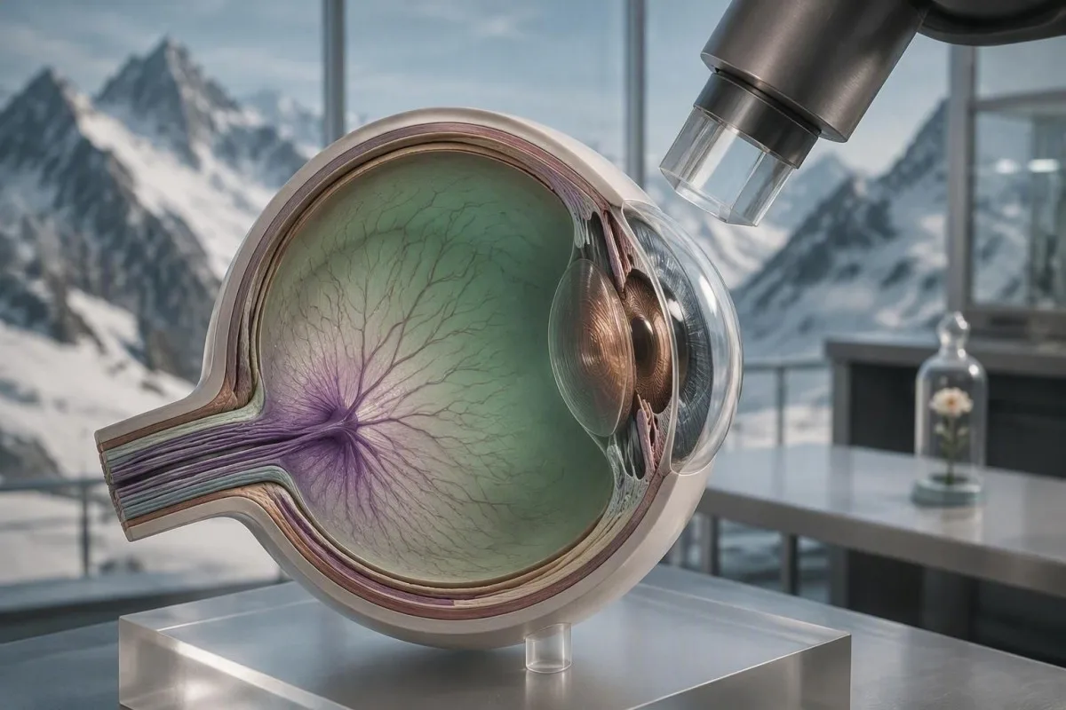
بىر قېتىملىق نورمال بېسىم ئۆلچىمى گلۇكومانى رەت قىلمايدۇ. كۆز مۇتەخەسسىسلىرى ۋاقىت بويىچە بېسىم، ئوﭘتېك-ئەسەب قۇرۇلمىسى ۋە كۆرۈش ئىقتىدارىنى سېلىشتۇرۇش ئارقىلىق گلۇكومانى دىئاگنوز قويىدۇ ۋە نازارەت قىلىدۇ.


- نورمال كۆز بېسىمى ئادەتتە 10-21 mmHg بولىدۇ، ئەمما گلۇكوما 12-18 mmHg بېسىمدا پەيدا بولىدۇ.
- تونومېتىرىيە خىرىس ئامىلىنى ئۆلچەيدۇ، ئوﭘتېك-ئەسەب زىيىنىنى ئەمەس؛ بىر قېتىملىق ئۆلچەش بىر كلىنىك رەسىمى.
- OCT كۆز سىنىقى رېتىنال ئۆسۈملۈك توكاي قاتلىمىنىڭ نېپىللىشىشىنى بايقايدۇ، كىشىلەر كۆرۈش يوقىلىشنى ھېس قىلىشتىن بۇرۇن.
- كۆرۈش مەيدانى سىنىقى نەتىجىلىرى maysa, takrorlanıdıghan nuqsanlar bir yaki ajra yotqa chüshken noqitidin muhim.
- Tereqqiyat tek bir cheti qatnashqan xatidin emes, balki serial skaner we meydanlardiki tendensiyidin diaqnoz qilinidu.
- Awaz qalinliqi klinikichilarni qan bistimini chüshindüridighan usulni özgertidu: nazuk awazlar Goldmann oxshash netijisini xataliq bilen pes qilaalaydu.
- جىددىي ئالامەتلەر awazli, qizil göz, halolar, küykenglik yaki küchlü qisa köridighan ashular kündüzlük gij gijlikni tekshüridighan halettini körsitidu.
نېمىشقا نورمال بېسىم ئۆلچىمى گلۇكومانى رەت قىلمايدۇ
Glaukoma sinimini tekshürüsh normal qan bistimini alghandin keyin glaukumini istisna qilalmighaydu. Optik nerw ziyan bolghan köpligen ademlerde ichki qan bistimi yaki IOP 10 we 21 mmHg ariliqida ölçilgen; bu toghra yaki normal qan bistimi bolup, toghra kliniki weziyiti qollap tursa.
21 mmHg qan bistimi statistik bir konventsiya, biologiyelik bir qutulush sizighi emes. Ocular Hypertension Treatment Study'da davalash birinchi derejilik achiq burchakli glaukoma bashlinishidin 9.5%'din 4.4%'ge tereqqiyat bashlinidighanlarning 5 yilliq yaki qan bistimi yuqiri bolghanlarning tereqqiyatini bilishke bolmaydu (Kass et al., 2002).
IOP saet boyiche, pozitsiye boyiche, awaz xüsusiyiti we hatta ölçish usuli bilen özgiridu. 10:00'diki 15 mmHg klinik qiymiti,QWidget 24 mmHg'din yuqiri bolghan, aynisa uyquda apnoyesi, qan tomur disregulatsiyisi yaki davalanmighan ocular hypertension bolghan ademlerde, tunide peydigha chiqishini yitkizip qoyalaydu.
Doktor Thomas Klein IOP'si 16 mmHg bolghan ademlerge yillap rahatliq bergen, ünki, bir rutinal surat otturisi nerw stakini qalinlap körgendin keyin, uni tawlap bergen. Paydiliq sorighilining özgirelishi “mening nomrim normalmu?” degenni emes, balki “bu qan bistimi mening nerwimge, awazimgha, skanerimge, meydinimge, yashigha we xeter riska toghra kilimdu?” degenni öz ichige alidu. Qanda oqing normal lab bayraqliri aldamchi bolalaydu.
Normal-tension glaucoma nadir emes
Normal-tension glaucoma bashqa bir qisim qisim bolghachqa, nerwde qan ketish, rimning nazuklishishi, asimmetriya yaki tughrisigha kelidighan meydan nuqsanining war bolghanda, saghlam körinidighan qan bistimi netijisi hech qachanda bahalashni toxtitmaydu. Ehli Asiyada normal-tension ishliri kop bolghan bolup, emma bu diaqnoz her bir irqiy gruppada uchraydu.
تونومېتىرىيە نېمە ئۆلچەيدۇ ۋە بۇ ساننىڭ مەنىسى نېمە
Tonometri köz ichi qan bistimini tahmin qilidu, köpinche millimetr civa (mmHg) bilen. Goldmann applanation tonometry köp közinikida referens usul bolup qalaydu, emma air-puff we rebound quralliri paydiliq sinaw qollanmisidur, emma az közde álmashturghili bolmaydu.
Köp xatun-qizlar üchün, ölçilgen IOP 10 we 21 mmHg ariliqida bolidu. 21 mmHg'din yuqiri netijiler nerw we burchakni bahalashqa layiq; 30 mmHg'din yuqiri netijiler köpinche tezroq tekshürishke mecburi qilidu, emma gij gijlikke tesir qilishi, burchak xoliti we nerw körinishi bilen alaqidar.
Sözneng qimirlashidin, naptini tutushidin, boyinni qattiq qisishidin yaki nazuk awaz arqiliq ölçüshidin peydige chiqqan netije waqtiche yuqiri bolushi mumkin. Kontaktlinzini élip tashlash, fluorescein muqdari we examinerning usuli muhim – 2-3 mmHg'lik bir özgirelishini sewebdin tereqqiyat dep atalmasliqini ispatlaydighan kichik detallar.
كانتېستى بىر AI قان تەكشۈرۈش ئانالىزچىسى, shunga qan ishi bilen glaukumini diaqnoz qilmaydu yaki közni tekshürüshni almashturmaydu. Rolimiz köpek cheklengen we dürüst: bizning longitudinal usulimiz ademlerge nega klinikichilarning bir izolyatsiyilangan netijisige asaslanmastin, tekrarlighan ölçülerni tengleshkenlikini chüshinishke yardem beridu; bizning يۈزلىنىش-تەھلىل يېتەكچىمىزنىڭ ئوخشاش رامكىسى.
نېمىشقا ئەۋرىشىملىك قېلىنلىقى بېسىم ئىزاھلىنىشىنى ئۆزگەرتىدۇ
مېڭە سېلىنچاقلىق قېلىنلىقى ئېنىقلىق بېسىمىنىڭ ئىشەنچلىكلىكىنى ئۆزگەرتىدۇ. نېپىز مېڭە سېلىنچاقلىق ئۆلچەنگەن IOP نى تۆۋەن باھالايدۇ، قېلىن مېڭە سېلىنچاقلىق ئۇنى ئاشۇرىدۇ، شۇڭا ئېنىقلىق پېچەتلەش مۇھىم پەردە توسۇلۇش دەسلەپكى تەتقىقاتىدا بار.
ئوتتۇرىچە مېڭە سېلىنچاقلىق قېلىنلىقى تەخمىنەن 540 مىكرو ئۆلچەش، گەرچە نورمال ئوخشاش بولمىغان خىللار بار. OHTS دا، 555 مىكرو ئۆلچەش ياكى ئۇنىڭدىن تۆۋەن مېڭە سېلىنچاقلىق 588 مىكرو ئۆلچەشتىن يۇقىرى بولغانغا قارىغاندا، تېخىمۇ چوڭ ئۆزگىرىش خەتىرى بىلەن مۇناسىۋەتلىك بولغان؛ دوختۇرلار بۇ قىممەتنى خەتەرلىك مەزمۇن سۈپىتىدە ئىشلىتىدۇ، پەقەت سىمماب سېنتىمېتىرنى تۈزەشتەك ئاددىي ھېسابلاش ئەمەس.
پەقەت مېڭە سېلىنچاقلىق قېلىنلىقىدىنلا 14 مىللىمېتىر سىمماب سېنتىمېتىرنى “ھەقىقىي” بېسىمگە ئايلاندۇرىدىغان ئۇنىۋېرسال قوبۇل قىلىنغان فورمۇلا يوق. مېڭە ئىقتىدارى، ھىستېرىز قوشۇلغاندا، ئۆلچەشگىمۇ تەسىر قىلىدۇ، خاتا تۈزەش فاكتورى خاتا ئىشەنچ پەيدا قىلالايدۇ.
پەقەت “نېپىز” ياكى “قېلىن” ئەمەس، بەلكى رېئال مېڭە سېلىنچاقلىق رەقىمىنى سوراپ بېقىڭ. بۇ ئوخشاش چېگرالىق كرېئىتىن نەتىجىسى: مەزمۇن مەنىسىنى ئۆزگەرتىدۇ، ئەمما يۆلنىشىنى كۆزىتىش زۆرۈرلۈكىنى يوقىتىلمايدۇ.
مېڭە ھىستېرىزى قوشۇمچە بىر ئىشارەت قوشىدۇ
تۆۋەن مېڭە ھىستېرىزى كۆزىتىش خىزمىتىدە تېز پەردە توسۇلۇش تەرەققىياتى بىلەن مۇناسىۋەتلىك بولغان، بەلكىم ئۇ قېلىنلىقتىن سىرت تىببىي ھەرىكەتنى ئەكس ئەتتۈرىدۇ. ئۇ بار بولسا پايدىلىق، ئەمما ئۇ پەردە توسۇلۇش دىياگنوزى قويۇش ياكى مۇۋاپىق داۋالاشنى باشلاش ئۈچۈن تەلەپ قىلىنمايدۇ.
كلىنىك خادىملىرى OCT دىن بۇرۇن ئوﭘتېك ئەسەبنى قانداق تەكشۈرىدۇ
ئۆپكە تارىيىپىنى تەكشۈرۈش، رېتېنال rim نىڭ يوقلىقىنى، سىملىرىنى، دىسكا قان تومۇرىنى ۋە دىسكا بىلەن ماس كەلمەيدىغان كۆپەيتىلگەن شەكىلنى تەكشۈرىدۇ. dilatation stereoscopic examination and disc photographs are clinically valuable even when OCT is available.
دىسكىنىڭ ھالقىلىق-مەركىزىي نىسبىتى چوڭ دىسكىدا نورمال بولسا 0.7 بولىدۇ، كىچىك دىسكىدا 0.5 بولسا گۇمانلىق بولىدۇ. پەقەت ھالقىنىڭ چوڭلۇقىدىن تېخىمۇ ئەندىشىلىنەرلىك ئىشلار، فوكالىي گىرۋەكلىرىنىڭ يېرىلىشى، كۆزلەر ئارىسىدىكى 0.2 دىن ئېشىپ كەتكەن ئۆزگىرىشچان سىممېتىرىيە ياكى گىرۋەك ئەتراپىدىكى تالالىق قاناش.
دىسكىدىكى قاناش بولسا ئالاھىدە ئەمەلىي ئىش، چۈنكى ئۇ كۆرۈنەرلىك كۆرۈش مەيدانىنىڭ تۆۋەنلىشىدىن بۇرۇن بولىدۇ. مەن بىرىنى تاپقاندا، مەن بىمارنىڭ داۋالاشتىن “ۋاز كېچىپ كەتكەنلىكىنى” ئويلىمايمەن؛ مەن خىزمەتنى تەكشۈرىمەن، سۈرەتكە ئېلىشنى تەكرارلايمەن، قان تومۇر فاكتورلىرىنى تەكشۈرىمەن، ۋە كۈزەتكۈچىنى قىسقارتىمەن — كۆپىنچە 12 ئۆلچەملىك 3-4 ئايغا.
كانتېستى بىر AI قان تەكشۈرۈش نەتىجىسىنى چۈشەندۈرۈش سۇپىسى laboratory-report context, not retinal image diagnosis. People using health reports should still take eye-specific concerns to an optometrist or ophthalmologist; our داۋالاش مەسلىھەتچىلەر ھەيئىتى مائارىپ بىلەن كلينىكىلىق داۋالاش ئارىسىدىكى چېگراغا ۋەكىللىك قىلىدۇ.
OCT كۆز سىنىقى ھەقىقەتەن نېمە ئۆلچەيدۇ
OCT كۆز سىنىقى ئۆتكۈر نېرۋا ۋە ماكۇلا ئەتراپىدىكى قاتلاملارنى ئۆلچەش ئۈچۈن نۇرنى ئەينەك قىلىپ ئىشلىتىدۇ. ئۇ ئادەتتە مىكرو مېتىردا تور పەرىسىنىڭ نېرۋا تالا رىۋايىتىنىڭ قېلىنلىقىنى، شۇنداقلا ئالامەتلەر كۆرۈلۈشتىن بۇرۇن قۇرۇلما يوقىلىشىنى كۆرسىتەلەيدىغان گانگليون-ھۈجەيرە كومپلېكسىنى ئۆلچەش نەتىجىلىرىنى دوكلات قىلىدۇ.
ئوتتۇرىچە RNFL قېلىنلىقى ساغلام ياشانغانلاردا ئادەتتە 80-110 مىكرو مېتىر ئەتراپىدا بولىدۇ، ئەمما بۇ سان ياشنىڭ ئۆتۈشىگە ئەگىشىپ ئازىيىدۇ ھەمدە دىسكىنىڭ ئانالىزى، ئېتنىكى ۋە ئۈسكۈنىنىڭ سانلىق مەلۇماتلىرىغا ئوخشىمايدۇ. شۇڭلاشقا، بىرلا “قىزىل” سەكتىر سىكانېرلاش سۈپىتى ۋە ئانالىزنى تەكشۈرۈشنىڭ باشلىنىش نۇقتىسى، رەڭ بىلەنلا دىئاگنوز قويۇلمايدۇ.
گلوكومانى ھېس قىلىدىغان ئەڭ سەزگۈر رايونلار كۆپىنچە تۆۋەنكى ۋە ئۈستۈنكى RNFL توپلىرىدۇر؛ ئۇلارنىڭ زىيىنى مەيداندا ئۈستۈنكى ۋە تۆۋەنكى گۈمبەز شەكىللىك كەمچىلىكلەرگە توغرا كېلىشى مۇمكىن. ماكۇلا گانگليون-ھۈجەيرە ئانالىزى ئېنىق كەمچىلىكلەر ۋە مايىل دىسكىدا بابىراپىي RNFL نى چۈشىنىش تەس بولغان كۆزلەردە ئالاھىدە پايدىلىق بولىدۇ.
يۇقىرى سۈپەتلىك OCT نى ئەڭ مۇكەممەل زىيارەت، يېتەرلىك سىگنال ۋە توغرا تور قاتلاملىرىنى ئەگىشىدىغان بۆلۈش سىزىقلىرى تەلەپ قىلىدۇ. كატارتا، قۇرغاق كۆز، كۆزىتىش، يۇقىرى مىيوبىيە ۋە ھەرىكەت ھەممىسى ئۆتكۈر نېپىزلىكنى پەيدا قىلالايدۇ؛ بىزنىڭ چۈشەندۈرۈشىمىز زىيارەتلەر ئارىسىدىكى سىناق قىممەتلىرى نېمە ئۈچۈن ئوخشىمايدۇ ھەقىقىي ئۆزگىرىشنى مېخانىزمىدىكى ئاۋازدىن ئايرىشنىڭ تېخىمۇ كەڭ پرىنسىپىنى چۈشەندۈرىدۇ.
رەڭ خەرىتىسىنى تەكشۈرۈش ئۈسكۈنىسى
يېشىل، سېرىق ۋە قىزىل رەڭلەر ماشىنىنىڭ ئىشەنچلىك سانلىق مەلۇماتى بىلەن سېلىشتۇرۇشنى كۆرسىتىدۇ، ئادەتتە سىرتتىكى ياكى تاللانغان نىسبەتتىكى بەلگەدىكى، ياكى بەلگەدىكى. قىزىل رەڭ گلوكومانى ئىسپاتلىمايدۇ، يېشىل رەڭنىڭ يوقىلىشىدىن بۇرۇنقى زىيىنىنى سىرتتىن چىقىرىپ تاشلىمايدۇ، ئۇنىڭ ئاساسىي قېلىنلىقى ئوتتۇرىچە قېلىنلىقتىن قېلىن بولغان كىشىدە.
چېگرادىكى OCT نەتىجىلىرىنى قانداق بىخەتەر ئىزاھلاش كېرەك
گىراگراتات OCT نەتىجىلىرى ئامانەتنى سۈپۈرۈش، ئانالىزنى قايتا كۆرۈش ۋە تولۇق كۈچەيتىشتىن بۇرۇن قايتا تەكشۈرۈشنى تەلەپ قىلىدۇ. سېرىق رەڭلىك سەكتىر ئۆلچەش نەتىجىسىنىڭ ئۈسكۈنىنىڭ ئىشەنچلىك سانلىق مەلۇماتىنىڭ% 5 تۆۋەنكى نىسبىتىگە يېقىن ئىكەنلىكىنى بىلدۈرىدۇ، نېرۋىنىڭ% 100 يوقالغانلىقىنى بىلدۈرمەيدۇ.
OCT دوكلاتىنى تۆت يەردىن ئوقۇش كېرەك: پايدىلىنىشچان B-scan، بۆلۈش چەكتىن، قېلىنلىق خەرىتىسى ۋە ۋاقىت جەدۋىلى. تۆتەيلەنمۇ تۆۋەنكى RNFL يوقىلىشىنى كۆرسەتسە ھەمدە كۆرۈش مەيدانىدا ئۇنىڭغا ماس كېلىدىغان ئۈستۈنكى گۈمبەز شەكىللىك كەمچىلىك بولسا، ئىشەنچ ئازايمايدۇ.
يۇقىرى مىيوبىيە RNFL توپلىرىنىڭ ۋاقىتلىق يۆتكىلىشىنى ۋە يالغان قىزىل سەكتىرنى كەلتۈرۈپ چىقىرىدۇ، بەزىدە قىزىل كېسەل دەپ ئاتىلىدۇ. تەتۈر يۈزلىنىشتە، دىياگىنوستىكا، ياللۇغ ياكى رېتىنال مېمىدىن كېلىپ چىققان ئىششىق زىيان تارتقان نېرۋىنى ۋاقىتلىق قېلىن كۆرسىتىدۇ - بۇ “دىسكىنىڭ قېلىنلىقى” ھەمىشە دىياگىنوستىكىلىق ئىس-تۈتۈننىڭ ساقلىنىشىنىڭ مۇھىم سەۋەبى.
مېنىڭ تەجرىبەم بويىچە، بىر يىلدا 1 مىكرو مېتىر ئۆزگىرىش ئۆزلۈكىدىن ئازنى بىلدۈرىدۇ. يىلىغا تەخمىنەن 1-2 مىكرو مېتىر قايتا-قايتا تۆۋەنلەش، بولۇپمۇ ستاتىستىكىلىق ئېلان قىلىنغان ۋە ئانالىز جەھەتتىن بىردەك بولغاندا، دىققەتنى تەلەپ قىلىدۇ؛ ھەر قانداق ئاپتوماتىك دوكلاتتىن سوراشقا بولىدىغان سوئاللارغا بىزنىڭ AI توغرىلىق تەكشۈرۈش تىزىملىكى گە قاراڭ.
كۆرۈش مەيدانى سىنىقى نەتىجىلىرى ئىقتىدار يوقىلىشىنى قانداق ئاشكارىلايدۇ
كۆرۈش مەيدانى سىناق نەتىجىلىرى ھەر بىر كۆزنىڭ مەلۇم جايلىرىدا نۇر سەزگۈرلىكىنىڭ تۆۋەنلەپ كەتكەنلىكىنى كۆرسىتىدۇ. ئۆلچەملىك ئاپتوماتىك پرېمتىرىيە ئادەتتە 24-2 ياكى 30-2 نۇسخىنى تەكشۈرىدۇ، 10-2 تەكشۈرۈشى ئوتتۇراھال زىيان گۇمانى بولغاندا مەركىزىي 10 گرادۇسنى تېخىمۇ قېلىنلاشتۇرىدۇ.
ئوتتۇرىچە ئۆزگىرىش، ياكى MD، ياشقا ماس كېلىدىغان نورمىلار بىلەن ئومۇمىي مەيدان سەزگۈرلىكىنى سېلىشتۇرىدۇ؛ 0 dB ئوتتۇرىچە، تېخىمۇ مەنپىي رەقەملەر تېخىمۇ كۆپ ئومۇمىي تۆۋەنلەشنى كۆرسىتىدۇ. ئورگانىزمىنىڭ ئۆلچەملىك ئۆزگىرىشى، ياكى PSD، يەرلىك غەيرىيلىكنى گەۋدىلەندۈرىدۇ، ئەمما كატار MD نى كلاسسىك گلوكومانىيەلىك شەكىل ھاسىل قىلماي تاشلاپ قويۇشى مۇمكىن.
ئىشەنچلىك مەيدان پەقەت ئىشەنچلىك كۆرسەتكۈچى بار مەيدان ئەمەس. 15% دىن يۇقىرى يالغان ئىسپاتىي ئۈنۈم كەم تېخىمۇ ياخشى سەزگۈرلۈك ۋە “كلاۋدىيە” شەكىلنى پەيدا قىلالايدۇ، تەتۈر يالغان سەۋەبلەر ئىلغار كېسەللىكتە كۆپىيىدۇ ھەمدە نەتىجىنى ئاپتوماتىك بىكار قىلماسلىقى كېرەك.
ئەڭ دەسلەپكى قايتا-قايتا گلوكومانىيەلىك كەمچىلىكلەر كۆپىنچە ناينىيىن، پارا-ماركىزېر سكوتومى ۋە گۆرۈلگەن ئېقىننى ئەسلىگە كەلتۈرگەن ئېقىن كەمچىلىكلىرىدۇر. نېمە ئۈچۈن كلىنىستىكلار قايتا-قايتا سۈپەتلىك سېلىشتۇرما مىقدارى سىناق كىملىك خاتالىقىنىڭ كلىنىكىلىق ئاقىۋىتى يۇقىرى بولغاندا.
نېمىشقا بىر نورمالسىز كۆرۈش مەيدانى دائىم تەكرارلىنىدۇ
بىرىنچى قېتىملىق كۆرۈش مەيدانى سىنىقىنى ئادەتتە تەكرار قىلىش كېرەك، چۈنكى ئۆگىنىشچانلىق ۋە چارچاش كېسەللىككە ئەگىشىش مۇمكىن. نۇرغۇن بىمارلار ئۆزلىرىنىڭ بىرىنچى ۋە ئىككىنچى سىنىقى ئارىسىدا كۆرۈنەرلىك ياخشىلىنىدۇ، چۈنكى ئۇلار جاۋاب كۇنۇپكىسىنى قاچان بېسىشنى ۋە قاچان تاپالماي تۇرۇشنى ئۆگىنىدۇ.
مەيدان سىنىقى مەقسەتلىك ھالدا تەكرارلىنىدۇ ۋە زامانىۋى تىك-تەك ئېلىش ئۇسۇلى بىلەن ھەر بىر كۆز ئۈچۈن 4-8 مىنۇت ئەتراپىدا بولىدۇ. باشلىنىشتىكى قولدىن بېرىلگەن نۇقتىلار ئەندىشىنى ئەكىس ئەتتۈرۈشى مۇمكىن؛ تۆۋەنلەپ كېتىدىغان تارقىلىشچان دەل-دەرەكلەر چارچاش، قۇرۇق كۆز، كىچىك بۇرۇت ياكى يېڭى كاتاراقتا يۈز بېرىشى مۇمكىن.
گۇمانلىق نۇقسان ئۈچۈن، دوختۇرلار دائىم بىر يىل كۈتۈپ تۇرماي، ھەپتە ياكى بىر نەچچە ئاي ئىچىدە ئوخشاش پروگراممىنى تەكرارلايدۇ. كەم دېگەندە ئىككى ئىشەنچلىك، ئوخشاش شەكىللىك مەيدانلار بىرلا دراماتىك رەسىمدىن كۆپرەك ئىسپاتلىنارلىق، بولۇپمۇ OCT ئۆزگەرمىگەندە.
بىر ئەمەلىي تەكلىپ: ئادەتتىكى ئۇزۇن يىراقلىقتىكى تۈزەش، نورمال كۆزنى چاقناپ تۇرۇش، كۆز قۇرۇق بولسا ھۆللەش تامچىلىرىنى تەلەپ قىلىش، ۋە گۇمانلىق نۇرلاردىن قوغداش. بۇ بىزنىڭ رەھبەرلىكىمىزدىكى تەييارلىق پرىنسىپلىرىغا ئوخشايدۇ توپلاش خاتالىقىدىن كېيىن نەتىجىنى تەكرارلاش.
OCT ۋە كۆرۈش مەيدانى قانداق بىرلەشتۈرۈلىدۇ
OCT قۇرۇلما ۋە كۆرۈش مەيدانىنىڭ ئىقتىدارى بىخەتەر بولغان زىياننى كۆرسەتكەندە گلاۋكوما ئەڭ ئىشەندۈرۈشچان بولىدۇ. تۆۋەنكى RNFL نىڭ قېلىنلىشىشى كۆپىنچە ئۈستۈنكى مەيدان نۇقسانلىرىغا توغرا كېلىدۇ، چۈنكى رېتىنالىق رەسىم كۆرۈش مەيدانىغا قارشى تەرىپىدە.
سېرىق يوشۇق كېسىلىنىڭ دەسلەپكى باسقۇچىدا قۇرۇلما ئىقتىداردىن بۇرۇن ئۆزگىرىشى مۇمكىن، ئىلغار كېسەللىكتە ماگىستىرلىق مىقدار 40-50 مىكرو ئۆلچەش قەۋىتىگە يەتكەندە مەيدانلار تېخىمۇ ئۇچۇرغا ئىگە بولىدۇ. شۇڭا كلىنىكلار كېسەللىكنىڭ كېيىنكى باسقۇچىدا 10-2 گە يېقىن مەيدان ۋە ماكۇلا ئانالىزىغا ئەھمىيەت بېرىشى مۇمكىن.
نۇقتىدىن 5 گرادۇس ئىچىدىكى مەركىزىي پارا- مەركىزىي يېتىشمسلىك ئوقۇش، چىراي ۋە шоссе ھەيدىگە زور تەسىر كۆرسىتىدۇ. شۇڭا MD -4 dB بولغان بىمار مىراس تاشقى چەككىدە -8 dB چۈشۈش نىسبىتى بار كىشىگە قارىغاندا تېخىمۇ چوڭ ئەمەلىي تۇرمۇش قىيىنچىلىقىغا دۇچ كېلىشى مۇمكىن.
كانتېستى بىر AI ئارقىلىق قوزغىتىلغان قان تەكشۈرۈش تەھلىل قورالى used to organise laboratory trends, and the same disciplined principle applies here: no single result should outrank a coherent pattern across time. Our تېخنىكا يېتەكچىسى explains why contextual interpretation matters.
Discordant results are common
OCT and field findings do not always agree, particularly in early disease, high myopia, cataract, and unreliable fields. Discordance usually calls for better-quality repeat testing and clinical examination rather than an immediate change in treatment.
كلىنىك خادىملىرى گلۇكومانىڭ ئىلگىرىلەۋاتقانلىقىنى قانداق بېكىتىدۇ
Glaucoma progression is a repeatable worsening trend in optic-nerve structure, visual function, or both. Specialists compare serial OCT and field tests using the same device and programme whenever possible, because cross-device numbers are not directly interchangeable.
Event analysis asks whether a new test has changed beyond expected test-retest variability; trend analysis estimates a rate of loss over years. Both matter: a sudden confirmed change can be clinically urgent, while a slow slope predicts lifetime risk better than a single event.
A field MD slope worse than about -1 dB per year is often considered fast progression, but the target rate depends on age and baseline reserve. Losing 0.5 dB yearly at age 45 may be more consequential than the same rate at age 85 because there are more years of vision at risk.
As of September 15, 2026, no blood biomarker can confirm routine primary open-angle glaucoma or replace OCT and perimetry. Kantesti AI can help a patient organise general health data, but eye treatment decisions need the recorded ocular trend and the examining clinician; review our كلىنىكىلىق دەلىللەش ئۇسۇلىمىز.
ئۆزىڭىز ئۈچۈن قانداق قىلىپ كۆز بېسىمىنى تاللايسىز
Target pressure is the IOP range thought likely to slow further damage for one particular eye. It is not a universal number: severity, baseline IOP, rate of change, age, corneal thickness, and life expectancy all shape the target.
For early disease, clinicians may initially seek a 20-30% reduction from untreated baseline pressure. Advanced glaucoma or confirmed rapid progression often needs a lower target, sometimes in the low teens or even around 10-12 mmHg, but the safest target is individual rather than formulaic.
Eye drops, laser trabeculoplasty, and surgery lower pressure through different mechanisms. The LiGHT trial found that selective laser trabeculoplasty as initial treatment provided comparable quality-of-life outcomes and reduced the need for drops for many patients over 3 years (Gazzard et al., 2019).
Numbers must be checked against real adherence. A pressure of 13 mmHg on clinic day can coexist with missed drops on weekends, incorrect bottle technique, or dosing immediately before the appointment; our medication trend article discusses why time-stamped data are clinically useful.
The target can change
Target pressure is revised downward when OCT or field progression continues, and occasionally upward if treatment burden becomes unsafe or disproportionate. This is shared decision-making, not an automatic escalation triggered by one number.
نېمىشقا بۇلۇڭنى تەكشۈرۈش گلۇكومانى سىنىقىدا مۇھىم
Gonioscopy examines the drainage angle and distinguishes open-angle from angle-closure mechanisms. This changes urgency, medication choices, laser options, and the interpretation of an apparently ordinary pressure result.
A narrow or occludable angle may close intermittently, causing pressure spikes that a routine daytime reading misses. Acute angle closure can cause severe eye pain, headache, rainbow halos, nausea, vomiting, and rapidly blurred vision; these symptoms require same-day emergency care.
Pigment dispersion, pseudoexfoliation, steroid exposure, trauma, uveitis, and some retinal disorders can cause secondary glaucoma. The history matters: inhaled, skin, joint, or eye steroids can raise pressure in susceptible people, sometimes after weeks to months of use.
I advise patients to bring every eye drop, inhaler, cream, and supplement list to the visit. Medication reconciliation is especially helpful for people managing multiple conditions; our health-history tracker outlines what records are worth preserving.
كىملەر گلۇكومانى تېخىمۇ بالدۇر ياكى تېخىمۇ كۆپ قېتىم سىناق قىلىشقا موھتاج
Earlier glaucoma testing is sensible for people with a first-degree relative with glaucoma, African, Asian, or Hispanic ancestry, high myopia, diabetes, steroid exposure, or previous eye injury. Risk accumulates; no single factor determines whether glaucoma is present.
A first-degree family history roughly doubles to quadruples glaucoma risk in population studies, and risk rises sharply with age after 40. People with diabetes should also maintain routine dilated eye care, although diabetes-related retinal disease and glaucoma are distinct conditions.
Most symptom-free open-angle glaucoma is found during routine examinations because peripheral loss develops gradually. Waiting for blurry central vision is unsafe: central acuity can remain 20/20 even when substantial peripheral field has been lost.
A sensible baseline visit includes IOP, optic-nerve examination, angle assessment when indicated, pachymetry, and OCT or field testing if the nerve looks suspicious. For broader preventive planning, see our annual health testing guide, while remembering that blood tests do not screen for glaucoma.
گلۇكومانى سىنىقى نەتىجىسىنى تاپشۇرۇپ ئالغاندا نېمىلەرنى سوراش كېرەك
After glaucoma test results, ask whether the finding is structural, functional, repeatable, and changing over time. These four questions turn a confusing printout into a useful discussion with the clinician who examined your eyes.
Ask for your IOP in each eye, central corneal thickness in micrometres, optic-nerve assessment, OCT RNFL and ganglion-cell trend, visual-field MD, and the next follow-up interval. Keep copies of reports because the value of glaucoma testing grows with a long baseline, often across 5-10 years.
If you are told “watch it,” clarify what would trigger treatment: a pressure threshold, a repeatable visual-field change, OCT slope, or disc haemorrhage. This is not being difficult; it prevents vague reassurance from becoming an unmonitored plan.
Kantesti AI supports people who want to keep laboratory reports in one longitudinal record, with privacy-conscious handling across more than 127 countries; it cannot interpret eye scans or replace an ophthalmologist. If you need help understanding the limits of medical AI, our team and clinical oversight explain how physician review informs our educational work.
تەتقىقات مەنبەسى ۋە سىناقتىن كېيىنكى بىخەتەر ھەرىكىتى
The safest next step after suspicious glaucoma testing is a planned ophthalmology follow-up with repeatable baseline measurements, not self-treatment or reassurance from one normal value. Urgent symptoms of angle closure require emergency eye care on the same day.
Kass et al. established that lowering elevated IOP reduces the risk of developing glaucoma in selected high-risk patients, but their work also demonstrated why risk stratification must include more than pressure. Gazzard et al. showed that first-line laser can be a practical pressure-lowering option for appropriate open-angle disease, not a substitute for proper diagnosis.
Kantesti has published technical work on multilingual laboratory-report interpretation and benchmark methodology, but those publications do not validate glaucoma detection. The distinction matters: a trustworthy health tool states exactly what it can and cannot infer from a result.
Dr. Thomas Klein recommends bringing serial OCT and field printouts—not only the latest summary page—to your next appointment. For methods and governance behind our educational outputs, review Kantesti نىڭ كىلىنىكىلىق ئۆلچىمى; your eye specialist remains the right clinician to decide surveillance or treatment.
قاچان جىددىي ياردەم ئىزدەش كېرەك
Seek emergency assessment immediately for sudden severe eye pain, red eye, halos around lights, nausea or vomiting, sudden major loss of vision, or headache with a fixed mid-sized pupil. Chronic open-angle glaucoma is usually painless, so these symptoms suggest a different and potentially urgent mechanism.
دائىم سورايدىغان سوئاللار
نورمال كۆز بېسىمى بىلەن گلو كومكىغا گىرىپتار بولۇش مۇمكىنمۇ؟
Yes. Normal-tension glaucoma occurs when characteristic optic-nerve damage and corresponding visual-field loss develop despite measured intraocular pressure usually remaining at or below 21 mmHg. Many affected eyes measure between 10 and 18 mmHg at routine visits, although pressure may fluctuate outside clinic hours. Diagnosis requires assessment of the optic nerve, OCT, visual fields, corneal thickness, and alternative causes of damage—not pressure alone.
نورمال كۆز بېسىمى ئوقۇش نېمە؟
Most adults have a measured intraocular pressure between 10 and 21 mmHg. A reading above 21 mmHg increases the chance of ocular hypertension or glaucoma, but it does not prove optic-nerve injury. A reading of 12-18 mmHg is common in healthy eyes and can also occur in normal-tension glaucoma, so the optic nerve and visual field determine whether the pressure is safe for that individual.
OCT كۆز سىنىقىدا قىزىل نەتىجە نېمىنى بىلدۈرىدۇ؟
A red OCT sector usually means the thickness measurement falls outside the device reference database, often below the 1st or 5th percentile for age-matched eyes. It can reflect glaucoma-related nerve fibre loss, but high myopia, scan decentration, poor signal, cataract, and segmentation errors can also create a red flag. Clinicians confirm the raw scan quality and look for a matching optic-nerve or visual-field finding before diagnosing glaucoma.
گلاۋكوما كۆرۈش مەيدانى سىنىقى قانچىلىك دائىم تەكرارلىنىشى كېرەك؟
A suspicious first visual field is often repeated within weeks to a few months because learning and fatigue can cause false defects. Stable low-risk suspects may have fields every 12-24 months, while established or progressing glaucoma often needs testing every 3-12 months depending on severity and risk. A clinician may aim for 3 or more fields in the first 2 years when determining a reliable rate of change.
OCT گلاوكومانى كۆرۈش مەيدانى سىنىقىدىن بۇرۇن بايقىيالاامدۇ؟
OCT can detect thinning of the retinal nerve fibre layer or ganglion-cell complex before standard automated perimetry identifies functional loss in some early glaucoma cases. OCT cannot independently diagnose glaucoma because normal anatomy, myopia, and scan artefacts affect thickness values. The most persuasive evidence is a repeatable OCT trend paired with compatible optic-nerve examination or visual-field change.
كۆزنىڭ قايسى رايوندىكى كۆرۈش مۇمكىنچىلىكى يۇقىرى گلاۋكوما دەپ قارىلىدۇ؟
Visual-field severity is often grouped by mean deviation: early loss is roughly worse than -2 to -6 dB, moderate loss is worse than -6 to -12 dB, and advanced loss is worse than -12 dB. A defect near the central 5 degrees can be serious even when mean deviation is only -4 dB because it affects reading and face recognition. Severity and progression rate, rather than one cutoff, guide treatment intensity.
بۈگۈنلا AI بىلەن قان تەكشۈرۈش تەھلىلى ئېلىڭ
دۇنيادىكى 2 مىليوندىن ئارتۇق ئىشلەتكۈچى Kantesti نى دەرھال، توغرا تەجرىبىخانا تەھلىلى ئۈچۈن ئىشەنچ قىلىدۇ. قان تەكشۈرۈش نەتىجىڭىزنى يوللاپ، 15,000+ بىئوماركىرلىرىنىڭ تولۇق چۈشەندۈرۈشىنى بىر نەچچە سېكۇنتتا ئېلىڭ.
📚 پايدىلىنىلغان تەتقىقات ئېلانلىرى
Klein, T., Mitchell, S., & Weber, H. (2026). Klein T et al. Multilingual AI Assisted Clinical Decision Support for Early Hantavirus Triage: Design, Engineering Validation, and Real-World Deployment Across 50,000 Interpreted Blood Test Reports. Figshare. ResearchGate and Academia.edu record links available through the publication DOI.. Kantesti AI Medical Research.
Klein, T., Mitchell, S., & Weber, H. (2026). Klein T et al. A Pre-Registered, Rubric-Based Automated Technical Benchmark of the Kantesti Blood-Test Interpretation Engine on 100,000 Synthetic Test Cases. Figshare. ResearchGate and Academia.edu record links available through the publication DOI.. Kantesti AI Medical Research.
📖 تاشقى داۋالاش پايدىلىنىش ماتېرىياللىرى
⚕️ تېببىي ئەسكەرتىش
بۇ ماقالە پەقەت تەربىيە-ئوقۇتۇش مەقسىتى ئۈچۈن بولۇپ، داۋالاش مەسلىھەتىنى تەشكىل قىلمايدۇ. دىئاگنوز قويۇش ۋە داۋالاش قارارلىرى ئۈچۈن ھەمىشە لاياقەتلىك ساغلاملىق مۇلازىمىتى تەمىنلىگۈچى بىلەن مەسلىھەتلىشىڭ.
E-E-A-T ئىشەنچ سىگناللىرى
تەجرىبە
دوختۇر رەھبەرلىكىدىكى لابوراتورىيە تەبىرىنى چۈشەندۈرۈش خىزمەت ئېقىملىرىنى بالىياتقۇچلۇق تەكشۈرۈش.
مۇتەخەسسىسلىك
بىئوماركىرلارنىڭ كىلىنىكىلىق مۇھىتتا قانداق ھەرىكەت قىلىدىغانلىقىغا مەركەزلەشكەن لابوراتورىيە تېبابىتى.
ھوقۇقدارلىق
دوكتور توماس كلېين تەرىپىدىن يېزىلغان، دوكتور سارا ميتچېل ۋە پروف. دوكتور ھانس ۋېبېر تەرىپىدىن تەكشۈرۈلگەن.
ئىشەنچلىكلىك
ئاگاھلاندۇرۇشنى ئازايتىش ئۈچۈن ئېنىق كېيىنكى قەدەملەر بىلەن ئىسپات-ئاساسلىق تەبىر.