
Ja—endokarditis może wystąpić mimo ujemnych posiewów krwi. Rozpoznanie zależy ôd tego, jak pobrano próbki, ôd stosowanio antybiotykōw, ôd ôbrazowanio serca i ôd starannie dobranych testōw na drobnoustroje, kere umykajō rutynowym posiewōm.

Najważniejsze informacje
- Ujemne posiewy niy wykluczajō endokarditis; wynik pokazujōncy brak wzrostu po 24 godzinach to tyż co inkszego niż kōńcowy wynik ujemny.
- Antybiotyki podane przedtym mogō zahamować wyhodowanie drobnoustrojōw, nawet kej zakażono zastawka nadal je; nigdy niy przestŏwej samodzielnie przepisanego leczenio, coby poprawić wyniki posiewu.
- Pobiyraniy prōbki zwykle ôbejmuje 2–3 zestawy posiewōw, ze ôkoło 8–10 mL we kożdej butelce dlo dorosłych, przed antybiotykami, kej to niy ôpóźnio pilnej ôpieki.
- Echo-kardiografia szuko zajynćio zastawek niezależnie ôd wzrostu w posiewie; kej podejrzenie je nadal wysokie, ujemne pierwsze badanie może wymagać powtōrzenio po 5–7 dniach.
- Celowano serologia może wykryć zakażynie Coxiella burnetii abo Bartonella; wybrane miana przeciwciał ôkoło 1:800 majō znaczenie diagnostyczne ino we prawidłowym kontekście klinicznym i testowym.
- Badania molekularne mōg wykryć mikrobowe DNA we krwi abo tkance zastawki, ale ujemny PCR niy wykluczo zapalenio wsierdzio, a dodotni DNA niy musi oznaczać, że żywe mikroorganizmy sóm ôbecne.
- Pilne objawy ôbejmujóm nagłōm słabość, problymy z godaniym, srogōm duszność, zapaść abo nowy zamōnt we gowie; dzwoń po pogotowie, a niy czekej na dalszy test.
- AI interpretacyjo mogōm wyjaśnić towarzyszōnce wzory wynikōw laboratoryjnych, ale ani raport AI, ani normalny wynik CRP niy mogōm potwierdzić abo wykluczyć zapalenio wsierdzio.
Czamu ujemny posiew niy wykluczo endokarditis
Jo, mozesz miec zapalenie wsierdzio z ujemnymi posiewami krwi. Antybiotyki mogōm hamować wzrost, niykere mikroorganizmy potrzebujōm specjalistycznego wykrywanio, a niydostateczne pobranie próbek może sprawić, że mikroorganizmy niy bydōm wykryte; lekarze łōnczōm posiewy z echokardiografiōm i inkszymi dowodami, zamiast traktować 1 ujemny wynik jako test wykluczający.
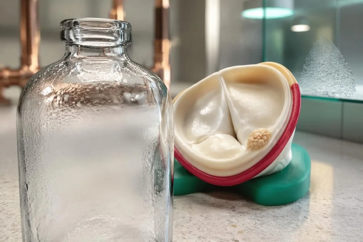
Zapalenie wsierdzio z ujemnymi posiewami krwi ôznaczo podejrzewane abo potwierdzone infekcyjne zapalenie wsierdzio, przi kerym rutynowe posiewy niy wykrywajōm prziczyny. Stanowisko American Heart Association podowo, że posiewy mogōm być ujemne w nawet 30% przipadkōw, chocioż ôdsetek mocno sie zmiynio w zależności ôd ekspozycyje na antybiotyki, ścieżek skierowań i dostympu do laboratoryjōw (DeSimone i in., 2025).
Ujemny posiew ôznaczo, że laboratorium niy wyhodowało mikroorganizmu w użytych warunkach; niy ôznaczo to, że zbadano zastawka serca. Raport wydany po 24 godzinach może być ino wstympny, podwiela echokardiogram może wykryć zmiany strukturalne bez wzglyndu na to, czy mikroorganizmy urosnōm w butelce.
Na przikład wyobraźcie sie 58-latka ze sztucznōm zastawkōm, 10 dniami niewyjaśnionej gorōnczki i posiewami pobranymi po leczeniu podejrzewanej infekcyje klatki piersiowej. Taki zestaw ôbjawōw zasługuje na diagnostyka prowadzōnōm przez lekarza, nawet jeźli piyrsze butelki pozostajōm ujemne; tyn sam ujemny wynik u ôsoby bez ôbjawōw i ryzyka zwiōnzanego z zastawkōm mo inksze znaczynie.
Kantesti je Analizatōr podszukowań krwi sztucznyj inteligyncyje kery wyjaśnio wyniki laboratoryjne, ale niy ôbraco 1 ujemnego posiewu w zapewniynie, że z zastawkōm serca je wszystko w porzōndku. Nasz organizacyjô i cel wyjaśniajōm tyn edukacyjny cel: podejrzewane zapalenie wsierdzio wymogo badanio, konsultacyje z mikrobiologiym i obrazowanio serca, a niy zautomatyzowanego werdyktu laboratoryjnego.
Jak antybiotyki podane przed posiewami krwi ôgraniczajō wykrywanie
Antybiotyki przed posiewami krwi mogōm zmniejszyć liczba żywych mikroorganizmōw, kere mogōm urosnōć, co powoduje ujemny wynik mimo utrzymujōncego sie zajynćio zastawki. Nawet 1 dawka może wpłynōńć na wykrycie, ale efekt zależy ôd mikroorganizmu, leku, dawki i czasu miyndzy podaniym a pobraniym.

Wcześniejsze leczenie antybiotykami je w wielu placōwkach ochrony zdrowio jednōm z głōwnych prziczyn zapalenio wsierdzio z ujemnymi posiewami. Stanowisko AHA ôddzielo pacjyntōw leczonych już wcześnij ôd tych, u kerych posiewy sōm ujemne bez ekspozycyje na antybiotyki, bo te 2 grupy wskazujōm na inksze prawdopodobne mikroorganizmy i inksze poczōntkowe rozważania dotyczōnce leczenio (DeSimone i in., 2025).
Oś czasu podawanio lekōw je bardziej informacyjno niż prosta ôdpowiedź „tak” abo „niy” na pytanie ô antybiotyki. Weź ze sobōm dokładno nazwa leku, informacyjo, czy był podawany doustnie czy dożylnie, daty piyrszej i ôstatniej dawki oraz godzina pobranio; dawka podano 2 godziny przed pobraniym stwarzo inkszy problym interpretacyjny niż kuracyjo zakōńczōno tydnie wcześnij.
Czasowe wstrzimanie leczenio je wyjōntkowo decyzjo specjalisty, a niy metodōm do stosowanio w doma. Wytyczne ESC z 2023 roku dopuszczajōm rozważynie tego u wybranych stabilnych pacjyntōw z podostrymi ôbjawami, bardzo krótkōm wcześniejszōm ekspozycyjōm i bez powikłań, ale niy ma uniwersalnego i bezpiecznego 48-godzinnego abo 72-godzinnego okresu wypłukiwanio, jaki pacjynci powinni próbować stosować sami.
U ôsoby z sepsōm, wstrzōnsym abo szybko pogarszajōncōm sie czynnościōm zastawki niy wolno ôpōźniać leczenio ino po to, coby uzyskać lepsze posiewy. Nasze wyjaśniynie czasu pobranio posiewu przi durze brzusznym ôbrazuje szerszo zasada pobieranio próbek, ale podejrzewane zapalenie wsierdzio mo swojōm pilnōm ścieżka obrazowanio i leczenio.
Co czyni pobranie próbek krwi do posiewu wystarczajōcym?
Odpowiednie pobranie próbek u dorosłych ôgōlnie ôznaczo 2–3 zestawy posiewōw krwi z odpowiednim ôbjyntościōm krwi, nojlepiej przed leczeniym. Zestaw ôbejmuje ôbi tlenowe i beztlenowe butelki; jakość pobranio, stan inkubacyje i kontakt z laboratorium majōm wpływ na to, jak mocno ujemny wynik może uspokajać.
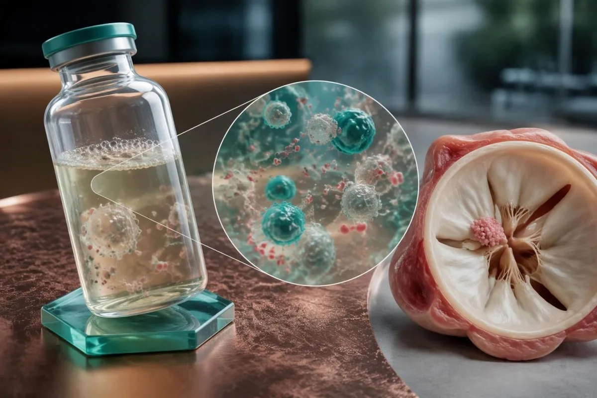
The AHA statement recommends at least 2 sets, ideally 3, before empirical antibiotics when feasible, with approximately 8–10 mL per adult bottle. Underfilling bottles reduces the amount of material examined, so several small samples do not necessarily provide the same diagnostic opportunity as properly filled sets.
The 2023 ESC guideline recommends 3 sets at approximately 30-minute intervals before antibiotics in the appropriate clinical setting (Delgado et al., 2023). The AHA approach emphasizes adequate volume and prompt collection; neither recommendation means an unstable patient should wait through a prolonged sampling schedule before receiving necessary treatment.
Routine automated cultures commonly reach a final report after about 5 days, although local protocols and organism-specific investigations differ. Ask whether no growth means preliminary, final or final for routine incubation only; specialized cultures may be separate orders rather than an automatic extension of the original test.
Fever spikes are not required for useful sampling because endocarditis often produces sustained bloodstream seeding. Kantesti AI can help distinguish a report's collection date from its reporting date when those details are supplied, while our przewodnik po osi czasu wynikōw laboratoryjnych explains why results taken at different treatment stages are not directly interchangeable.
Czamu Coxiella i Bartonella potrzebujō celowanych badań
Coxiella burnetii i Bartonella species are major considerations when routine cultures remain negative, particularly without prior antibiotics. Targeted antibody testing and molecular assays can provide evidence that standard culture bottles miss; exposure history helps prioritize these 2 groups.
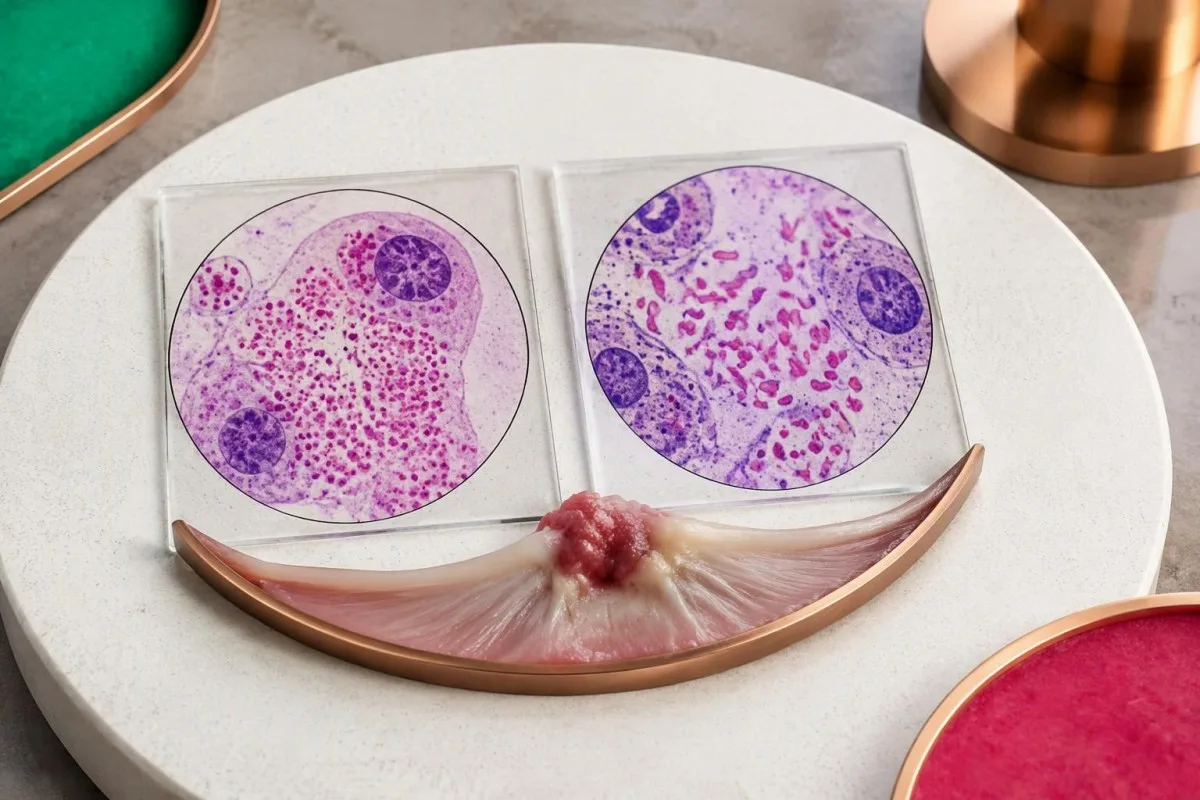
Coxiella burnetii causes Q fever and can produce persistent infection involving a damaged or prosthetic valve. Exposure to sheep, goats or cattle is useful history, but aerosol spread means direct animal handling is not required; a person may lack an obvious exposure despite months of otherwise unexplained symptoms.
The 2023 Duke-ISCVID criteria recognize Coxiella phase I IgG greater than 1:800 as a major microbiological criterion when the specified testing framework applies. Phase I and phase II results are not interchangeable, and our Q fever antibody guide explains why a generic positive Q fever antibody report is incomplete.
Bartonella henselae is associated with cats and cat fleas, while Bartonella quintana is associated with body lice and circumstances that increase exposure to them. These associations guide testing without establishing a diagnosis; 1 cat scratch or a remote history of cat ownership does not make an antibody result proof of valve involvement.
Bartonella henselae or quintana IgG of at least 1:800 by the specified immunofluorescence method can fulfill a major Duke-ISCVID criterion (Fowler et al., 2023). Our wyjaśniynie miana Bartonella addresses antibody interpretation, but endocarditis assessment additionally requires cardiac findings and consideration of cross-reactivity.
Kere inksze drobnoustroje mogō umknōńć rutynowym posiewōm?
Other causes include Tropheryma whipplei, Brucella and selected fungi, with testing guided by exposures, immune status and valve history. Modern culture systems recover many previously difficult organisms within about 5 days, so culture negativity should not automatically be blamed on slow growth.
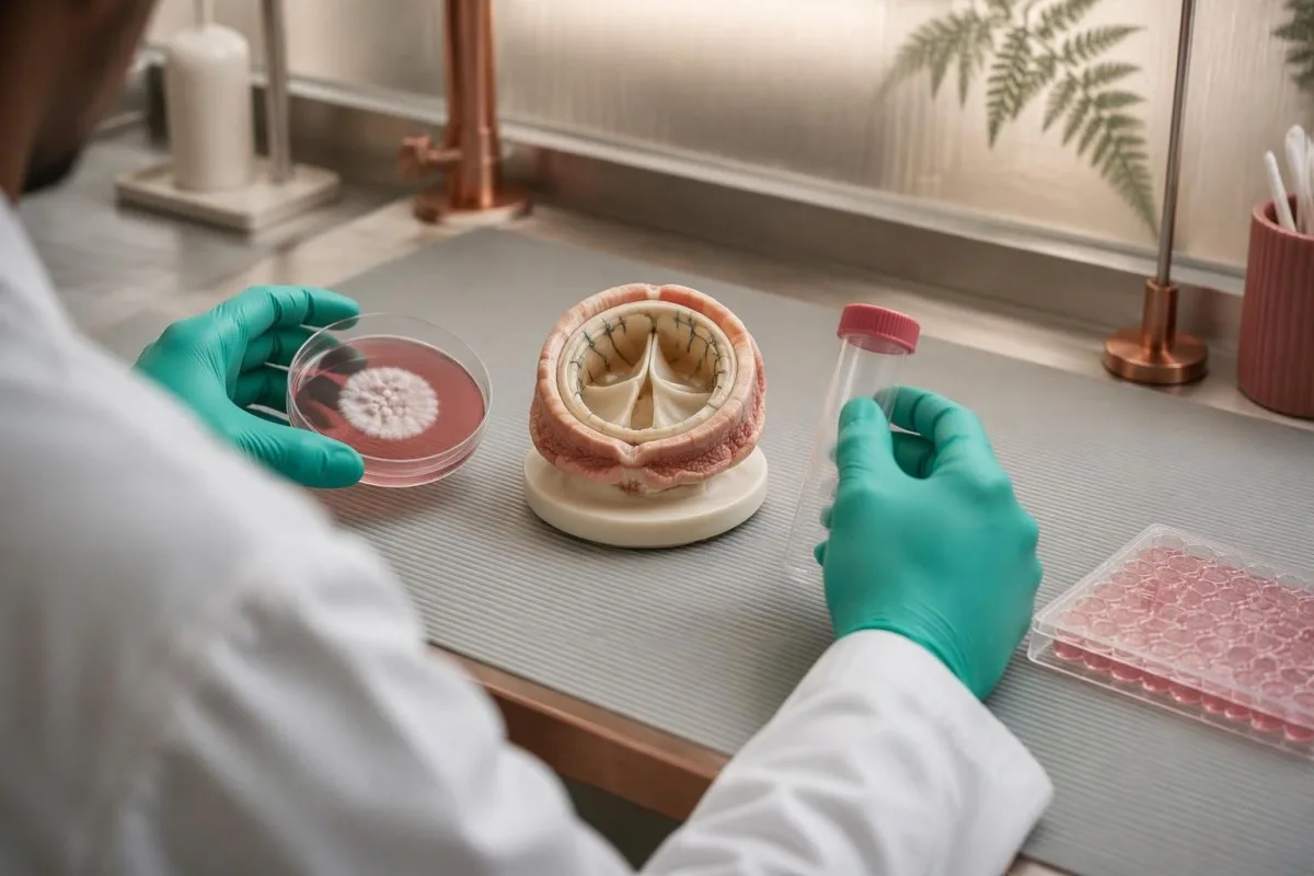
HACEK organisms historically required prolonged incubation, but modern automated systems usually detect them during routine incubation. The group contains 5 named categories of oral-associated bacteria; asking for weeks of incubation solely because HACEK is suspected may add less value than reviewing antibiotic exposure, sample volume and the actual laboratory system.
Tropheryma whipplei endocarditis may occur without classic diarrhea or obvious gastrointestinal disease. A history of unexplained joint symptoms over months or years can be a clue, but a stool result alone does not establish valve involvement; targeted PCR on suitable material, especially valve tissue when available, is more informative.
Brucella testing becomes more relevant after unpasteurized dairy consumption, livestock exposure or travel to an endemic area. Our Brucella culture and PCR guide explains these complementary methods, and suspected exposure should be communicated before specimen processing because the laboratory needs appropriate handling precautions.
Aspergillus endocarditis often has negative routine blood cultures, whereas Candida can grow but may still be missed. Tests such as galactomannan or beta-D-glucan are supporting evidence rather than valve-specific confirmation; our galactomannan interpretation guide explains why 1 positive fungal marker requires context and confirmation.
Jak echokardiografia pōmogo, kej posiewy sōm ujemne
Echo-kardiografia can identify vegetations, new valve leakage and complications despite negative cultures. Transthoracic echocardiography, or TTE, is usually the first scan; transesophageal echocardiography, or TEE, provides closer views when TTE is inconclusive or the clinical suspicion remains high.
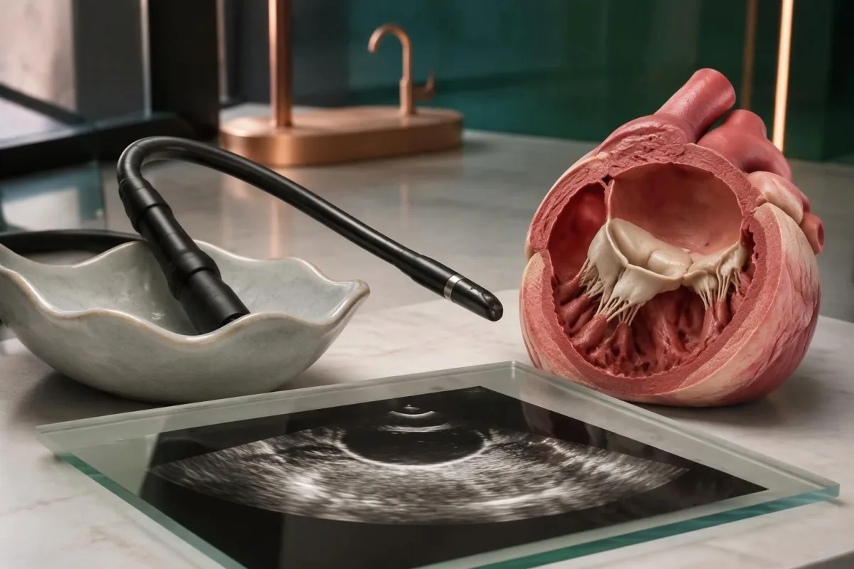
A vegetation is a valve-associated mass, but its appearance alone does not identify the organism. In a hypothetical 62-year-old with persistent fever and negative cultures, a mobile valve mass plus newly severe regurgitation changes the diagnostic assessment more than an isolated nonspecific rise in an inflammatory marker.
TEE is particularly useful with prosthetic valves, intracardiac devices, limited TTE image quality or concern about an abscess around the valve. The 2023 ESC guideline generally recommends TEE in suspected endocarditis even after a positive TTE, except selected unequivocal right-sided native-valve cases with good-quality images (Delgado et al., 2023).
A negative TTE does not carry the same exclusion value in every patient. For example, a 4 mm abnormality may be harder to visualize than a large mobile mass, and prosthetic material can obscure adjacent structures; clinicians interpret the scan together with valve type, symptoms and the technical quality of the examination.
Endocarditis involves the inner heart lining and valves, not primarily the heart muscle. Our przewodnik po badaniach laboratoryjnych przy zapalyniu miynśnia sercowego explains that distinction: 1 normal troponin result cannot exclude a valve infection, because troponin answers a different question about myocardial injury.
Kedy powtōrzne echo, tomografia komputerowo serca abo PET dostarczō wiyncyj informacyji
If suspicion remains high, the 2023 ESC guideline recommends repeating echocardiography after 5–7 days following an initially normal or inconclusive study. Cardiac CT and selected nuclear imaging can add evidence, especially around prosthetic valves or when complications extend beyond what ultrasound shows.
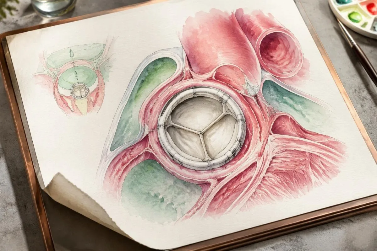
Repeat imaging is not simply repetition for reassurance; a small early abnormality may become visible, or a different imaging angle may clarify an uncertain finding. The 5–7-day interval is a guideline framework, not a reason to wait if breathlessness, conduction abnormalities or hemodynamic deterioration develops sooner.
Cardiac CT is especially useful for evaluating perivalvular anatomy, such as abscesses, pseudoaneurysms or fistulas. TEE can be better for small mobile vegetations and leaflet perforations, so 2 imaging methods may be complementary rather than interchangeable; CT planning also considers kidney function, contrast exposure and radiation.
FDG-PET/CT can support prosthetic-valve or device-associated endocarditis and identify distant sites needing investigation. Recent procedures and sterile tissue responses can also cause uptake; the 2023 ESC and Duke-ISCVID frameworks handle postoperative timing differently, which is why a scan should be interpreted by specialists familiar with the applicable criteria.
There is no single scan that rules out every presentation, particularly early native-valve disease. If chest discomfort accompanies worsening breathlessness, our przewodnik po pilnych badaniŏch przi bōlu w klatce piersiowyj explains why 1 reassuring laboratory result should not replace an immediate clinical assessment.
Jak interpretować celowanō serologię, coby niy wyciōngać zbyt dalekich wnioskōw
Celowano serologia measures antibodies to selected organisms and can help establish culture-negative endocarditis, but antibodies are not a direct image of the valve. The 1:800 thresholds used for Coxiella and Bartonella have specific organism, antibody-phase and assay requirements.
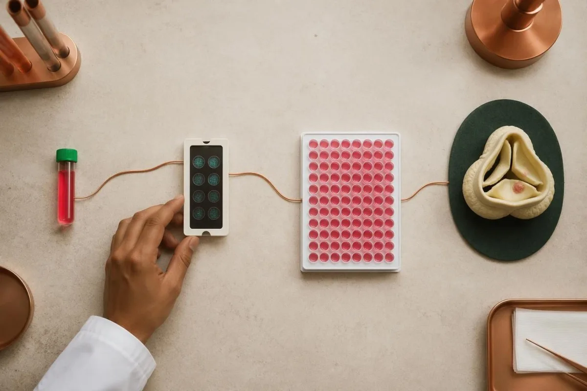
A titer of 1:800 means reactivity remains detectable after the serum has been diluted to that ratio. Coxiella phase I IgG greater than 1:800 and Bartonella IgG at least 1:800 are separate Duke-ISCVID provisions; rounding greater than into greater than or equal to can incorrectly change how a borderline report is classified.
Assay methods and reporting increments differ between laboratories. A result expressed as an index value or units per milliliter cannot automatically be converted to a 1:800 immunofluorescence titer, so clinicians may request reference-laboratory confirmation rather than applying a published cutoff to a non-equivalent test.
Cross-reactivity, previous exposure and delayed antibody formation can complicate interpretation in opposite directions. Our guide to pozytywnych wyników przeciwciał explains why 1 positive result may not indicate active disease, while an early negative result can also be insufficient when the exposure history and imaging remain concerning.
Broad untargeted panels can generate incidental positives and make the diagnosis less clear. When cultures remain negative at approximately 72 hours, the AHA statement supports expanding the investigation with microbiology input, commonly including Coxiella and Bartonella serology and exposure-directed tests—not treating every detected antibody as a separate infection.
Kedy PCR abo sekwencjonowanie mogō ôkreślić przyczynę
PCR and sequencing can detect microbial genetic material when cultures cannot recover living organisms. Targeted blood PCR for Coxiella, Bartonella or Tropheryma whipplei has a recognized diagnostic role; valve tissue can also undergo organism-specific PCR, broad-range testing and histological examination.
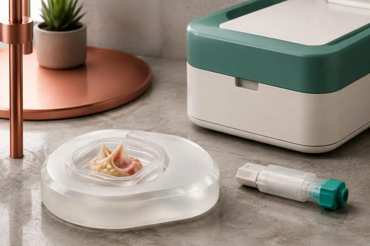
The 2023 Duke-ISCVID criteria recognize positive blood nucleic-acid tests for these 3 organisms as major microbiological evidence in the specified framework (Fowler et al., 2023). A standard bloodstream multiplex panel may not include them, so a negative panel should not be described as negative testing for all endocarditis organisms.
Valve tissue obtained during clinically indicated surgery can be especially valuable after antibiotic exposure. Broad-range bacterial 16S rRNA gene testing and fungal 18S or ITS testing may detect organisms missed by culture, but neither approach has 100% sensitivity and results still require contamination-aware interpretation.
Specimen handling should be planned before a valve procedure whenever possible. Separate material may be needed for culture, molecular testing and formalin-fixed histology; placing the entire specimen in 1 container can limit later options, and tissue should never be obtained solely because a patient wants a more definitive laboratory answer.
Microbial cell-free DNA sequencing may help selected difficult cases, but access, contamination, cost and clinical interpretation remain variable. Our PCR sample timing explanation illustrates why specimen timing matters; for endocarditis, 1 negative molecular result does not cancel convincing cardiac or pathological evidence.
Jak lekarze łōnczō dowody, coby postawić rozpoznanie
Blood culture negative endocarditis diagnosis depends on the combination of microbiology, imaging, clinical features and sometimes tissue findings. Under the 2023 Duke-ISCVID clinical criteria, definite endocarditis can be classified by 2 major criteria, 1 major plus 3 minor criteria, or 5 minor criteria.

The Duke-ISCVID criteria standardize evidence; they are not a home scoring tool or a substitute for specialist judgment. Fever above 38°C is 1 minor criterion, but a person can have serious endocarditis without a measured fever, particularly after treatment or when immune responses are altered.
For a hypothetical example, consider a 55-year-old with negative routine cultures, imaging that meets a major endocarditis criterion and Bartonella IgG of 1:1600 by an appropriate assay. Those 2 major criteria can establish the clinical classification, provided specialists verify the findings and assess alternatives; the negative cultures do not erase the other evidence.
Nonbacterial thrombotic endocarditis is an important alternative when a valve mass occurs with negative cultures. Cancer, antiphospholipid syndrome and lupus can produce sterile valve deposits, and echocardiography alone may not distinguish them from infection; antibiotics before sampling can make that distinction harder rather than resolving it.
Kantesti's laboratory explanations should remain separate from claims of diagnostic certainty about endocarditis. Our techniczne i kliniczne standardy describe the boundaries of interpretation: 1 apparently compatible laboratory pattern cannot replace an endocarditis team's assessment of scans, exposures, complications and competing diagnoses.
Co mogō pedzieć wyniki CRP, białych krwinek i nerek
CRP, white blood cell counts and kidney tests can reveal the body's response and complications, but none can confirm or exclude endocarditis. A CRP of 80 mg/L indicates substantial inflammation in many laboratories; it does not identify a valve source or distinguish every infectious from noninfectious cause.
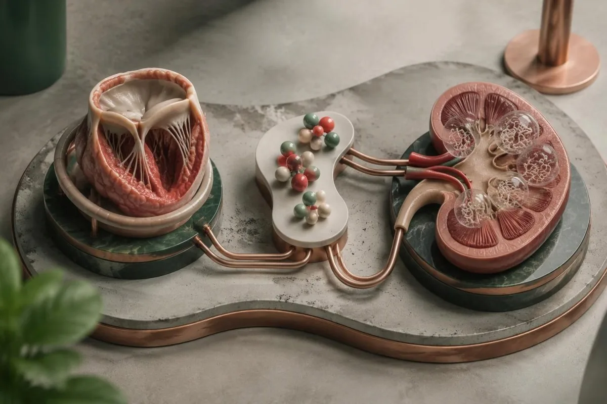
CRP reference limits commonly fall below 5 mg/L or 10 mg/L, depending on the laboratory, while adult WBC intervals often approximate 4–11 × 10⁹/L. Normal results can occur in subacute or previously treated disease, and our porównanie WBC i CRP explains why disagreement between these markers is not inherently an error.
Kantesti je platforma do interpretacji wyników badań krwi AI that can place a supplied CRP of 80 mg/L alongside the CBC, creatinine and treatment timeline without assigning the inflammation to a valve. A falling CRP may support improvement, but 1 falling marker does not prove that an abscess, vegetation or embolic risk has resolved.
New blood or protein in urine can reflect immune-complex kidney involvement, embolic injury or an unrelated urinary problem. Our przewodniku po osadzie moczu explains why microscopy matters: red cell casts, if present, carry different implications from 1 isolated positive dipstick result and warrant clinician assessment.
Reduced complement can support an immune-complex process but is not specific for infection. Our przewodnik C3 i C4 explains that overlap; for example, a creatinine rise from 90 to 140 µmol/L requires assessment for kidney injury, medication effects and other causes rather than attribution to endocarditis alone.
Kere ôbjawy wymagajō pilnej pomocy abo badania jeszcze tego samego dnia?
Sudden neurological symptoms, severe breathlessness, collapse, chest pain or new confusion require emergency assessment, whether cultures are positive or negative. In the UK, call 999 or 112; elsewhere, use the local emergency number rather than waiting for another culture or an AI interpretation.
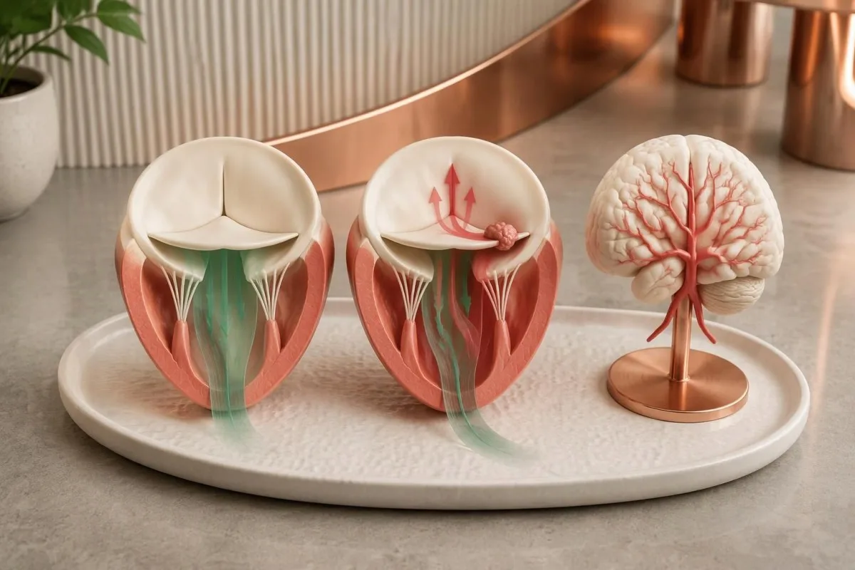
Sudden facial weakness, arm weakness, speech difficulty, loss of vision or major imbalance can indicate a stroke caused by embolization or another emergency. Our stroke warning sign guide explains immediate action; record the last time the person was known well, even if the symptoms disappear within 10 minutes.
Severe breathlessness at rest, inability to speak comfortably, fainting or rapidly worsening swelling can indicate acute heart failure or circulatory compromise. A home oxygen reading of 98% does not exclude serious valve dysfunction, so a normal pulse oximeter result should not delay emergency help when symptoms are severe.
Persistent unexplained fever with a prosthetic valve, previous endocarditis, an intracardiac device or recent bloodstream infection deserves prompt clinician contact, often the same day. A temperature above 38°C adds concern, but older adults and immunocompromised people may become seriously unwell without reaching that threshold.
Sudden severe abdominal or flank pain, a cold painful limb or rapidly reduced urine output also needs urgent assessment because embolic and kidney complications are possible. Small skin spots or splinter-like nail marks alone are nonspecific; 1 such finding should prompt context-sensitive medical advice, not self-diagnosis from internet photographs.
Jak przebiogo leczenie bez dodatniego posiewu
Treatment can begin before the cause is identified when the clinical situation warrants it. Clinicians select initial therapy using valve type, illness severity, previous antibiotics and exposure history, then adjust it as targeted results arrive; many regimens last approximately 4–6 weeks, with important organism-specific exceptions.

Culture-negative endocarditis does not have 1 universal antibiotic regimen. Previously treated disease may still involve common staphylococci, streptococci or enterococci, whereas genuinely culture-negative disease without treatment can require coverage for different organisms; some causes, such as Coxiella infection, need therapy far longer than 6 weeks.
Drug monitoring targets cannot simply be copied from another infection. Our vancomycin AUC explanation describes the commonly used 400–600 mg·h/L exposure framework for serious MRSA infections under specified assumptions, but the treating team decides whether that framework and that drug apply to the individual endocarditis case.
Aminoglycosides are not required for every presentation and can cause kidney or hearing toxicity. Our gentamicin monitoring guide explains why a level collected 1 hour too early or late may mislead; doses must not be adjusted from a random concentration without the regimen and collection timing.
Kantesti can help explain supplied laboratory trends during prescribed treatment, but medication changes and decisions about surgery remain with the clinical team. Heart failure, uncontrolled infection or selected embolic-risk situations can require a procedure even when cultures are negative; 1 favorable CRP trend cannot overrule those structural or clinical indications.
Bezpieczne korzystanie z ôpisōw laboratoryjnych i badań naukowych
A useful consultation record includes culture dates, antibiotic timing, valve history and imaging reports, not just a list of negative tests. Bring these 4 categories together so clinicians can distinguish an incomplete investigation from genuinely reassuring evidence and avoid repeating tests that answer the wrong question.

Kantesti je Narzędzie do analizy badań krwi oparte na AI that supports understanding of accompanying results, not a replacement for urgent cardiac assessment. Our u wyjasniyniy technologii AI describes interpretation workflows; an explanation produced in approximately 60 seconds should never become a reason to postpone emergency or same-day care.
Ask 3 practical questions: Were the cultures collected before antibiotics, was the first echocardiogram sufficient for this valve type, and which targeted organism tests are justified by the history? Our zasady ôdpowiedzialnego używanio AI support checking source documents and escalating clinical uncertainty rather than accepting an automated conclusion at face value.
Kantesti's 2 supporting Figshare publications listed below are background educational materials, not endocarditis diagnostic studies or evidence validating detection of this condition. The related hematology marker explanation may help readers understand associated anemia, but blood group, LDH and reticulocytes cannot establish valve infection.
The endocarditis recommendations in this article instead rely on 3 directly relevant clinical publications: the 2023 ESC guideline, the 2023 Duke-ISCVID criteria and the 2025 AHA scientific statement. These sources address different tasks—clinical management, diagnostic classification and culture-negative investigation—so apparent differences should be interpreted within their intended scope.
Często zadawane pytania
Czy można mieć zapalenie wsierdzia, jeśli wszystkie posiewy krwi są ujemne?
Ja, endokarditis może wystōmpić, nawet kej wszyjske rutynowe posiewy krwi sōm ujemne. Wcześniyjsze podowanie antybiotykōw, drobnoustroje, kere ciynżko wykryć, a tyż niydostateczne pobieranie prōbek mogōm zmniejszyć szansa na ich wyhodowanie, tōż lekarze łōnczōm posiewy z ôbrazowaniem serca i ukierunkowanymi badaniami. Wynik pokazujōncy brak wzrostu po 24 godzinach może tyż być wstympny, a niy ôstateczny. Ujemne posiewy niy usprawiedliwiajōm ignorowanio utrzymujōncych sie ôbjawōw ani niepokojōncych zmian na zastawkach.
Ile posiewōw krwie trza wykōnać przi podejrzeniu zapalenio wsierdzio?
U dorosłych we diagnostice stosuje sie zwykle 2–3 zestawy posiewów krwi, nojlepi przed podaniym antybiotykōw, kej niy ôpōźnio to pilnego leczenio. Kożdy zestaw zawiyro zwykle jednã butelka tlenowõ i jednõ beztlenowõ, po ôkoło 8–10 mL w kożdyj butelce dlo dorosłych. Wytyczne ESC z 2023 r. zalecajōm 3 zestawy pobiyrane w ôdstympach ôkoło 30 minut, kej je to właściwe, a stanowisko AHA podkreślo co nojmynij 2 zestawy, nojlepi 3. U dzieci ôbjyntości muszōm być dostosowane do wieku i masy ciała, a niy do ôbjyntości butelek dlo dorosłych.
Czy mom przestać brać antybiotyki przed powtórnym pobraniem posiewów krwi?
Niy przestowej samowolnie brać przepisanych antybiotykōw, coby polepszyć wyniki posiewu krwi. Już jedna dawka może wpłynōńć na wykrycie drobnoustrojōw, ale niy ma uniwersalnie bezpiecznego 48-godzinnego ani 72-godzinnego ôkresu bez antybiotykōw przy podejrzyniu zapalenio wsierdzio. Specjaliści mogō rozważyć wstrzimanie leczenio ino u wybranych, stabilnych pacjentōw z niewielkōm wcześniejszōm ekspozycyjōm na antybiotyki i bez powikłań. Sepsa, niewydolność serca abo szybkie pogorszenie stanu czyniō ôpóźniynie leczenio szczegōlnie niebezpiecznym.
Czy prawidłowe echo serca wyklucza zapalenie wsierdzia?
Prawidłowy piyrszy echokardiogram niy zawdy wykluczo zapalenie wsierdzio, ôsobliwie kej badanie je ôgraniczone technicznie abo materyjoł protetyczny zasłanio zastawka. Echokardiografia przełykowo może dostarczyć dodatkowych informacyji po ujemnym abo niyjednoznacznym badaniu przezklatkowym. Wytyczne ESC z 2023 roku zalecajōm powtōrzyć echokardiografijo po 5–7 dniach, kej podejrzywanie wciąż je wysokie. Nowe, ciynżkie ôbjawy wymogajōm wcześniejszego ôcynianio, a niy czekanio na upływ tego czasu.
Kere testy djiagnozujōm endokardyt bezdodatni w posiewie?
Endokarditis bez wzrostu drobnoustrojów w posiewach ocynio sie z użyciem echokardiografije, odpowiednich posiewōw, ukierunkowanej serologije i wybranych testōw molekularnych, a niy jednego uniwersalnego badanio krwi. IgG przeciwko fazie I Coxiella powyżyj 1:800 oraz prawidłowo zmierzōne IgG przeciwko Bartonella wynoszōnce co najmniyj 1:800 majōm określōne zastosowanie w kryteriach Duke-ISCVID z 2023 roku. Ukierunkowany PCR z krwi i badanie tkanki zastawki pobranej podwiela klinicznie wskazanyj operacyje mogōm wykryć niykere drobnoustroje, kere inaczyj mogłyby pozostać niewykryte. Wyniki trza interpretować społem, bo przeciwciała, DNA i obrazy zastawek majōm roztōmajte ograniczynia.
Mogōm być CRP i biołe krwinki normalne przi endokardyjitydzie?
CRP i liczba białych krwinek mogōm być prawidłowe przi endokarditis, ôsobliwie w podostrym abo wczéśnij leczōnym przebiygu. Wiela laboratorijōw używo limitōw CRP niżyj 5 abo 10 mg/L i przedziałōw WBC dlo dorosłych ôkoło 4–11 × 10⁹/L, ale wartości w tych przedziałach niy wykluczajōm infekcyje zastawki. Podwyższōne wyniki tyż niy mogōm wskazać, że endokarditis je jich prziczynōm. Utrzymujōnco sie gorōnczka przi ryzyku choroby zastawki, ciynżkie duszności abo nagłe objawy neurologiczne wymagajōm ôceny pod nadzorzym lekarza, niyależnie ôd tych liczb.
Dowiydz się, jak czytać wyniki badańo krwi
Wgraj raport laboratoryjny, a AI Kantesti wyjaśni kożdy biomarker prostym jynzykiym w pora sekund. Używane bezma 2 miliony ludzi na cołkim świecie.
Ôdniesiynia
- Klein, T., Mitchell, S., & Weber, H. (2026). Przewodnik ô grupie krwi B Rh−, badaniu krwi LDH i liczbie retikulocytōw. Badania medyczne AI Kantesti. DOI
- Klein, T., Mitchell, S., & Weber, H. (2026). Biegunka po poście, czorne drobinki we stolcu i przewodnik GI 2026. Badania medyczne AI Kantesti. DOI
- Delgado V et al. (2023). 2023 ESC Guidelines for the management of endocarditis. Europejski Czasopismo Kardiologiczne. DOI
- Fowler VG Jr et al. (2023). The 2023 Duke-International Society for Cardiovascular Infectious Diseases Criteria for Infective Endocarditis: Updating the Modified Duke Criteria. Clinical Infectious Diseases. DOI
- DeSimone DC et al. (2025). Blood Culture–Negative Endocarditis: A Scientific Statement From the American Heart Association: Endorsed by the International Society for Cardiovascular Infectious Diseases. Journal of the American Heart Association.
- 2M+Ônalyzowane badania
- 127+Kroje
- 100+Jynzyki
Ô zespole medycznym


Sygnały zaufanio E-E-A-T
Doświadczynie
Kliniczny przeglōnd prowadzōny przez lekarza w ramach procydur interpretacyje wynikōw laboratorijnych.
Ekspertyza
Skupiyńce na medycynie laboratorijnej: jak biomarkery zachowujōm sie w klinicznym kontekście.
Autorytetność
Zweryfikowane medycznie przez dr. Thomasa Kleina, przy współudziale dr Sarah Mitchell i prof. dr. Hansa Webera.
Godność
Interpretacyja na bazie dowodōw z jasnymi ścieżkami dalszego postępowania, coby zredukujōć alarm.



Zastrzeżynie medyczne
Ten artykuł je wyłōncznie do celōw edukacyjnych i nie stanowi porady medyczno. Zawsze skonsultuj się z wykwalifikowanym pracownikiem ochrony zdrowio w sprawie decyzji o diagnozie i leczeniu.
Opublikowane · Skontaktuj sie z redakcyjōm
Opublikowane przez Kantesti Ltd, zarejestrowane w Anglii i Walii, numer firmy. 17090423, Londyn, Zjednoczone Królestwo.
