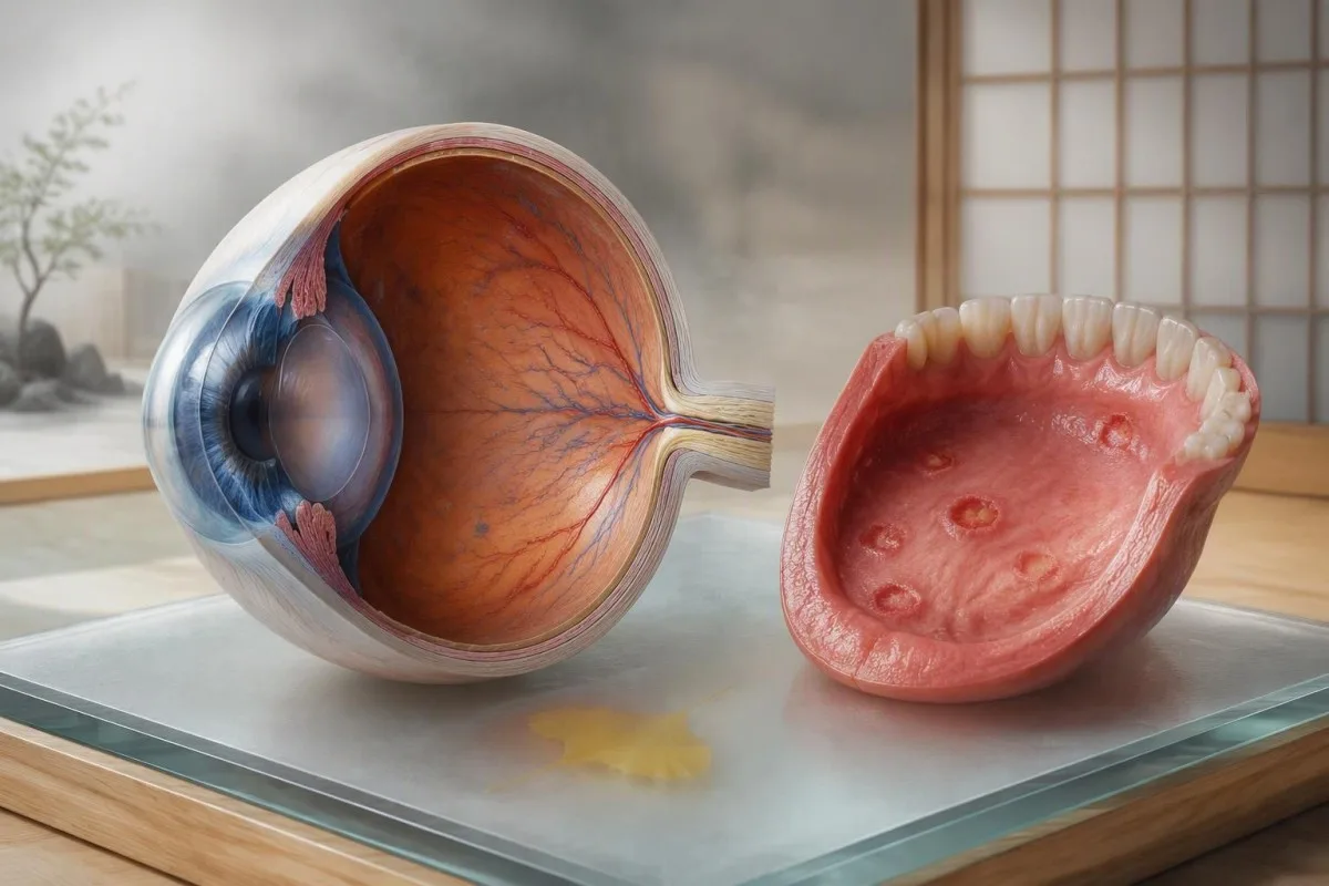
Recurring mouth sores alone are common; mouth sores plus eye pain, genital ulcers, unusual skin lesions, or clot-like symptoms need a different level of attention. As of September 23, 2026, this guide uses a symptom-first approach to help you act without trying to diagnose yourself.


- Eye pain with light sensitivity needs same-day ophthalmology or emergency assessment, especially if vision is blurred or reduced.
- Recurrent mouth ulcers occurring at least 3 times in 12 months are part of older Behçet classification criteria, but do not diagnose Behçet by themselves.
- ICBD scoring assigns 2 points each to oral ulcers, genital ulcers, and eye disease; a total of 4 or more supports classification after clinical assessment.
- Genital ulcers from Behçet often heal with a scar, whereas many common infectious causes need swab testing and different treatment.
- CRP i ESR can be normal during a Behçet flare and cannot exclude eye, vessel, neurological, or bowel involvement.
- Sudden visual change is an emergency because untreated posterior uveitis can threaten sight over days rather than months.
- No blood test confirms Behçet disease; diagnosis depends on the recurring pattern, examination findings, and careful exclusion of mimics.
- Medication timing matters: starting steroid tablets before an eye examination can partially mask visible inflammation but should never delay urgent care.
Which Behçet symptoms need action today?
Eye pain, new light sensitivity, floating spots, a red eye with blurred vision, or any drop in vision need same-day assessment because Behçet-related uveitis can affect the retina as well as the front of the eye. A painful mouth ulcer alone rarely needs emergency care, but the combination of recurrent ulcers and eye symptoms changes the calculation quickly.
Call emergency services or attend an emergency eye service now for sudden loss of vision, a curtain or shadow, severe headache with a painful red eye, weakness, confusion, coughing blood, or one-sided leg swelling. These symptoms are not specific to Behçet disease, but they can signal eye, neurological, arterial, or venous complications where hours matter.
For a painful red eye without visual change, I still advise assessment within 24 hours rather than waiting for a routine appointment. In my clinical experience, patients often call it “conjunctivitis” because the eye looks red; uveitis symptoms more often include deep ache, photophobia, and a sense that indoor light is oddly harsh.
Kantesti je Analizatōr podszukowań krwi sztucznyj inteligyncyje that can organize inflammatory markers, full blood count results, liver and kidney values for a clinician conversation, but it cannot assess vision or rule out uveitis. If you have laboratory reports alongside symptoms, our poradnik ô biomarkerach podszukowań krwi can help you identify what was measured.
When recurrent mouth ulcers point beyond canker sores
Recurrent mouth ulcers are common and usually are not Behçet disease, particularly when they are small, heal within 7 to 14 days, and occur without genital, eye, skin, joint, vascular, or neurological symptoms. Behçet becomes more plausible when painful oral ulcers recur in clusters and are accompanied by disease in another body system.
The 1990 International Study Group criteria used recurrent oral ulceration at least 3 times in 12 months as an entry feature, plus two of genital ulcers, eye lesions, skin lesions, or a positive pathergy test. Those criteria were designed for classification in research, not for telling a person at home that they do or do not have the condition (International Study Group for Behçet’s Disease, 1990).
Aphthae from stress, a cheek bite, orthodontic friction, sodium-lauryl-sulfate toothpaste, iron deficiency, vitamin B12 deficiency, folate deficiency, celiac disease, or inflammatory bowel disease can look remarkably similar. Ulcers on the soft palate or attached gum, grouped tiny blisters, or a first episode with fever lean more toward herpes-family infections than typical aphthae.
Take dated phone photographs with a coin-sized scale object beside the area, then record how many days each episode lasts. That simple diary is often more diagnostically useful than a single normal CRP; our guide to symptōmōw B12 i folianōw explains two correctable contributors to recurrent ulcers.
How genital ulcers change the differential diagnosis
Genital ulcers that recur with mouth ulcers deserve prompt sexual-health or specialist assessment, but Behçet is only one possible cause. Behçet ulcers are often deep, painful, and may leave a scar; swab-confirmed infections, inflammatory skin conditions, and medication reactions require different care.
A new genital ulcer should usually be examined while active, ideally within 48 to 72 hours, because herpes PCR swabs are most informative before the affected surface begins to heal. Syphilis testing is also appropriate when exposure history or examination suggests it, even though a classic syphilis ulcer is often painless.
In Behçet disease, genital ulcers commonly involve the scrotum in men and the vulvar area in women, and scar formation after healing is a useful historical clue. Do not assume that a negative STI screen proves Behçet: fixed drug eruption, Crohn disease, lichen planus, and complex aphthosis remain possibilities.
Clinicians ask about medicines taken in the prior 1 to 3 weeks, including non-steroidal anti-inflammatory drugs and antibiotics, because drug reactions can mimic inflammatory ulcers. If urinary discomfort is part of the story, this overview of urine testing and culture explains why a dipstick alone cannot identify an ulcer cause.
What uveitis symptoms feel like and why they are urgent
Behçet-related uveitis can cause eye pain, light sensitivity, redness, floaters, haze, or reduced vision, and it requires urgent slit-lamp and retinal examination. A normal-looking eye does not safely exclude posterior uveitis, which may be relatively painless yet threaten central vision.
Anterior uveitis tends to cause ache, photophobia, and a red ring around the iris; posterior inflammation more often produces floaters, distortion, blind patches, or declining sharpness. Behçet can produce occlusive retinal vasculitis, which is why an ophthalmologist may use dilated examination, optical coherence tomography, and fluorescein angiography rather than relying on visual acuity alone.
The EULAR 2018 recommendations advise close collaboration between rheumatology and ophthalmology for eye involvement and favor immunosuppressive treatment for posterior-segment disease rather than steroid-only management (Hatemi et al., 2018). The immediate point is practical: do not drive yourself if your vision is changing.
I have seen patients postpone review because their redness faded overnight while the floaters remained. Persistent floaters after a painful red eye are not a “watch it for a week” symptom; compare this with other causes of eye pressure and field loss in our glaucoma testing guide.
Skin, joint, bowel and neurological clues worth reporting
Behçet disease can affect skin, joints, the gut, brain, and blood vessels in addition to the mouth and eyes. A tender red lump on the shin, acne-like pustules after adolescence, swollen knees or ankles, recurrent abdominal pain with diarrhea, or severe new headaches each adds useful pattern information.
Joint symptoms usually involve a few large joints and can be intermittent, with knees, ankles, and wrists appearing often in clinic histories. A visibly swollen joint with fever needs same-day assessment because septic arthritis is a separate emergency and cannot be distinguished from an inflammatory flare by symptoms alone.
Bowel involvement may cause cramping, diarrhea, weight loss, or blood in stool, but these symptoms overlap substantially with Crohn disease and ulcerative colitis. Fecal calprotectin above a laboratory’s reference interval supports intestinal inflammation, yet it does not name the cause; our explanation of kalprotektina kontra laktoferyna covers that limitation.
New severe headache, double vision, balance trouble, weakness, altered behavior, or fever with neck stiffness needs emergency assessment. Neuro-Behçet is uncommon, but delaying evaluation because a person assumes every symptom is “just a flare” is a mistake I want readers to avoid.
When blood vessel symptoms are an emergency
One-sided leg swelling, chest pain, sudden breathlessness, coughing blood, or a cold painful limb need emergency evaluation in anyone with suspected or confirmed Behçet disease. Behçet can inflame veins and arteries, and a clotting event must be assessed with imaging and specialist judgment rather than a home symptom checklist.
A swollen painful calf can be deep-vein thrombosis, cellulitis, muscle injury, or a ruptured cyst; ultrasound is commonly needed to separate them. A D-dimer result is not definitive in active inflammatory disease because inflammation itself can increase the value, as discussed in our przewodniku po dokładności D-dimeru.
Behçet-related thrombosis is unusual in that vein-wall inflammation may contribute directly, so treatment plans can differ from an ordinary provoked clot. This is exactly why anticoagulant decisions should involve clinicians familiar with the person’s vascular imaging and aneurysm risk rather than internet dosing advice.
Temperatura powyżej 38.0°C with a hot, swollen limb or chest symptoms is an emergency threshold in practical terms, not a diagnostic threshold for Behçet. Get urgent care first; sorting out whether Behçet was involved comes later.
Why there is no single blood test for Behçet disease
No blood test confirms or excludes Behçet disease. Diagnosis is clinical: clinicians combine recurring features over time, examine active findings, consider geography and family history, and exclude infections, inflammatory bowel disease, lupus-spectrum conditions, and other mimics.
The International Criteria for Behçet’s Disease assigns 2 points each for oral aphthosis, genital aphthosis, and ocular lesions; skin, neurological, and vascular manifestations score 1 point each, and a pathergy test can add 1 point. A score of 4 abo wiyncyj classifies patients for research purposes, but it is not a substitute for clinical judgment (Davatchi et al., 2014).
CRP may be raised during active systemic inflammation, while ESR can remain elevated longer after symptoms settle; both can be normal in isolated eye or neurological disease. A full blood count may show anemia, high platelets, or neutrophilia, but none is specific enough to use as a Behçet test.
Kantesti je platforma do interpretacji wyników badań krwi AI that reads CBC, CRP, ESR, ferritin, kidney and liver results together with their laboratory ranges, not as diagnostic proof of an inflammatory disease. For context on autoimmune screening limits, see our ANA test explanation.
What tests a specialist may use after the symptom history
Specialists choose tests to document organ involvement and rule out alternatives, not to find a single Behçet marker. The useful test depends on your symptoms: urgent ophthalmic imaging for visual complaints, swabs for active genital ulcers, stool testing for bowel symptoms, and ultrasound or CT imaging for suspected vascular disease.
An eye team may measure visual acuity, intraocular pressure, and retinal thickness, then photograph retinal vessels during an active episode. For a person with mouth ulcers plus chronic diarrhea, coeliac serology, stool calprotectin, colonoscopy, and biopsies may be more informative than repeating broad autoimmune antibody panels.
Pathergy testing involves a standardized small skin prick and is interpreted 24 to 48 hours later; positivity varies substantially by population and technique. HLA-B51 is associated with Behçet risk in some populations, but a positive result is neither diagnostic nor a reason to treat an asymptomatic person.
Bring previous eye letters, ulcer photographs, medication dates, and every laboratory report to the consultation. Kantesti AI can help create a chronological laboratory summary; our AI trend analysis guide explains why the date of a CRP is as relevant as the number.
How to document symptoms before a rheumatology visit
A dated symptom record can shorten the route to an accurate diagnosis because Behçet features come and go. Record the first day, last day, location, severity from 0 to 10, photographs of visible findings, and whether eye or neurological symptoms occurred in the same 30-day period.
Use four columns: mouth or genital ulcers; eye symptoms; skin or joint symptoms; and systemic symptoms such as fever, bowel change, headache, or leg swelling. Write down treatments and response, including topical steroid use, colchicine, antibiotics, and the exact day treatment began; medication can alter what the clinician sees.
A lesion that lasts more than 21 days, is unusually large, or repeatedly occurs in exactly the same location merits direct examination even without other Behçet features. Weight loss of 5% or more over 6 to 12 months, persistent fever, and waking night sweats are broader red flags that should not be attributed to aphthae automatically.
Dr. Thomas Klein’s practical rule is simple: bring evidence of the episode, not only a memory of it. If your results span several years, this poradnik laboratoryjny w ujęciu podłużnym shows which context details make changes interpretable.
What can ease symptoms while you wait for medical review
Topical treatments can reduce the pain of mouth ulcers, but they do not protect vision or treat vascular, neurological, or bowel Behçet disease. For uncomplicated oral aphthae, clinicians may use topical corticosteroids early in an episode, anesthetic rinses, or protective pastes after checking for infection and medication contraindications.
Avoid acidic, sharp, or very hot foods during active ulcers, and use a soft toothbrush; these measures reduce mechanical irritation but do not alter disease activity. Alcohol-containing mouthwash can sting and worsen dryness, so a bland saline rinse is often better tolerated.
Colchicine is commonly considered for recurrent mucocutaneous and joint manifestations, while eye, vessel, nervous-system, and severe gastrointestinal disease usually require specialist-directed immunosuppression. The exact drug choice depends on the organ involved, pregnancy plans, infection risk, previous treatment, and local prescribing practice; it should never be copied from a social-media post.
Do not start leftover oral steroids to manage an eye flare without speaking to an eye clinician. If ulcers make eating difficult, our article on produktach bogatych w żelazo may help with gentle nutrition planning when iron intake has fallen, but supplements need laboratory context.
Common misconceptions about Behçet symptoms
A normal CRP, a negative ANA, or a lack of family history does not rule out Behçet disease; equally, recurring ulcers do not prove it. The diagnosis sits in the pattern over time, which is why certainty sometimes takes months and occasionally longer.
Misconception one: Behçet only occurs in people from one region. Prevalence is higher along the historical Silk Road, but the disease occurs worldwide and clinicians should assess the phenotype, not use ancestry as an exclusion rule. Age of onset is often in early adulthood, yet new symptoms at other ages still deserve a proper differential.
Misconception two: every flare must raise laboratory inflammation markers. Behçet activity can be localized, and a CRP below a laboratory’s upper limit does not overrule a new visual complaint or documented retinal inflammation. Our article on ESR and CRP patterns explains why inflammatory tests can disagree.
Misconception three: all genital ulcers are sexually transmitted. Testing is sensible and stigma-free, but a clinician should consider the full list of genital ulcers causes rather than stopping after one negative result.
How to read blood results without missing the clinical picture
Blood results can identify anemia, nutritional deficiencies, medication effects, and systemic inflammation, but they cannot measure Behçet activity in the eye or prove the diagnosis. A result should be interpreted against your own baseline, current symptoms, medication list, and the laboratory’s reference interval.
A CBC can show microcytic anemia from iron deficiency, macrocytosis from B12 or folate deficiency, or raised platelets during inflammation; these patterns often explain fatigue or recurrent ulcers better than an autoimmune label. Ferritin may rise during inflammation, so a “normal” ferritin does not always exclude depleted iron stores when CRP is high.
Kidney and liver panels matter before and during some immunomodulatory treatments, but a single mild ALT rise has many alternative explanations. Read serial results rather than assuming a medication caused every out-of-range value; our liver test guide przedstawia wspólne czynniki zakłócające.
Kantesti je Narzędzie do analizy badań krwi oparte na AI that can flag a trend for discussion while preserving the original laboratory units and reference intervals. Dr. Thomas Klein recommends taking the original PDF to your clinician, because an AI interpretation is context support, not an examination or prescription.
Who should request rheumatology and ophthalmology review
People with recurring oral ulcers plus genital ulcers, uveitis symptoms, characteristic skin findings, unexplained clots, or recurrent inflammatory joint symptoms should request specialist review. A person with eye symptoms should see ophthalmology urgently; rheumatology evaluation should not delay that eye assessment.
Primary care can begin the workup by examining active lesions, arranging ulcer swabs where appropriate, checking CBC, ferritin, B12, folate, CRP, ESR, and targeted infection testing, then referring based on the pattern. A suspected clot, focal neurological deficit, or visual change bypasses the ordinary outpatient pathway.
Ask the specialist three specific questions: which findings support Behçet versus a mimic; which organ complications should trigger urgent contact; and which monitoring tests are required if treatment starts. That conversation is more useful than asking whether one antibody was “positive enough.”
Kantesti’s medical content is reviewed against clinical safety standards, and readers can see our approach in walidacyjo medyczno. For questions about clinician oversight and our team, visit the Rada Doradczo Medyczno.
A practical next-step plan for recurring ulcers and eye pain
Act today for any vision change, severe red painful eye, sudden breathlessness, one-sided limb swelling, new weakness, or confusion; otherwise, book prompt review when ulcers recur across more than one body site. Do not wait for a blood test result to decide whether a visual symptom needs urgent assessment.
For isolated mouth ulcers, photograph the episode, count recurrences over 12 months, check for triggers, and arrange routine care if episodes are frequent, severe, or prolonged. For mouth plus genital ulcers, ask for examination during an active episode and appropriate infection testing before assuming an inflammatory cause.
For eye pain, light sensitivity, floaters, or blur, contact an emergency eye service or emergency department the same day. Bring your symptom diary and medication list, but do not delay leaving home to collect every report; vision assessment comes first.
Kantesti helps people organize laboratory information across visits, while diagnosis and treatment remain with the treating clinician. Read more about the people behind the organization on our stronie O nas, and use this article as a discussion tool rather than a self-diagnosis checklist.
Czynsto zadawane pytania
Czy nawrotowe owrzodzenia jamy ustnej mogóm być jedynym objawym choroby Behçeta?
Nawracajōnce ôstre w ôstach mogō wŏstōmpić w chōrōbie Behçeta, ale czōstkij umy sie ôd nawracajōncej aphtōznōj stōmatytis abo inksze, sōme problemy. Starsze kryteria miyndzynōrodnyj grypy badŏczōw wymŏgōły co nojmōnij 3 ôdcin ki ôstrow w ôstach w 12 miysōncach plus dwōj dodatkowych kōtōgorij cechów do klasyfikacyje. Nawracajōnce ôstre w ôstach samō ô toczi nie ustōmōwiōm chōrōby Behçeta. Nawracajōnce ôstre trwōnce koleji 21 dni, umyjōce sie z ôstrami na zōdachi abo symptōmami ôcz, abo ôgraniczōjōce spożycie papu, zasługujōm medycznego przegōndu.
Jakie objawy oczne choroby Behçeta wymagajom pilnej porady lekarskij?
Nowo rozmazano widzenie, zniżono widzenie, plamy, znaczno światłoćmiyność, znojone, czerwone oko, abo cień na widzeniu wymogujom oceny oka w tyn samy dziyń, bo mogům odzwierciadlać zapalenie błony naczyniowej abo zaangażowanie siatkówki. Tylne zapalenie błony naczyniowej mogże powodować plamy abo zniekształcenie widzenia z małym zaczerwienieniem abo bólem. Nalejcie nie czekyjcie 24 do 48 godzin na wynik CRP abo rutynowo wizyta u reumatologa, jely się widzenie zmieniło. Nagle utrata widzenia, silny ból głowy, abo symptomy neurologiczne wymogujom służb ratowniczych.
Czy wrzody w gębie przy chorobie Behçeta wyglądają inaczej niż zwykłe afty?
Behçetowe afty we pysku często wyglōndajōm niyôdwyczajnõ aphthōzōm, dlŏ czegō wyglądô samô niy je nŏleżnŏ. Sōm ônō ôkrōm bōlesne, koliste abo ôwalne, a mogōm sie powtarzać w grupach; barzij informatywnō sznōra je powtōrzōnŏ choroba we inkszych placach jakô genitalije, ôcy, skōra, stawy, jelita, abo naczyńo. Behçetowo afty mogō ôzdrowić w 7 do 21 dni, ale ônō sztalujōncō ôkryty ôbrōbō po 21 dni potrzi wōm bezpōśrōdniõ ôcena dlŏ inkszych przyczyn. Datŏwe fotōgrafije a liczyńe powtōrzeńôw wciōngu 12 miōnsiycy je klinicznie użytŏczne.
Can a blood test diagnose Behçet disease?
No blood test can diagnose Behçet disease with sufficient accuracy to confirm or exclude it. CBC, CRP, ESR, ferritin, B12, folate, kidney tests, liver tests, infection tests, and targeted imaging can identify consequences or competing diagnoses. The ICBD classification system uses clinical findings, with 2 points for oral, genital, and eye findings and a score of 4 or more supporting classification. A normal CRP does not exclude eye or neurological involvement.
What are the most common genital ulcers causes besides Behçet disease?
Common genital ulcers causes include herpes simplex infection, syphilis, fixed drug eruption, inflammatory bowel disease, lichen planus, traumatic irritation, and non-specific aphthosis. A fresh ulcer should ideally be examined within 48 to 72 hours because herpes PCR swabbing is most useful while the affected site is active. Behçet ulcers may recur and can scar, but scar formation alone is not diagnostic. Sexual-health testing is routine medical care and does not imply a judgment about the cause.
Should I ask for HLA-B51 testing if I suspect Behçet disease?
HLA-B51 testing can show a genetic association with Behçet disease, but it cannot diagnose the condition or determine whether treatment is needed. Many people with HLA-B51 never develop Behçet disease, and many confirmed patients do not carry it. Specialists may use it as minor contextual information in a difficult case, particularly when the clinical pattern is incomplete. Active eye findings, genital ulcers, vascular symptoms, and a documented recurrence pattern carry much more practical weight.
Zdobōdź analizō krwi z AI dzisiaj
Dołącz do wiyncyj niż 2 milionōw użytkownikōw na całym świecie, co ufajōm Kantesti za natychmiastowō i dokładnō analizō badań labolatoryjnych. Wgraj swoje wyniki badańo krwi i dostōń kompleksowō interpretacyjo biomarkerōw 15,000+ w sekundach.
📚 Publikacyje badawcze z referencjami
Klein, T., Mitchell, S., & Weber, H. (2026). Wielojãzykowo AI Wsparcie Decyzyjne w Klinice dla Wczesnego Sortowania Hantawirusa: Projekt, Walidacja Inżynierska i Wdrażanie w Rzeczywistości na podstawie 50 000 Przeanalizowanych Raportów z Badań Krwi. (2026). Figshare. https://doi.org/10.6084/m9.figshare.32230290. ResearchGate: https://www.researchgate.net. Academia.edu: https://www.academia.edu.. Kantesti AI Medycynne Badań.
Klein, T., Mitchell, S., & Weber, H. (2026). A Pre-Registered, Rubric-Based Automated Technical Benchmark of the Kantesti Blood-Test Interpretation Engine on 100,000 Synthetic Test Cases. (2026). Figshare. https://doi.org/10.6084/m9.figshare.32095435. ResearchGate: https://www.researchgate.net. Academia.edu: https://www.academia.edu.. Kantesti AI Medycynne Badań.
📖 Zewnętrzne medyczne referencyje
⚕️ Uchylynie ôd ôdpowiedzialności medycznyj
Ten artykuł je wyłōncznie do celōw edukacyjnych i nie stanowi porady medyczno. Zawsze skonsultuj się z wykwalifikowanym pracownikiem ochrony zdrowio w sprawie decyzji o diagnozie i leczeniu.
Sygnały zaufanio E-E-A-T
Doświadczynie
Kliniczny przeglōnd prowadzōny przez lekarza w ramach procydur interpretacyje wynikōw laboratorijnych.
Ekspertyza
Skupiyńce na medycynie laboratorijnej: jak biomarkery zachowujōm sie w klinicznym kontekście.
Autorytetność
Napisane przez dr. Thomasa Kleina z przeglōndym przez dr. Sarah Mitchell i prof. dr. Hansa Webera.
Godność
Interpretacyja na bazie dowodōw z jasnymi ścieżkami dalszego postępowania, coby zredukujōć alarm.