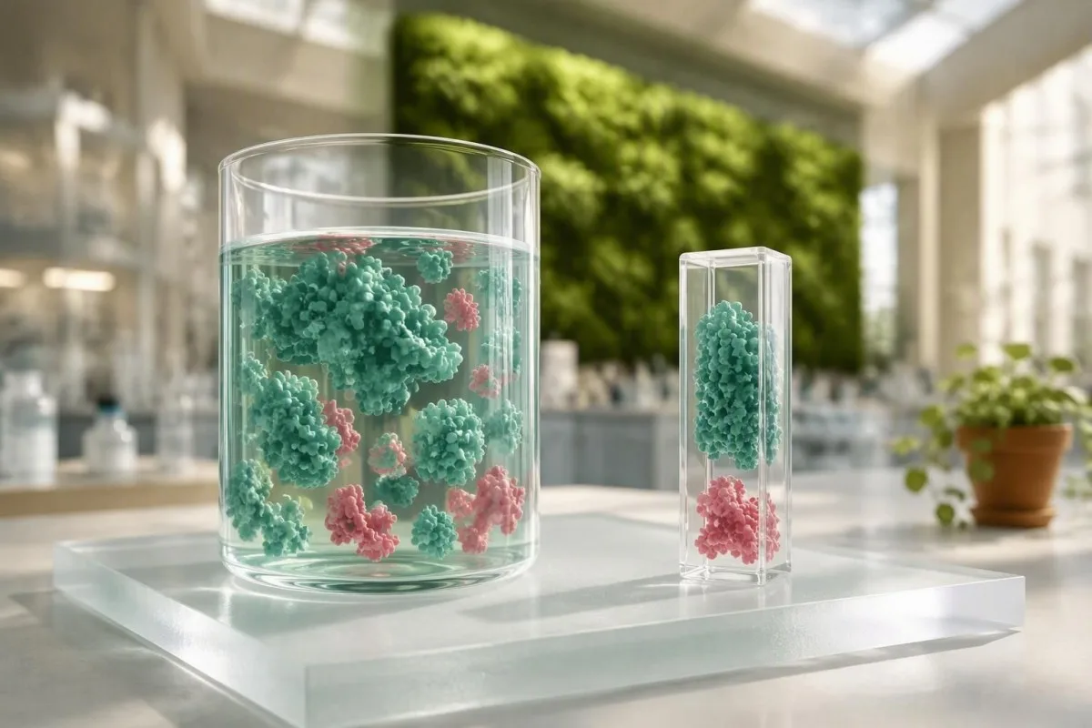
The gamma gap is the difference between total protein and albumin. It can point toward extra antibodies or other globulins, but it cannot diagnose an infection, liver condition, or plasma-cell disorder by itself.


- Gamma gap calculation is total protein minus albumin, using the same units from the same laboratory report.
- Typical value is roughly 2.0-3.5 g/dL (20-35 g/L), although laboratories rarely flag the calculated gap itself.
- Elevated gamma gap above 4.0 g/dL (40 g/L) has traditionally prompted follow-up, but this cutoff is not a diagnosis.
- Albumină contează because dehydration can raise total protein and albumin together, while low albumin can widen the gap without extra antibodies.
- Polyclonal patterns occur with chronic inflammation, autoimmune disease, liver disease, and persistent immune stimulation.
- Monoclonal proteins require confirmation with serum protein electrophoresis, immunofixation, and serum free light chains.
- revizuire urgentă is sensible when a high gap accompanies new anemia, kidney impairment, high calcium, bone pain, fever, or unintended weight loss.
- Repetarea testării after recovery from a short illness is often more informative than ordering an extensive panel from one borderline result.
What the gamma gap shows on a blood panel
A gamma gap blood test result is not a separate test: it is the amount of total serum protein left after albumin is subtracted. A widened gap suggests that non-albumin proteins, especially immunoglobulins, are relatively increased; it is a clue, not a diagnosis.
Total protein includes albumină plus globulins, a broad group containing antibodies, transport proteins, clotting-related proteins, and acute-phase reactants. Albumin usually accounts for about 55-65% of serum protein in well adults, so a changing albumin result can alter the protein gap even when antibody production is unchanged.
The phrase “gamma gap” is slightly imprecise. Laboratory chemistry cannot determine from this subtraction whether the additional protein sits in the gamma fraction, beta fraction, or several fractions; proteinele serice și raportul A/G explain that distinction in more depth.
Kantesti este o Analizor de teste de sânge AI that calculates the protein gap from matched total-protein and albumin values, then checks it against liver markers, kidney function, calcium, and the complete blood count. In my practice, that surrounding pattern has been far more useful than a single calculated number.
Why clinicians call it a clue
A gamma gap of 4.2 g/dL can reflect a resolving viral illness, chronic hepatitis, autoimmune activity, or a monoclonal immunoglobulin. Those possibilities have very different implications, which is why the gap should start a clinical question rather than end one.
Gamma gap calculation: total protein minus albumin
The gamma gap calculation is total protein minus albumin. If total protein is 7.8 g/dL and albumin is 4.1 g/dL, the gamma gap is 3.7 g/dL.
Use g/dL with g/dL or g/L with g/L; do not mix units. A value of 72 g/L total protein minus 41 g/L albumin equals 31 g/L, which is the same as 3.1 g/dL after dividing by 10.
Most routine reports list total protein and albumin but not the calculated gap. The analizele de sânge ghidează helps place a derived value alongside the laboratory’s own reference interval rather than applying a universal internet cutoff.
A calculation can be misleading when the two measurements were taken on different dates, after large fluid shifts, or by methods with different calibration. I have seen a “rising” gap disappear when we compare same-day results from the same laboratory—an unglamorous but clinically meaningful detail.
What is a normal protein gap and why ranges vary?
A gamma gap around 2,0-3,5 g/dL is common in adults, but no single reference range fits every laboratory or clinical setting. The often-cited 4.0 g/dL threshold is a screening convention, not a biological dividing line.
The 4.0 g/dL cutoff has modest sensitivity for the conditions people worry about most. In a nationally representative analysis, Liu et al. found that this threshold was highly specific but insensitive for HIV, hepatitis C, and monoclonal gammopathy, so a normal gap does not rule those conditions out (Liu et al., 2020).
Age adds nuance. Older adults have a higher background prevalence of monoclonal gammopathy of undetermined significance, or MGUS, while pregnancy and fluid expansion can lower albumin and create a wider calculated gap without a primary antibody disorder.
Total protein above 8.3 g/dL (83 g/L) deserves context, particularly if albumin is not elevated with it. Our guide to cauzele proteinelor totale ridicate separates concentration from genuinely increased immunoglobulin production.
How infection and immune activation raise the gap
Persistent immune stimulation can raise the gamma gap because B cells produce a broad mixture of antibodies, called polyclonal immunoglobulins. Viral infections, chronic bacterial inflammation, and some parasitic illnesses can all create this pattern.
Acute infections often change the gap only modestly, and the timing matters. A person tested on day 5 of mononucleosis or during a respiratory infection may have elevated globulins for weeks after fever and fatigue improve; EBV result patterns can clarify when Epstein-Barr testing is relevant.
A high gap alone is not a reason to test indiscriminately for every chronic infection. Exposure history, travel, sexual history discussed confidentially, abnormal liver enzymes, lymph-node findings, and symptoms such as prolonged fever should guide the next test.
In one familiar clinic scenario, a 29-year-old with a gap of 4.1 g/dL, reactive lymphocytes, and recent sore throat had a polyclonal pattern that normalized over 10 weeks. The repeat result mattered because her albumin recovered from 3.5 to 4.2 g/dL as the acute illness settled.
Liver disease patterns behind an elevated gamma gap
Chronic liver disease can widen the gamma gap through both increased immunoglobulin production and reduced albumin synthesis. A low albumin result without elevated globulins can also widen the gap, so liver interpretation needs more than one number.
Albumin is made in the liver, but it has a half-life of roughly 20 days. A low albumin value therefore reflects sustained illness, poor intake, protein loss, fluid status, or impaired synthesis more often than a sudden change over 24 hours.
In cirrhosis, a classic electrophoresis appearance is broad polyclonal gamma elevation with beta-gamma bridging, but this is neither universal nor diagnostic from chemistry alone. Review ALT, AST, alkaline phosphatase, bilirubin, platelet count, and INR together; our decodare a testelor hepatice explică acest tipar.
Dr. Thomas Klein’s practical rule is to pay attention when a rising gap occurs alongside falling albumin and platelets. That cluster carries more clinical weight than a stable 4.0 g/dL gap in someone whose liver enzymes, platelets, and prior results are unremarkable.
Autoimmune disease and chronic inflammation
Autoimmune and inflammatory conditions may elevate the gamma gap by producing broad, persistent antibody responses. Rheumatoid arthritis, Sjögren disease, lupus, inflammatory bowel disease, and chronic lung inflammation are examples, but symptoms and targeted testing determine relevance.
C-reactive protein and erythrocyte sedimentation rate can support an inflammatory explanation, although neither is specific. CRP often changes within 24-48 hours of a stimulus, while ESR can remain elevated longer because it is influenced by fibrinogen, anemia, age, and immunoglobulin concentration.
Kantesti este o platforma de interpretare analize sange AI that reads an elevated gap alongside CRP, ESR, blood counts, ferritin, and organ-function results rather than implying that one marker identifies an autoimmune disease. That distinction prevents a lot of unnecessary alarm.
Autoantibody panels should be ordered for a clinical reason. Dry eyes and mouth, inflammatory joint swelling, purpura, neuropathy, photosensitive rash, or kidney findings are more useful prompts than a gamma gap of 3.8 g/dL alone; immunoglobulin testing is often a more sensible first clarification.
When a gamma gap raises concern for monoclonal proteins
A persistent high gamma gap can occur with a monoclonal protein, but the gap cannot identify MGUS, myeloma, or related disorders on its own. Serum protein electrophoresis and immunofixation determine whether one immunoglobulin clone is present.
A monoclonal protein, sometimes called an M-protein or M-spike, is a concentrated immunoglobulin made by one plasma-cell clone. By contrast, infection and autoimmunity usually produce a wide, diffuse rise across many antibody types—one reason M-spike results cannot be inferred from the gap.
The International Myeloma Working Group defines MGUS by serum monoclonal protein below 3 g/dL, bone marrow clonal plasma cells below 10%, and no attributable end-organ damage. Its updated criteria also recognize specific biomarkers that identify myeloma before overt complications occur (Rajkumar et al., 2014).
A gap of 4.5 g/dL with normal hemoglobin, calcium, creatinine, and no symptoms is not automatically cancer. Still, persistence over 3-6 months, particularly in an older adult, is a reasonable reason to discuss electrophoresis, immunofixation, and serum free light chains with a clinician.
Low albumin, dehydration, and misleading gap results
The protein gap can widen because albumin falls, even if globulins stay unchanged. Dehydration often raises both albumin and total protein together, while fluid overload, inflammation, kidney loss, or liver dysfunction can lower albumin.
Consider two people with total protein of 7.6 g/dL: albumin 4.5 g/dL produces a 3.1 g/dL gap, whereas albumin 3.2 g/dL produces a 4.4 g/dL gap. The second result may reflect lost or reduced albumin rather than an excess of antibodies.
Protein-losing kidney conditions can lower albumin and may produce swelling around the eyes or ankles, foamy urine, or unexpectedly low total protein. A urine albumin-creatinine ratio and kidney assessment are more direct than repeatedly calculating the gap; FIB-4 liver risk is useful when liver disease is part of the pattern.
In my experience, overnight fasting is not needed for total protein or albumin, but an unusually hard endurance event, dehydration from gastroenteritis, or intravenous fluids before the draw should be recorded. Those details explain more “abnormal” protein calculations than patients expect.
Kidney function changes the meaning of protein results
Kidney disease can change protein results through urinary albumin loss, reduced clearance of light chains, or chronic inflammation. A high gamma gap paired with declining eGFR or protein in urine deserves more attention than an isolated gap.
An eGFR below 60 mL/min/1.73 m² for at least 3 months is one criterion for chronic kidney disease, but a one-time lower eGFR can reflect dehydration, medication effects, or a temporary illness. Creatinine, cystatin C where appropriate, and urine ACR add important context.
Free light-chain ratios need kidney-adjusted interpretation because both kappa and lambda light chains rise as filtration falls. A mildly abnormal ratio in advanced kidney disease is therefore not interchangeable with a monoclonal disorder diagnosis; BUN and creatinine differences can help prepare for that discussion.
Kantesti AI uses trend analysis to flag a widening gap with a falling eGFR or new anemia as a follow-up pattern. Our BUN/creatinine research guide also explains why hydration changes urea more readily than it changes antibody proteins.
Which follow-up tests are usually considered?
Follow-up for an elevated gamma gap usually starts with confirming the result and reviewing the whole panel. The next test depends on whether the dominant clue is low albumin, liver injury, inflammation, infection risk, kidney change, or possible monoclonal protein.
For a persistent gap of 4.0 g/dL or higher without an obvious explanation, clinicians commonly consider CBC, creatinine and eGFR, calcium, liver panel, urinalysis or urine ACR, quantitative IgG/IgA/IgM, serum protein electrophoresis, immunofixation, and serum free light chains. Not every patient needs every test.
When inflammatory symptoms are present, targeted CRP, ESR, hepatitis testing, HIV testing with consent, autoimmune studies, or imaging may be more useful than a broad unselected panel. ANCA testing patterns illustrate why antibody results need symptom-led interpretation.
The evidence is honestly mixed on using the gamma gap as a standalone screening gateway. Liu et al. (2020) showed that a 4.0 g/dL threshold misses many cases of the conditions it is meant to flag, which is why a clinician may test despite a smaller gap when the symptom pattern is persuasive.
When an elevated gap needs prompt medical review
An elevated gamma gap warrants prompt clinical review when it occurs with anemia, reduced kidney function, calcium above the laboratory range, persistent bone pain, unexplained weight loss, night sweats, or recurrent infections. The combination—not the gap alone—creates urgency.
Seek urgent care for confusion, severe weakness, shortness of breath at rest, markedly reduced urine output, fever with immunosuppression, or severe new back pain with weakness or bladder changes. Those symptoms need assessment regardless of the protein gap value.
The myeloma-related CRAB features are hyperCalcemia, Renal impairment, Anemia, and Bone involvement. They are not a home diagnosis checklist, but they explain why clinicians take a possible monoclonal protein more seriously when hemoglobin is falling or creatinine is rising; review the cale de testare pentru cancerul de sânge for the usual staged approach.
A stable gap of 4.1 g/dL in an otherwise well person does not usually mean an emergency department visit. A same-week appointment is reasonable if the result is new or persistent, while the pace should be faster when red flags or multiple abnormal results are present.
When to repeat total protein and albumin
For a mild, unexpected gamma gap elevation during a recent illness, repeating total protein and albumin in about 4-12 săptămâni is often reasonable if a clinician finds no red flags. A meaningful trend requires comparable timing, laboratory method, and clinical conditions.
Repeat sooner when albumin is falling, total protein is rising quickly, or the CBC, calcium, kidney markers, or liver tests have changed. A slow isolated drift of 0.2 g/dL can be ordinary analytical and biological variation; a sustained rise of 1.0 g/dL is harder to dismiss.
Record recent infections, vaccines, exercise, dehydration, new medicines, weight change, and intravenous fluids at each draw. This small timeline can distinguish a transient immune response from a reproducible pattern better than memory alone; momentul recoltării analizelor de sânge oferă un șablon practic.
Kantesti este o Instrument de analiză a analizelor de sânge bazat pe AI that compares previous total protein, albumin, and related markers so that a stable personal baseline is visible. A value outside a population range is less concerning when it has been unchanged for years and the surrounding panel remains reassuring.
Common mistakes after seeing a high protein gap
Do not assume a high gamma gap means cancer, start supplements to “lower proteins,” or try to correct the number with extreme hydration. The right response is to verify the calculation and interpret it with symptoms, medications, and the rest of the laboratory panel.
Extra water shortly before a retest can dilute albumin and total protein, making the result harder—not easier—to interpret. Normal hydration and ordinary meals are usually preferable unless the ordering clinician gives specific preparation instructions.
Do not stop prescribed immunosuppressants, liver medicines, or supplements solely because the gap changed. Some agents alter liver enzymes or renal function, but a medication review should consider dose, start date, indication, and risks; the Blogul Kantesti includes medication-safety trend discussions.
Dr. Thomas Klein advises patients to bring the actual numeric values and dates, not just an “H” flag. A laboratory flag identifies a statistical outlier, while clinical significance comes from magnitude, trajectory, and the company it keeps.
How to prepare for your clinician visit
Bring your total protein, albumin, calculated gap, prior results, medication list, and a short symptom timeline to the appointment. A focused history can make the difference between a sensible repeat test and an unnecessary cascade of investigations.
Useful questions include: Is my albumin low, my globulin high, or both? Has this pattern persisted? Do my blood count, calcium, kidney tests, liver markers, urine tests, or symptoms suggest electrophoresis or immunoglobulin testing?
Kantesti AI can organize a longitudinal report before a visit, but it does not replace examination, history taking, or diagnostic decisions. As of September 13, 2026, our clinical methodology and quality safeguards are available through our standardele noastre de validare medicală.
If a clinician recommends testing for monoclonal proteins, ask what result would change management and how follow-up will be scheduled. Our physician-led Consiliul consultativ medical emphasizes that patient-facing interpretation should clarify uncertainty, not manufacture certainty from a calculated gap.
Întrebări frecvente
Ce este un interval gamma ridicat la un test de sânge?
O diferență de gamma de 4,0 g/dL (40 g/L) sau mai mare este descrisă în mod obișnuit ca fiind crescută, deși laboratoarele nu folosesc un singur prag universal. Diferența este egală cu proteina totală minus albumina și reflectă proteinele non-albumine, în special globulinele. O valoare peste 4,0 g/dL poate apărea în caz de inflamație, infecție, boală hepatică, albumină scăzută sau proteină monoclonală, dar nu diagnostichează niciuna dintre aceste afecțiuni. CBC-ul din jur, funcția renală, calciul, testele hepatice, simptomele și valorile anterioare determină dacă testarea suplimentară este utilă.
Cum se calculează diferența gamma?
Calculați diferența gamma prin scăderea albuminei serice din proteina serică totală, utilizând unități de măsură corespondente și aceeași recoltare de sânge. De exemplu, o proteină totală de 8,1 g/dL minus o albumină de 3,9 g/dL este egală cu o diferență gamma de 4,2 g/dL. În unități SI, 81 g/L minus 39 g/L este egal cu 42 g/L, ceea ce se traduce în 4,2 g/dL. Nu calculați o tendință din rezultate măsurate în zile diferite sau cu unități de măsură necorespondente.
Un gamma gap crescut înseamnă cancer?
Nu, un gamma gap crescut nu înseamnă cancer. Mulți oameni cu un gap peste 4,0 g/dL au creșteri policlonale ale anticorpilor din cauza infecției, activității autoimune sau a bolilor hepatice, iar unii au o explicație bazată pe albumină scăzută. O elevație persistentă însoțită de anemie, afectare renală, calciu crescut, dureri osoase sau pierdere în greutate poate impune electroforeza proteinelor serice și teste conexe pentru o proteină monoclonală. Chiar și atunci, MGUS este mai frecvent decât mielomul și necesită interpretare de specialitate.
Deshidratarea poate cauza un decalaj gamma ridicat?
Deshidratarea crește mai des atât proteina totală, cât și albumina prin concentrare, deci este posibil să nu lărgească semnificativ intervalul gamma. Un interval lărgit este cauzat mai frecvent de globuline crescute, albumină redusă sau ambele. Schimbările severe de fluide pot face în continuare ca un rezultat calculat să fie nesigur, în special în caz de vărsături, diaree, exerciții fizice de anduranță sau fluide intravenoase. Repetarea proteinei totale și a albuminei atunci când organismul este normal hidratat poate clarifica o valoare limită, cum ar fi 3,8-4,2 g/dL.
Ce teste urmează după un decalaj proteic ridicat?
Urmărirea după un indice proteic seric crescut poate include repetarea unui panel metabolic complet, CBC, creatinină și eGFR, calciu, teste hepatice, sumar de urină sau raport albumină/creatinină urinară și imunoglobuline cantitative. Dacă o proteină monoclonală este plauzibilă, electroforeza proteinelor serice, imunofixarea și testarea lanțurilor ușoare libere serice sunt etape următoare comune. Alegerea depinde de gradul de creștere, simptome, vârstă și anomalii asociate, mai degrabă decât de un prag unic de 4,0 g/dL. Un clinician poate repeta în mod rezonabil testul mai întâi dacă o boală scurtă recentă oferă o explicație probabilă.
Can a normal gamma gap rule out myeloma or MGUS?
No, a normal gamma gap cannot rule out MGUS or myeloma. Some monoclonal proteins are small, some produce mainly light chains, and some patients have albumin values that mask a rise in globulins. Research by Liu et al. in 2020 found that the 4.0 g/dL threshold had low sensitivity for monoclonal gammopathy. Symptoms, anemia, renal changes, calcium results, and formal protein studies are more decisive than the gamma gap alone.
Obține astăzi analiză de sânge cu AI
Alătură-te a peste 2 milioane de utilizatori din întreaga lume care au încredere în Kantesti pentru analiza instantanee și precisă a analizelor de laborator. Încarcă rezultatele analizelor tale de sânge și primește o interpretare completă a biomarkerilor 15,000+ în câteva secunde.
📚 Publicații de cercetare citate
Klein, T., Mitchell, S., & Weber, H. (2026). Kantesti AI. (2026). BUN/Creatinine Ratio Explained: Kidney Function Test Guide. Zenodo. https://doi.org/10.5281/zenodo.18207872. ResearchGate: https://www.researchgate.net/; Academia.edu: https://www.academia.edu/. Kantesti AI Medical Research.
Klein, T., Mitchell, S., & Weber, H. (2026). Kantesti AI. (2026). Urobilinogen in Urine Test: Complete Urinalysis Guide 2026. Zenodo. https://doi.org/10.5281/zenodo.18226379. ResearchGate: https://www.researchgate.net/; Academia.edu: https://www.academia.edu/. Kantesti AI Medical Research.
📖 Referințe medicale externe
⚕️ Declarație medicală
Acest articol are doar scop educațional și nu constituie sfat medical. Consultă întotdeauna un furnizor calificat de servicii medicale pentru decizii privind diagnosticarea și tratamentul.
Semnale de încredere E-E-A-T
Experienţă
Revizuire clinică condusă de medici a fluxurilor de lucru pentru interpretarea analizelor.
Expertiză
Focalizare pe medicina de laborator asupra modului în care biomarkerii se comportă în context clinic.
Autoritate
Scris de dr. Thomas Klein, cu revizuire de dr. Sarah Mitchell și prof. dr. Hans Weber.
Încredere
Interpretare bazată pe dovezi, cu căi clare de urmărire pentru a reduce alarmele.