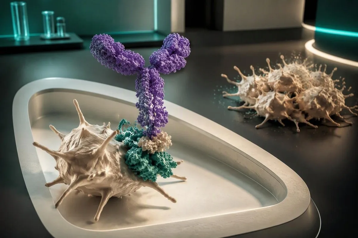
A positive PF4 antibody screen detects antibody binding—not necessarily platelet activation or HIT. Diagnosis depends on platelet-count changes, heparin timing, the clinician-assessed 4Ts score, assay strength and, usually, functional testing; a concerning clinical pattern needs urgent assessment before confirmation returns.


- Positive PF4 screen means antibodies were detected; it does not establish that those antibodies activate platelets or cause HIT.
- Platelet fall greater than 50% can support HIT even when the remaining count exceeds 150 × 10^9/L.
- Heparin timing is usually a platelet fall beginning 5–10 days after exposure; recent exposure can produce a much faster reaction.
- HIT 4Ts score classifies clinical probability as low at 0–3, intermediate at 4–5 and high at 6–8 points.
- Optical density is assay-specific: an ELISA OD of 0.6 and an OD above 2.0 do not carry the same evidential weight.
- Functional confirmation with a serotonin-release assay or another validated platelet-activation assay usually distinguishes binding antibodies from clinically relevant antibodies.
- Urgent suspected HIT requires immediate clinician assessment; heparin withdrawal and alternative anticoagulation may be needed before confirmatory results.
- د بیړني حالت نښې include new breathlessness, chest pain, one-sided leg swelling, a cold painful limb or sudden neurological changes.
What does a positive PF4 antibody screen actually mean?
A PF4 antibody test positive result means the assay detected antibodies that bind its PF4-containing target; it does not prove heparin-induced thrombocytopenia. Clinicians combine that result with platelet changes, exposure timing, the 4Ts score and, usually, a platelet-activation test before assigning the diagnosis.
PF4, or platelet factor 4, is a protein released from platelet granules that can form antigenic complexes with heparin. In immune HIT, certain antibodies—usually IgG—recognize those complexes and activate platelets through Fc receptors; a screening assay detects binding but cannot directly demonstrate that downstream behavior.
A positive result can therefore be analytically correct yet clinically misleading: the antibody exists, but the patient does not have HIT. That distinction is different from a laboratory error, and our explanation of مثبت انټي باډي پایلې helps separate antibody detection from disease diagnosis without treating every reactive result as harmless.
کانټیسټي یو د AI د وینې معاینې شنونکی that can help explain a PF4 report alongside dated platelet results, but it cannot establish HIT from an uploaded screen. Our platform serves more than 2 million users across 127 countries; for this particular result, missing medication dates matter more than the speed of an interpretation.
Why the percentage platelet fall matters more than one count
A platelet decrease greater than 50% is a major HIT clue even when the final count remains within the laboratory reference interval. The clinician needs the trajectory and an appropriate preceding peak—not simply whether today's platelet result has a low flag.
Consider a hypothetical patient whose platelets fall from 380 to 170 × 10^9/L after heparin begins: that is a 55% decrease, despite a final count above 150. Conversely, a stable count of 95 × 10^9/L in someone with longstanding thrombocytopenia supplies much less evidence of a new heparin-related process.
The calculation is 100 × (preceding peak − nadir) ÷ preceding peak, but choosing the peak requires clinical judgment. After surgery, the relevant comparison may be a recovering postoperative peak rather than the admission count; our guide to د وینې ازموینو ترمنځ بدلونونه explains why the sequence can change the interpretation.
A platelet nadir below ۱۰ × ۱۰^۹/L is unusual for uncomplicated HIT and should broaden the differential, although overlapping illnesses can produce very low counts. I would also check whether the decline began before heparin, whether a dilutional fall followed major fluid administration and whether the laboratory reported platelet clumping—three details a single PF4 result cannot answer.
Which heparin exposure dates make HIT more likely?
Typical immune HIT produces a platelet fall beginning 5–10 days after heparin starts. A fall within approximately 24 hours can fit rapid-onset HIT when the patient has circulating antibodies from recent exposure, especially heparin received within the preceding 30 days.
Heparin exposure includes more than a visible infusion. Low-molecular-weight heparin injections, catheter flushes, dialysis anticoagulation and procedural doses can all matter; a patient may remember only the daily injection while the medication administration record documents additional exposures. Ask for dates from the preceding 100 days, not just the current admission.
A fall on day 2 without recent heparin exposure is less typical for immune HIT than a fall on day 7, but the count history must be complete. The first abnormal count may have been measured days after the actual decline began, which is why post-discharge laboratory changes need to be matched to inpatient records rather than interpreted as a new outpatient event.
Delayed-onset HIT can become apparent after heparin has already stopped, sometimes with thrombosis after discharge. If a patient develops new leg swelling several days after leaving hospital, the absence of a current heparin prescription does not settle the question; the clinician needs the previous exposure, platelet trend and symptoms together before dismissing a positive screen.
How clinicians use the HIT 4Ts score before interpreting PF4
د HIT 4Ts score estimates clinical probability using thrombocytopenia, timing, thrombosis and other explanations for the platelet fall. Scores of 0–3 are low probability, 4–5 intermediate and 6–8 high; the score should be assessed by a clinician using reliable records.
Each of the 4 categories receives 0, 1 or 2 points. For thrombocytopenia, a fall greater than 50% with a nadir at least 20 × 10^9/L earns 2 points; a 30–50% fall or nadir of 10–19 earns 1, while a smaller fall or nadir below 10 earns 0 under the standard scoring system.
In their 2012 Blood meta-analysis, Cuker and colleagues reported a negative predictive value of 99.8% for a low 4Ts score; intermediate and high scores had substantially weaker positive predictive performance. That makes a correctly assessed low score useful for exclusion, not a reason to call every score of 4 a diagnosis (Cuker et al., 2012).
زه توماس کلاین، ایم ډي, Chief Medical Officer at Kantesti, and my approach to a score near the 3-versus-4 boundary is to identify which fact would change the category. Missing counts or an undocumented flush can shift the result; our بایومارکر تشریح لارښود provides background, but neither a patient calculator nor an AI-generated score replaces clinician adjudication.
Why the fourth T often causes disagreement
د other-causes category awards 2 points when no alternative cause is apparent, 1 when another cause is possible and 0 when another cause is definite. Sepsis, cardiopulmonary bypass, chemotherapy and mechanical circulatory support may coexist with HIT, so identifying an alternative is not automatically equivalent to proving it explains the entire platelet trajectory.
What PF4 antibody optical density can—and cannot—tell you
PF4 antibody optical density, or OD, measures signal strength in certain enzyme-linked immunosorbent assays. A stronger signal generally raises the likelihood of platelet-activating antibodies, but cutoffs and predictive values are assay-specific; OD is neither an antibody concentration nor a universal HIT severity scale.
یو OD of 0.6 just above one laboratory's cutoff is not equivalent to an OD of 2.4 on that same assay. In their quantitative study, Warkentin and colleagues found that increasing PF4 enzyme-immunoassay OD strongly tracked the probability of a strongly positive serotonin-release assay; their numerical relationships cannot be transferred unchanged to every laboratory method (Warkentin et al., 2008).
Some ELISAs use a positivity threshold around 0.4, while other laboratories use different cutoffs or reporting conventions. A chemiluminescent assay may report units instead of OD, and a rapid qualitative assay may report only positive or negative; our explanation of کیفی په پرتله کمي پایلې helps prevent false comparisons between unlike methods.
د assay name, antibody class, numeric result and laboratory cutoff before comparing two reports. A highly positive ELISA with a high 4Ts score can make HIT sufficiently likely that functional confirmation is not always required under the ASH pathway, but that exception belongs to the treating team—not to a patient interpreting an isolated OD above 2.
Why IgG-specific and polyspecific PF4 screens differ
IgG-specific PF4 assays focus on the antibody class usually responsible for immune HIT. Polyspecific assays may also detect IgA and IgM, which can increase positive results without establishing platelet activation; the report's antibody class therefore changes how a positive screen should be weighed.
The mechanism matters: pathogenic IgG can bridge PF4-containing complexes to platelet FcγRIIa receptors, promoting activation and thrombin generation. Detecting IgA or IgM binding alone does not demonstrate that mechanism, so two laboratories may produce different screening conclusions without either having made an obvious analytical mistake.
A positive antibody assay is not automatically a functional assay—a distinction also relevant to a positive direct Coombs result. The conditions are different, but the interpretive lesson is useful: identifying antibodies on a laboratory target and proving a clinically significant downstream effect are separate questions that require different evidence.
Neither IgG specificity nor a heparin-inhibition step turns an ELISA into proof of platelet activation. An inhibition step can support binding specificity, while functional testing examines behavior; I would document these separately rather than compress them into the phrase 'HIT test positive,' particularly when the platelet fall is only 20% or the timing is poorly documented.
How serotonin-release and other functional assays confirm HIT
A functional HIT assay tests whether the patient's antibodies activate donor platelets under controlled conditions. The serotonin-release assay, or SRA, and heparin-induced platelet activation assay, or HIPA, provide evidence that a positive PF4 binding screen reflects a platelet-activating antibody.
کلاسیک SRA measures serotonin released from prepared donor platelets after exposure to patient serum and different heparin concentrations. Many protocols look for release at low heparin concentrations with suppression at a high concentration, commonly د کولوریکټل سرطان له درملنې وروسته باید په وخت سره وکتل شي، په ځانګړي ډول که د تکرار ازموینې پر مهال لوړوالی ښيي. د CA 19-9 کچه له; the laboratory's complete validated criteria—not one percentage copied from a website—determine whether the pattern is positive.
HIPA assesses platelet aggregation, while other specialist methods use flow cytometry or PF4-enhanced activation systems. Donor platelet responsiveness, sample handling and method selection affect performance, so a report should identify the test rather than merely say 'confirmation negative'; turnaround may take several days when specimens travel to a reference laboratory.
کې پوښل شوې ده. یو نورمال aPTT or INR does not replace functional confirmation, and an elevated D-dimer does not confirm HIT. Our clotting test interpretation guide explains what these other tests measure; they can guide treatment monitoring or investigate complications, but they do not answer whether anti-PF4 antibodies activate platelets.
What if PF4 is positive but the functional test is negative?
A positive PF4 screen with a negative functional assay usually means HIT is unlikely, particularly when the clinical probability is low or the screening signal is weak. A high 4Ts score with a strongly positive immunoassay is a discordant pattern that warrants specialist review rather than automatic dismissal.
For example, a hypothetical patient with a 4Ts score of 2, an ELISA OD of 0.55 and a negative SRA has very different evidence from someone with a score of 7, an OD of 2.8 and a negative SRA. The first pattern strongly favors nonpathogenic antibody detection; the second raises questions about the clinical diagnosis and functional assay sensitivity.
False-negative functional assays can occur, including uncommon cases investigated with PF4-enhanced testing at specialist centers. Conversely, a strongly positive screen does not make every alternative diagnosis disappear; the clinician should review donor responsiveness, specimen collection relative to treatment and whether the suspected thrombosis was objectively confirmed before ordering another assay.
کانټیسټي یو دی AI د وینې ازموینې تشریح پلیټفارم that can help organize these contradictory findings, but it cannot overrule a hematologist or validate a reference laboratory assay. Our د کلینیکي تایید (validation) تګلاره describes the role of clinical standards, while our report source-checking guidance explains why the original numeric report and method should remain available.
When suspected HIT needs action before confirmation returns
Intermediate- or high-probability suspected HIT needs urgent clinician assessment without waiting for an SRA. The treating team generally stops heparin and considers a non-heparin anticoagulant while testing proceeds, because the immediate concern is thrombosis—not simply a low platelet count.
د 2018 American Society of Hematology guideline recommends stopping heparin in intermediate- or high-probability suspected HIT and selecting non-heparin anticoagulation according to bleeding risk and other indications. With an intermediate score, prophylactic intensity may be appropriate while awaiting the immunoassay when bleeding risk is high and no separate therapeutic indication exists; a positive immunoassay generally supports therapeutic-intensity treatment unless contraindicated (Cuker et al., 2018).
New chest pain, unexplained breathlessness, one-sided leg swelling, a cold painful limb or sudden neurological symptoms require emergency assessment. Someone with those symptoms should not spend the next hour uploading reports or waiting for an outpatient callback; our explanation of urgent chest pain testing makes the same distinction between laboratory interpretation and emergency evaluation.
A hospitalized patient should tell the nurse or treating doctor immediately; an outpatient with a concerning result should contact the prescribing team urgently, ideally before the next scheduled heparin dose. Do not substitute, restart or adjust anticoagulants yourself: an isolated positive screen can lead to unnecessary treatment, while true suspected HIT can make continued exposure dangerous.
How clinicians choose treatment while HIT testing is pending
Non-heparin anticoagulant selection depends on clinical stability, kidney and liver function, bleeding risk and planned procedures. Options include argatroban, bivalirudin, danaparoid where available, fondaparinux and selected direct oral anticoagulants; a positive PF4 result alone is not a prescribing instruction.
Short-acting intravenous agents are often favored when a patient is unstable or may need an urgent procedure, because their effects are easier to adjust. Argatroban requires particular attention to hepatic impairment, while several alternatives need renal consideration; our kidney function test guide explains why one apparently normal creatinine value may not settle drug clearance.
Warfarin should not be started during acute HIT before platelet recovery, usually to at least 150 × 10^9/L; premature use can worsen the prothrombotic imbalance. Argatroban can also raise the INR independently of warfarin, so our explanation of what INR measures should not be used as a self-directed transition protocol.
Kantesti cannot recommend an anticoagulant dose from a PF4 screen, and routine platelet transfusion is generally avoided in acute HIT unless bleeding or another compelling indication changes the balance. For HIT with thrombosis, treatment commonly continues for ۳–۶ میاشتې; isolated HIT usually requires a shorter clinician-directed course, at least through platelet recovery, rather than an automatic six-month prescription.
Why positive PF4 screens are tricky after cardiac surgery
له cardiopulmonary bypass, platelet counts often fall for nonimmune reasons, and PF4 antibodies may develop without clinical HIT. A recovery followed by a second platelet fall around days 5–10 is more concerning than an immediate postoperative decline that steadily improves.
د biphasic platelet pattern is the practical clue: an early fall after bypass, a partial recovery and then a new decline compatible with antibody-mediated timing. A persistently low count starting immediately after surgery is less characteristic of HIT, although complicated postoperative courses require careful review rather than a shortcut based on the graph alone.
Routine PF4 screening after surgery can find antibodies that would never have caused symptoms, creating a misleading HIT label and unnecessary alternative anticoagulation. An elevated D-dimer has the same context problem after a major procedure; our discussion of D-dimer غلط مثبتونه explains why recent surgery reduces the specificity of another commonly alarming laboratory result.
In intensive care, sepsis, extracorporeal circuits, transfusion and medications can each contribute to thrombocytopenia, while heparin exposure may be nearly continuous. I would reconstruct at least the first 10 treatment days where available and look for a new change in slope or confirmed thrombosis, not let the presence of an alternative cause erase a genuinely compatible second fall.
Which other conditions can explain falling platelets?
Sepsis, disseminated intravascular coagulation, drug-related thrombocytopenia, thrombotic microangiopathy and sample-related platelet clumping can mimic parts of the HIT picture. A positive PF4 screen does not exclude these diagnoses, and some require urgent treatment through a completely different pathway.
د EDTA پورې تړلې د پلیټلېټونو ټوټې کېدل (clumping) can create an artificially low automated count without a true fall in circulating platelets. The laboratory may examine a smear and repeat the count using an alternative collection method; our guide to د EDTA له امله د پلیټلیټونو ټوټې کېدل explains why the repeat method and any dilution correction should be documented rather than assumed.
Low platelets combined with red-cell fragmentation, hemolysis or neurological changes should prompt consideration of thrombotic microangiopathy. Severe ADAMTS13 deficiency, generally activity below 10%, supports TTP in the appropriate setting; our explanation of ADAMTS13 and TTP risk describes why suspected TTP also cannot wait for leisurely outpatient confirmation.
A manual smear can identify schistocytes, but their presence is not a diagnosis of HIT; our discussion of د عاجلو schistocyte موندنو په اړه covers the alternative reasoning. If platelets are 18 × 10^9/L with marked hemolysis, I would resist explaining everything with a weak PF4 positive and instead ask which process best accounts for the complete laboratory pattern.
What to record now—and what a positive result means later
Document the distinction between suspected HIT, confirmed HIT and a positive PF4 screen without HIT. Future prescribing decisions depend on that distinction, because a mistaken lifelong heparin label can complicate dialysis, surgery and thrombosis treatment long after the original platelet count has recovered.
Keep dated platelet counts, all heparin exposures, the clinician's 4Ts assessment, the screening method and numeric result, functional results and thrombosis imaging. A compact timeline is more useful than a folder of undated screenshots; our explanation of medication safety trend tracking shows how serial results can be organized without treating a trend tool as a prescribing service.
Antibodies may remain detectable for weeks to months after platelet recovery, so a repeat positive screen does not necessarily indicate ongoing acute HIT. Conversely, a later negative result does not authorize unsupervised heparin re-exposure after confirmed HIT; specialist decisions depend on the clinical phase, antibody status, procedure and availability of alternatives.
کانټیسټي یو دی د AI لاب ټېسټ د تفسیر خدمت that can help arrange serial reports into a discussion-ready timeline, provided the uploaded dates and units are correct. Our د AI ټکنالوژۍ لارښود explains the interpretation workflow; the clinician should still confirm the final chart label and whether a temporary heparin warning needs to become a documented long-term precaution.
Evidence, clinical review and questions for your next assessment
The safest next step is to ask how your PF4 result fits the clinical probability—not whether positive means certain HIT. د د اکتوبر ۱۱، ۲۰۲۶, this article's diagnostic and treatment framework is anchored to the ASH 2018 guideline and the cited 4Ts and optical-density research, not an invented newer guideline.
راوړي 5 questions to the treating team: What was my percentage platelet fall? Does the timing fit all known heparin exposures? What is my clinician-assessed 4Ts score? Which assay and cutoff produced the positive result? Is functional confirmation needed, and what is the safe treatment plan while it is pending?
زه توماس کلاین، ایم ډي, Chief Medical Officer at Kantesti Ltd, and the distinction I want patients to retain is straightforward: the test can support the diagnosis, but it cannot replace the clinical story. Our طبي مشورتي بورډ provides information about clinical expertise; this article is educational and cannot adjudicate an individual case or provide emergency monitoring.
د ۳ بهرنۍ طبي سرچینې below directly support the HIT discussion. The separately listed Figshare publications on digestive symptoms and women's health are publisher-requested educational resources, not evidence for PF4 assay accuracy or HIT treatment; their DOI identifiers establish where those resources are hosted, not that they are peer-reviewed clinical HIT studies.
پوښتل شوې پوښتنې
ایا د PF4 انټي باډي ازموینه دا معنی لري چې ما HIT لري؟
د PF4 انټي باډي مثبت ازموینه په خپله د هپارین-اړه د تخمدان کمښت تشخیص نه کوي. کلینیکي ډاکټران د پلیټلیټ شمیرو بدلونونه، د هپارین سره د تماس وخت، د 4Ts سکور، او ځانګړي سکرینینګ ازموینې ارزوي. د پلیټلیټ کمښت چې له 50% څخه زیات وي او مناسب وخت ولري، له ثابت پلیټلیټونو سره د یوازې ضعیف مثبت په پرتله ډیر اندیښمن دی. د فنکشنل ازموینې معمولا د دې معلومولو لپاره کارول کیږي چې ایا موندل شوي انټي باډي پلیټلیټونه فعالوي.
ایا کله چې زما د پلیټلیټونو شمیر نورمال وي، زه کولی شم HIT ولرم؟
د HIT پیښه حتی کله چې د پلیټلیټ شمیر له 150 × 10^9/L څخه پورته پاتې شي، ځکه چې فیصدي کمښت مهم دی. له 380 څخه 170 × 10^9/L ته راټیټیدل د 55% کمښت څرګندوي او په مناسبه کلینیکي ترتیب کې د HIT ملاتړ کولی شي. درملنه کونکي کلینیک باید اړونده مخکینۍ لوړوالی غوره کړي او وخت بیاکتنه وکړي. له همدې امله عادي ښکاري وروستی شمیر د HIT احتمال رد نه کوي.
د PF4 انتی باډي اوپټیکل کثافت قوي مثبت څه دی؟
د ELISA آپټیکل کثافت له 2.0 څخه پورته یا لوړ ارزول کیدای شي اکثرا قوي مثبت ګڼل کیږي، مګر تفسیر د دقیقې ازموینې او د لابراتوار د پرېکړې پورې اړه لري. 0.6 ارزښت او 2.4 ارزښت په ورته طریقه مختلف ثبوت وزن لري، مګر د مختلفو میتودونو پایلې ممکن په مستقیم ډول د پرتله کیدو وړ نه وي. د آپټیکل کثافت د ټوټې کیدو شدت نه اندازه کوي او په خپله درملنه نه ټاکي. لوړ 4Ts سکور د قوي مثبت ELISA سره یوځای کیدل ممکن کله ناکله د کلینیکي لارښود تشخیصي لاره کې د فعال تایید غیر ضروري کړي.
What does a positive PF4 test with a negative SRA mean?
A positive PF4 screen with a negative serotonin-release assay usually makes HIT unlikely, especially with a low 4Ts score of 0–3 or a weak screening signal. Most such results reflect antibodies that bind the assay target without activating platelets. A high 4Ts score of 6–8 and a strongly positive immunoassay deserve specialist review despite a negative SRA. Rare discordant cases may require review of assay performance or additional specialist functional testing.
Should treatment wait until the HIT confirmation test comes back?
Treatment should not wait for functional confirmation when the clinician assesses intermediate- or high-probability suspected HIT. A 4Ts score of 4–8 generally triggers urgent assessment, heparin withdrawal and consideration of non-heparin anticoagulation according to bleeding risk and other clinical factors. An isolated positive screen with a properly assessed low score does not automatically require the same treatment. Patients should contact their treating team urgently rather than change anticoagulants themselves.
How long after heparin exposure can HIT develop?
Typical immune HIT causes a platelet fall beginning 5–10 days after heparin starts. Rapid-onset HIT can develop within approximately 24 hours when antibodies persist from recent exposure, especially within the previous 30 days. Exposure 30–100 days earlier can also affect the clinician's timing assessment. Delayed-onset HIT may become apparent after heparin has stopped, so recent hospitalization and procedural doses remain relevant.
همدا نن د AI په مرسته د وینې ازموینې تحلیل ترلاسه کړئ
له 2M+ څخه زیات کاروونکي په ټوله نړۍ کې زموږ په Kantesti باور لري چې د لابراتوار ازموینو تحلیل په فوري او دقیق ډول کوي. خپل د وینې ازموینې پایلې اپلوډ کړئ او په ثانیو کې د 15,000+ بایومارکرونو بشپړه تشریح ترلاسه کړئ.
📚 د څېړنې خپرونې چې حواله شوې دي
کلین، ټي، مېچېل، ایس، او وېبر، ایچ. (2026). د روژې وروسته اسهال، په غایطه موادو کې تور داغونه او د معدې ناروغي لارښود ۲۰۲۶. Kantesti د AI طبي څېړنه.
کلین، ټي، مېچېل، ایس، او وېبر، ایچ. (2026). د ښځو د روغتیا لارښود: تخمدان، مینوپاز او هورمونل نښې. Kantesti د AI طبي څېړنه.
📖 بهرني طبي مراجع
Cuker A et al. (2018). American Society of Hematology 2018 guidelines for management of venous thromboembolism: heparin-induced thrombocytopenia. Blood Advances.
Cuker A et al. (2012). Predictive value of the 4Ts scoring system for heparin-induced thrombocytopenia: a systematic review and meta-analysis. Blood.
Warkentin TE et al. (2008). Quantitative interpretation of optical density measurements using PF4-dependent enzyme-immunoassays. د Thrombosis او Haemostasis ژورنال.
⚕️ طبي ردونه
دا مقاله یوازې د زده کړې لپاره ده او طبي مشوره نه جوړوي. د تشخیص او درملنې د پرېکړو لپاره تل د وړ روغتیايي خدمت وړاندې کوونکي سره سلا وکړئ.
د E-E-A-T باور نښې
تجربه
د ډاکټر تر مشرۍ لاندې کلینیکي بیاکتنه د لابراتواري تفسیر د کاري بهیرونو لپاره.
تخصص
د لابراتواري طب تمرکز پر دې چې بایومارکرونه په کلینیکي شرایطو کې څنګه چلند کوي.
واک ورکول
د ډاکټر توماس کلاین له خوا لیکل شوی، د ډاکټر سارا میچل او پروف. ډاکټر هانس ویبر له خوا بیاکتنه.
اعتبار
د شواهدو پر بنسټ تفسیر د روښانه تعقیبي لارو چارو سره، تر څو اندیښنه کمه شي.