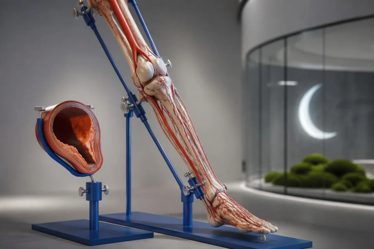
Walking-related calf discomfort can be a circulation warning—even when routine blood tests look reassuring. Foot wounds, nighttime toe pain, and unexpectedly short walking distances deserve a closer look.


- Walking pain from peripheral artery disease often returns with similar effort and improves within about 10 minutes of stopping.
- Quiet symptoms do not exclude arterial disease: reduced activity and diabetic neuropathy can conceal walking-related discomfort.
- Foot wounds that are not healing need prompt assessment; do not wait 2 weeks before seeking help when diabetes or poor circulation is possible.
- Rest pain in the toes or forefoot, especially at night, can indicate severely reduced circulation and warrants urgent assessment.
- Abnormal ABI is an ankle-brachial index of 0.90 or lower, supporting a diagnosis of peripheral artery disease.
- Normal-range ABI is 1.00–1.40, but persistent exertional symptoms may require an exercise ABI or additional vascular testing.
- Noncompressible arteries can produce an ABI above 1.40; a toe-brachial index of 0.70 or lower is abnormal.
- Sudden limb changes—new pain, coldness, pallor, numbness, or weakness—require emergency care immediately.
- خوێن-ئاڵوگۆڕەکان (Blood tests) assess cholesterol, glucose, and kidney-related risk; no routine laboratory result can rule out a blocked leg artery.
What are the early signs of peripheral artery disease?
Peripheral artery disease symptoms include calf, thigh, or buttock discomfort during walking that eases with rest, reduced walking endurance, and foot wounds that heal poorly. Some people have few symptoms. Sudden leg or foot pain with coldness, numbness, or weakness is an emergency—not something to monitor overnight.
نەخۆشی شەریانەکانی ئەندامە دەرەکییەکان, usually shortened to PAD, develops when arteries supplying the legs become narrowed, most often by atherosclerotic plaque. As of October 12, 2026, this article uses the 2024 multisociety ACC/AHA guideline as its main clinical reference; the calendar date does not imply that a newer PAD guideline exists (Gornik et al., 2024).
An illustrative 62-year-old who stops twice on a familiar shopping route may describe a tired calf rather than pain. The useful question is whether the same muscle discomfort returns with comparable effort and disappears after stopping; a change from last month's walking ability can be more revealing than the adjective used to describe it.
Kantestî yek e Analyzerê testa xwînê ya AI that helps explain cholesterol, glucose, and kidney results relevant to vascular risk, but laboratory interpretation cannot establish leg-artery patency. I'm Thomas Klein, MD, writing in the Chief Medical Officer role: my practical priority is matching symptoms to the right examination, not finding a reassuring number; our پسپۆڕی ڕێکخراوەکەمان explains the service behind that distinction.
Why does leg pain when walking improve with rest?
Intermittent claudication symptoms occur when working leg muscles need more oxygen than narrowed arteries can deliver. Discomfort usually develops during walking and improves within about 10 minutes of stopping, often sooner; the patient does not necessarily need to sit down for vascular pain to settle.
Exertional muscle discomfort can feel like cramping, tightness, aching, heaviness, or fatigue rather than a sharp pain. A person who develops calf tightness after roughly 200 meters on level ground may notice it earlier uphill because the oxygen requirement increases; the distance is a personal comparison, not a diagnostic cutoff.
A 3-detail symptom diary is useful: record the activity that brings symptoms on, the muscle affected, and how long recovery takes after stopping. Keep pace and route reasonably similar when comparing days; a shorter distance during faster walking does not automatically mean the artery has become more narrowed, and deliberately provoking severe symptoms is unnecessary.
Nighttime cramps are not the same pattern as exertional claudication, and magnesium supplements do not open narrowed arteries. Our discussion of magnesium for muscle cramps explains why treating every calf complaint as a mineral problem is unhelpful; recurrent activity-related discomfort deserves clinical assessment even when a magnesium result falls within the laboratory range.
Where does PAD pain occur, and what does its location suggest?
PAD-related discomfort commonly affects the calf, but narrowing higher in the arterial system can cause thigh or buttock symptoms. Location offers a clue to the affected circulation, not a precise diagnosis; 1 person can have arterial narrowing at several levels and more than one cause of pain.
Calf claudication often reflects reduced flow through arteries above the calf, including the superficial femoral or popliteal artery. Thigh or buttock discomfort can suggest aortoiliac disease, particularly when walking triggers it consistently; imaging and pressure measurements are needed before assigning the problem to a specific artery rather than guessing from 1 painful area.
Bilateral symptoms do not rule out PAD, and unilateral symptoms do not prove it. In an illustrative 55-year-old with buttock aching, the distinction between lumbar spinal stenosis and arterial disease depends more on standing tolerance, posture, pulses, and physiological testing than on whether the patient points to the hip or the buttock.
Muscle disease can also limit walking, especially when weakness is more prominent than a repeatable exertional ache. A clinician may select creatine kinase or other investigations if the examination suggests that direction; our guide to muscle-related aldolase results explains a different diagnostic pathway, not a substitute for an ABI when the symptom pattern suggests arterial insufficiency.
Why can peripheral artery disease cause few symptoms?
PAD can be clinically quiet because people walk too little to trigger symptoms, gradually adapt their activity, or have neuropathy that reduces pain perception. Absence of classic claudication does not establish normal circulation; the 2001 JAMA study by McDermott and colleagues linked nonclassic symptom patterns with meaningful functional impairment.
Activity adaptation can hide deterioration: someone may choose a nearer parking space, stop using stairs, or lean on a shopping trolley without recognizing those changes as symptoms. Comparing 2 concrete activities—such as reaching the bus stop and climbing one flight of stairs—often gives a clearer history than asking only whether the legs hurt.
Diabetic neuropathy can reduce protective sensation while PAD reduces the oxygen supply needed for repair, creating a particularly risky combination for the feet. A painless blister is therefore not automatically a harmless blister; checking both feet daily and inspecting footwear matters more than waiting for pain to announce a problem that may already be affecting tissue.
Screening is risk-based, not a reason for every well adult to request every vascular test. The 2024 ACC/AHA guideline considers resting ABI screening reasonable in people at increased PAD risk, while advising against it in people without increased risk or suggestive findings; our approach to risk-based annual blood work similarly separates useful assessment from indiscriminate testing.
Which foot wounds and skin changes suggest poor circulation?
Nonhealing toe or foot wounds, a persistently cooler foot, and color changes can suggest reduced arterial circulation, especially with diabetes or smoking exposure. Tissue loss with confirmed PAD may indicate chronic limb-threatening ischemia; the 2-week duration used in its clinical definition is not advice to delay seeking care.
Wound location helps the examination: arterial problems often affect toes, pressure points, or the outer foot, while venous ulcers more often appear around the ankle with swelling. These are tendencies, not rules; 2 processes can coexist, and a photograph cannot reliably establish whether an apparently small wound has adequate circulation beneath it.
Color and temperature changes deserve comparison between both feet rather than judgment in isolation. A foot that becomes pale when raised and reddish when lowered can show an arterial pattern, but hair loss or shiny skin alone is nonspecific; new black discoloration, spreading redness, drainage, or rapidly increasing discomfort needs urgent assessment rather than a home-care experiment.
Normal infection markers do not demonstrate that a wound is safe or adequately perfused. Conte and colleagues' 2019 Global Vascular Guidelines recommend urgent specialist referral for suspected chronic limb-threatening ischemia; our explanation of WBC and CRP disagreements clarifies why clinicians assess the foot directly instead of relying on 1 laboratory result to exclude a serious local problem.
When does nighttime foot pain suggest advanced PAD?
Ischemic rest pain typically affects the toes or forefoot when circulation is inadequate even without exercise. It may worsen when the leg is elevated in bed and ease when the foot hangs down; new persistent symptoms need urgent, preferably same-day assessment rather than waiting 2 weeks to satisfy a diagnostic definition.
Position-dependent relief occurs because lowering the foot can increase the pressure available to move blood toward distal tissues. That observation is a clinical clue, not a treatment plan: sleeping in a chair for several nights may temporarily reduce discomfort while swelling develops and the underlying arterial problem remains unresolved, sometimes with little visible change at first.
Night pain has several causes, including neuropathy, arthritis, cramps, and pressure from footwear or bedding. Burning in both feet with reduced sensation points toward a nerve contribution, whereas persistent forefoot pain with a cool foot or a wound raises greater vascular concern; 2 conditions can occur together, so a neuropathy diagnosis should not end the circulation assessment.
Diabetes changes urgency, because pain may underestimate tissue damage and glycemic control affects healing. Our guide to نیشانە هۆشدارییەکانی زۆربوونی شەکری خوێن بکە explains the metabolic side, but a current glucose of 110 mg/dL does not rule out impaired foot circulation; clinicians still need pulses, vascular measurements, sensation testing, and inspection of the affected area.
How can you distinguish vascular pain from nerve or joint pain?
Vascular claudication usually follows muscle effort and settles after stopping; spinal stenosis often improves with sitting or bending forward, while arthritis centers on a joint. These patterns overlap. Comparing 3 features—trigger, relief, and location—helps direct testing, but symptoms alone cannot safely exclude PAD.
Neurogenic claudication can occur during standing as well as walking, and relief may depend on flexing the spine rather than merely resting the leg muscles. Someone who can cycle comfortably but struggles to stand upright for 5 minutes may have spinal stenosis; this is a useful clue, not proof, because cycling workload and concurrent PAD can complicate that comparison.
Peripheral neuropathy more often produces burning, tingling, numbness, or altered sensation, sometimes when no exercise has occurred. Vitamin B12 deficiency is one possible contributor, and our borderline B12 interpretation explains why the clinical examination matters alongside the number; Kantesti cannot determine whether an artery is narrowed by interpreting a B12 result or a symptom description.
Êşa movikan often comes with stiffness, swelling, reduced movement, or tenderness at a specific joint, whereas vascular discomfort is typically felt in a muscle group. Our discussion of پشکنینی ئازاری ناڕوون covers selective laboratory assessment; in a patient with 2 plausible diagnoses, checking circulation and investigating the joint can both be necessary rather than choosing one explanation prematurely.
Who should take possible PAD symptoms especially seriously?
Smoking exposure, diabetes, older age, chronic kidney disease, hypertension, and abnormal cholesterol increase the likelihood of PAD. The 2024 ACC/AHA guideline identifies age 65 or older as increased risk; younger people also qualify when additional risk factors or atherosclerosis in another arterial territory are present.
مێژووی جگەرەکێشان remains relevant after cessation, although stopping substantially benefits cardiovascular and limb health. A 48-year-old with substantial tobacco exposure and predictable calf discomfort should not be dismissed as too young for PAD; clinicians weigh cumulative exposure, symptom pattern, and examination findings rather than using a birthday as a yes-or-no diagnostic boundary.
Inherited lipid risk can exist despite a sensible diet and an ordinary-looking routine cholesterol result. Our explanation of پشکنینی لیپۆپڕۆتین(a) discusses why a one-time measurement may help cardiovascular risk assessment; no lipoprotein(a) value, however, establishes whether 1 particular leg artery is open, narrowed, or responsible for today's walking symptoms.
Hypertension and kidney disease often cluster with arterial disease, so the evaluation should include current medicines and established diagnoses rather than ordering a large panel without a question. Our guide to hypertension-related laboratory clues describes targeted testing; a patient with known coronary disease plus a new walking limitation deserves attention even if their last 2 blood-pressure readings were controlled.
How does an ankle-brachial index test diagnose PAD?
An ankle-brachial index, or ABI, compares systolic pressure at the ankle with systolic pressure in the arm. For each leg, clinicians generally divide the higher of its 2 ankle-artery pressures by the higher brachial pressure; a resting ABI of 0.90 or lower supports PAD.
ABI measurement usually uses pressure cuffs and a handheld Doppler after the patient has rested supine for roughly 5–10 minutes. Both arms and both ankles are measured because the denominator and the individual leg result matter; cuff size, technique, movement, and failure to allow rest can alter readings enough to confuse an otherwise sensible clinical interpretation.
An ABI is a physiological test, not an ordinary laboratory test and not something calculated from a lipid panel. An ankle systolic pressure of 100 mmHg divided by an arm pressure of 140 mmHg gives an ABI of approximately 0.71; comparing the correct pressures helps avoid an artificially reassuring ratio when one arm pressure is unusually low.
Kantestî yek e پلاتفۆرمی تێکست/وەشاندنی تاقیکردنی خوێنی AI that helps organize the laboratory part of vascular risk assessment, not the cuff-and-Doppler examination. Our ڕێبەری نیشانەی تاقیگە explains that complementary role; when leg symptoms suggest PAD, a normal cholesterol result should not postpone an ABI, and an ABI result should not replace assessment of diabetes, kidney function, or overall cardiovascular risk.
What do ABI results mean, and when can they mislead?
Resting ABI results are abnormal at 0.90 or lower, borderline at 0.91–0.99, normal at 1.00–1.40, and noncompressible above 1.40. A normal resting result does not exclude exercise-related arterial insufficiency, and a high result can reflect stiff arteries rather than unusually good circulation (Gornik et al., 2024).
Arterial calcification can prevent a cuff from compressing an ankle artery adequately, particularly in diabetes or chronic kidney disease. The resulting pressure may be falsely high, making the ABI harder to interpret; our guide to kidney disease staging explains the accompanying kidney assessment, but the vascular solution is often toe pressure testing rather than reassurance from 1 ankle measurement.
A borderline ABI is not a diagnosis by itself and should be interpreted alongside the history and Doppler waveforms. For example, an ABI of 0.96 in someone with reproducible calf symptoms generally prompts consideration of exercise testing, while the same number in a person without symptoms requires a different discussion about risk and whether further evaluation would change management.
ABI severity does not equal limb urgency: wound status, rest pain, sensation, and the speed of symptom onset also matter. An ABI of 0.70 without tissue loss is a different clinical situation from a similar ratio in a person with a deteriorating toe wound; urgent care decisions should never rest on a threshold alone or wait for an especially low result.
When are toe pressures, exercise ABI, or scans needed?
Exercise ABI helps evaluate exertional symptoms when resting ABI is normal or borderline, while toe testing is useful when ankle arteries are noncompressible. A toe-brachial index of 0.70 or lower is abnormal; toe pressure below 30 mmHg indicates severe ischemia and poorer healing prospects in the appropriate clinical setting.
Exercise testing measures ankle pressures after a controlled walking challenge rather than assuming resting circulation will predict performance during activity. Laboratories use different validated protocols, and a single percentage-drop rule is not universally sufficient; the 2024 ACC/AHA guideline supports exercise ABI for suspected chronic symptomatic PAD with resting ABI above 0.90 and no higher than 1.40.
Wound-focused perfusion tests may include toe pressure, transcutaneous oxygen pressure, or skin perfusion pressure because healing depends on local circulation, not only the ankle ratio. Toe measurements are influenced by temperature and technique, so a cold room can matter; a result below 30 mmHg deserves vascular interpretation rather than being treated as an exact prediction that a particular wound cannot heal.
Duplex ultrasound, CT angiography, and MR angiography answer anatomical questions, particularly when revascularization is being considered; not every abnormal ABI requires a contrast scan. Kantesti can help explain kidney laboratory results before that discussion, and our BUN-to-creatinine interpretation supplies background, but contrast decisions depend on the broader clinical context, current eGFR, and whether acute kidney injury is present.
Which sudden leg or foot symptoms require emergency care?
Sudden limb pain, coldness, pallor, numbness, or weakness can signal acute limb ischemia and require emergency assessment immediately. Call your local emergency number or go to an emergency department; do not wait for all 6 traditional warning signs or for a routine appointment, laboratory result, or online interpretation.
The traditional 6 Ps are pain, pallor, pulselessness, paresthesia, paralysis, and poikilothermia—meaning an unusually cold limb. Patients are not expected to assess pulses reliably, and weakness or loss of sensation is particularly concerning; the emergency instruction is based on a sudden change, not on successfully completing a checklist or comparing the foot with a photograph.
Do not attempt home treatment by walking through the symptoms, applying tight compression, using strong heat, or massaging the limb. Note when the change began and bring a medication list if doing so does not delay departure; new weakness developing over 1 hour is not the same problem as stable calf tightness recurring over several months.
A warm, swollen leg suggests a different differential, including deep vein thrombosis, but it still needs prompt assessment; accompanying chest pain, breathlessness, or collapse is an emergency. Our explanation of سنوورەکانی تاقیکردنەوەی ئاساییی خوێنبوون clarifies why a recent normal PT or aPTT cannot exclude a new arterial or venous clot, and blood testing must not delay emergency vascular evaluation.
What helps after PAD is diagnosed?
PAD treatment combines cardiovascular risk reduction, smoking cessation, foot care, and structured exercise when the limb is stable. Supervised exercise programs commonly use 30–45 minutes of active exercise at least 3 times weekly for at least 12 weeks; rest pain, tissue loss, or sudden deterioration requires a different, urgent pathway.
High-intensity statins are recommended for PAD, usually aiming for at least a 50% LDL-C reduction; atorvastatin 40–80 mg and rosuvastatin 20–40 mg are common high-intensity regimens. Treatment is individualized for tolerance and interactions, and our discussion of risk-based LDL targets explains why an ordinary laboratory reference range is not necessarily the right treatment goal after PAD is diagnosed.
Antithrombotic treatment depends on symptoms, procedures, and bleeding risk; clinicians commonly prescribe an antiplatelet drug for symptomatic PAD and may choose additional therapy for selected patients. Do not start aspirin or an anticoagulant on the strength of leg discomfort alone; our هەڵبژاردەی خۆراکی لەسەر بنەمای بەڵگە can support prevention, but no food or supplement replaces indicated medication or restores an abruptly blocked artery.
Follow-up should track function and the feet, not just a better lipid result: compare a familiar walking route, inspect both feet, and report a new wound or rest pain promptly. Kantesti AI can help organize laboratory trends for that discussion, while our ڕێبازی تفسیرکردنی AI explains its boundaries; 1 improved panel is encouraging but does not prove that leg circulation or tissue healing has normalized.
Research publications and the limits of blood-test interpretation
PAD diagnosis requires clinical and vascular assessment, while blood tests clarify associated cardiovascular and kidney risk. The 2 repository publications listed below are background laboratory education, not PAD clinical trials or evidence that AI can diagnose arterial obstruction; the directly relevant medical evidence appears separately in the external references.
Source quality depends on what was tested, not whether a document has a DOI: a repository identifier does not establish peer review, clinical validation, or effectiveness in patients. Kantesti's kidney and urinalysis publications are therefore kept separate from the 2024 PAD guideline, the JAMA symptom study, and the Global Vascular Guidelines; ResearchGate and Academia.edu links below are search links, not claims of verified publication listings.
Kantestî yek e ئامێری توێژینەوەی تاقیکردنەوەی خوێن بە پشتبەستن بە AI for explaining uploaded laboratory information, not measuring ABI or examining wounds. Our ستانداردە کلینیکییەکان و سنووردارێتییەکان describe how to assess interpretation claims; as Thomas Klein, MD, my editorial rule is straightforward: if 1 symptom pattern suggests a threatened limb, the next step is medical care, regardless of what an uploaded report appears to say.
Physician oversight should be verifiable, with authorship, review status, and supporting sources checked before publication rather than inferred from a medical-sounding byline. Our تیمی ڕێنمایی پزیشکی provides organizational context; for an appointment, bring 3 useful items—a symptom timeline, your medication list, and recent laboratory results—but leave immediately for emergency care if sudden limb warning signs develop.
Pirsên Pir tên Pirsîn
نیشانەکانی سەرەتایی نەخۆشیی شریانەکانی ئەندامەکان چین؟
نیشانەکانی سەرەتایی نەخۆشیی شەریانەکانی ئەندامەکانی دەرەوە زۆرجار بریتین لە ئازار، گرژبوون یان ماندووبوونی قاچ، ڕان یان ناوچەی کونگ لە کاتی ڕۆیشتندا، کە دوای وەستان باشتر دەبێت، بە گشتی لە ماوەی نزیکەی ١٠ خولەکدا. هەندێک کەس تەنها کەمبوونەوەی توانای ڕۆیشتن یان برینی پێیەک تێبینی دەکەن کە بە خراپی چاک دەبێتەوە. نێورۆپاتیی پەیوەندیدار بە شەکرە و چاڵاکیی سنووردار دەتوانن شێوازی ئازاری ئاسایی بشارنەوە. دووبارەبوونەوەی نیشانەکانی کاتی چاڵاکی پێویستی بە پشکنینی کلینیکی و زۆرجار پشکنینی پێوەری ئەنکل-براکیال هەیە.
Can you have peripheral artery disease without leg pain?
Peripheral artery disease can occur without recognizable leg pain, particularly when a person is inactive, adapts their walking habits, or has reduced sensation from neuropathy. The absence of pain does not establish normal arterial circulation or eliminate cardiovascular risk. An ABI of 0.90 or lower supports PAD even when classic claudication is absent. Testing decisions depend on risk factors, examination findings, and whether symptoms or wounds suggest impaired circulation.
How is PAD leg pain different from sciatica or arthritis?
PAD-related claudication usually affects a muscle during activity and improves after stopping, often within about 10 minutes without requiring a change in posture. Spinal stenosis may improve mainly with sitting or bending forward, while sciatica can cause radiating pain and tingling. Arthritis more often produces pain, stiffness, or reduced movement around a specific joint. These conditions can coexist, so symptom patterns guide testing rather than definitively excluding PAD.
What is a normal ankle-brachial index?
A normal resting ankle-brachial index is 1.00–1.40 under the 2024 ACC/AHA PAD guideline classification. An ABI of 0.90 or lower is abnormal, and 0.91–0.99 is borderline. Values above 1.40 suggest noncompressible ankle arteries and usually require toe-brachial testing. Persistent walking-related symptoms may require exercise ABI even when the resting result is normal.
Can normal blood tests rule out peripheral artery disease?
Normal routine blood tests cannot rule out peripheral artery disease because they do not measure pressure or flow through the leg arteries. Cholesterol, glucose, and kidney tests help assess associated risk and plan treatment. A person can have PAD with an LDL cholesterol result inside the laboratory reference range. A resting ABI of 0.90 or lower supports PAD, while some symptomatic patients need exercise ABI, toe testing, or imaging.
When is a nonhealing foot wound urgent?
A foot wound that is worsening or failing to heal needs prompt assessment, particularly in a person with diabetes, known PAD, or reduced foot sensation. The 2-week duration used in definitions of chronic limb-threatening ischemia is not a reason to wait before seeking care. Black discoloration, spreading redness, drainage, fever, or associated rest pain increases urgency. Sudden coldness, numbness, weakness, or severe limb pain requires emergency assessment immediately.
Which sudden PAD symptoms mean I should go to the emergency department?
Sudden leg or foot pain with new coldness, pallor, numbness, or weakness can indicate acute limb ischemia and requires emergency care immediately. Do not wait for all 6 traditional signs—pain, pallor, pulselessness, paresthesia, paralysis, and poikilothermia—to develop. Call your local emergency number or go to an emergency department without waiting for an ABI appointment or blood test. Do not try to treat the problem with vigorous walking, massage, strong heat, or tight compression.
ئەمڕۆ AI-پاوەرد لەسەر تاقیکردنەوەی خوێن بەدەست بهێنە
بە یارمەتی زیاتر لە 2 ملیۆن بەکارهێنەر لە هەموو جیهاندا کە Kantesti دەستپێدەکەن بۆ تاقیکردنەوەی لابراتۆری ڕاست و بەهێز لە کاتێکی کەم. ڕەخنەی تاقیکردنەوەی خوێنت بنێرە و تفسیرێکی تەواو لە 15,000+ نیشانەی زیستی (biomarkers) لە ماوەی چرکەکاندا وەرگرە.
📚 توێژینەوە سەرچاوە پەیوەندیدارەکان
کلاین، ت.، میچێڵ، س.، & وێبەر، ه. (2026). Rêjeya BUN/Kreatînîn Şirovekirî: Rêbernameya Testa Fonksiyona Gurçikan. Kantesti توێژینەوەی پزیشکی AI.
کلاین، ت.، میچێڵ، س.، & وێبەر، ه. (2026). Urobilinogen لە تاقیکردنەوەی پیشاب: ڕێنمای گشتی Urinalysis 2026. Kantesti توێژینەوەی پزیشکی AI.
📖 سەرچاوەی پزیشکی دەرەکی
Gornik HL et al. (2024). 2024 ACC/AHA/AACVPR/APMA/ABC/SCAI/SVM/SVN/SVS/SIR/VESS Guideline for the Management of Lower Extremity Peripheral Artery Disease. .
McDermott MM et al. (2001). Leg symptoms in peripheral arterial disease: Associated clinical characteristics and functional impairment. JAMA.
Conte MS et al. (2019). Global vascular guidelines on the management of chronic limb-threatening ischemia. Journal of Vascular Surgery.
⚕️ Daxuyaniya Bijîşkî
ئەم مادەیە تەنها بۆ. I think I must continue but user expects all items.
سەنگەری باوەڕپێکردن E-E-A-T
Tecribe
ڕەوی پشکنینی کلینیکی لەلایەن پزیشکەوە بۆ ڕێکخستنی ڕووداوەکانی لێکدانەوەی ئازمایش.
Pisporî
گرێدانی لابراتۆرییەی پزیشکی بەوەی چۆن بایۆمارکەرەکان لە کۆنتێکستی کلینیکی دەگۆڕن.
Desthilatdarî
نووسراوە لەلایەن د. توماس کلاین بە پشکنینی لەلایەن د. سارا میچێڵ و پڕۆف. د. هانس وێبەر.
Bawerî
لێکدانەوە بە بنەمای دڵنیابوون (Evidence-based) بە ڕێڕەوی دوایینەوەی ڕوون بۆ کەمکردنەوەی هەست بە ترس/هەڵوەشاندن.