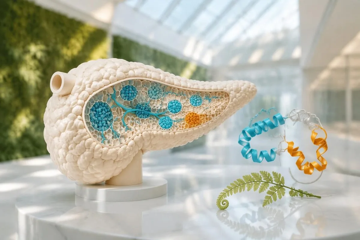
Elevated proinsulin cannot diagnose insulinoma by itself. Clinicians need a sample taken during documented hypoglycemia, interpreted with glucose, insulin, C-peptide and medication screening—not simply a high result from routine metabolic testing.

Негізгі тұжырымдар
- Proinsulin test results become diagnostically useful when measured alongside laboratory-confirmed low glucose, commonly below 55 mg/dL (3.0 mmol/L) in an adult hypoglycemia evaluation.
- High proinsulin levels alone do not establish insulinoma; insulin resistance, medication effects, kidney impairment and assay differences can change interpretation.
- Endogenous insulin secretion is supported by insulin at least 3 µIU/mL, C-peptide at least 0.6 ng/mL and proinsulin at least 5 pmol/L during hypoglycemia, interpreted as a pattern.
- The 5 pmol/L threshold is a guideline decision point during low glucose, not a universal upper reference limit or a tumor-specific cutoff.
- Medication screening must include relevant sulfonylureas and meglitinides because these drugs can reproduce an insulinoma-like laboratory pattern.
- A supervised fast may last up to 72 hours when suspected fasting hypoglycemia cannot be captured spontaneously; never recreate this test at home.
- Meal-related episodes may require a supervised mixed-meal assessment, sometimes extending to 5 hours, rather than a prolonged fast.
- Immediate safety comes first: an alert person who can swallow can take 15–20 g of fast-acting carbohydrate and recheck glucose after 15 minutes.
Does a high proinsulin test result mean insulinoma?
A high proinsulin test result does not diagnose insulinoma. The clinically meaningful question is whether pancreatic insulin secretion remains active when laboratory plasma glucose is low—often below 55 mg/dL (3.0 mmol/L)—and whether medications or other explanations account for that pattern.
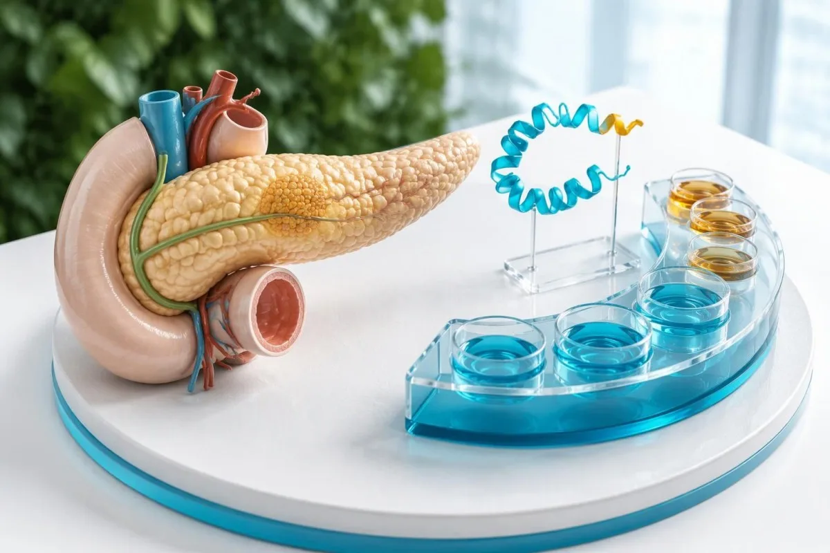
Ан insulinoma is an insulin-secreting pancreatic neuroendocrine growth; the proinsulin result cannot establish its location, behavior or presence. A value of 18 pmol/L drawn with glucose of 104 mg/dL answers a different question from the same value drawn with glucose of 42 mg/dL, because normal pancreatic secretion should become strongly suppressed during hypoglycemia.
Кантести - бұл AI қан анализінің анализаторы that helps explain why proinsulin at 18 pmol/L cannot be interpreted independently of the simultaneous glucose value. Our биомаркерді интерпретациялау нұсқаулығы supports reading laboratory results in context, but an AI explanation cannot collect a critical sample, exclude medication exposure or confirm a pancreatic diagnosis.
The Endocrine Society guideline recommends evaluating an adult hypoglycemic disorder when Уиппл триадасы is documented: compatible symptoms, low plasma glucose and improvement after glucose rises (Cryer et al., 2009). Those 3 elements prevent an isolated laboratory flag or a sensor alarm from becoming a premature insulinoma label; severe symptoms still require treatment before every diagnostic detail is available.
What does proinsulin measure in pancreatic secretion?
Proinsulin is the precursor from which pancreatic beta cells produce insulin and C-peptide. Processing leaves mature insulin with 2 peptide chains, while C-peptide is released separately; circulating proinsulin reflects both secretion and how efficiently that precursor is processed.
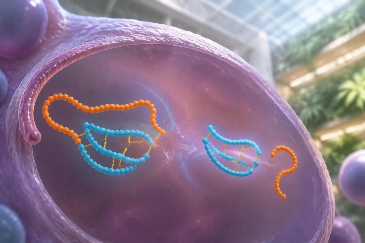
Proinsulin is not simply another name for insulin, and the 2 tests are not interchangeable. Most precursor processing occurs inside beta-cell secretory granules before release; increased secretory demand or abnormal processing can increase circulating precursor molecules, so a high result may reflect beta-cell workload rather than autonomous secretion from a pancreatic growth.
C-peptide helps identify endogenous insulin production because injected insulin does not contain C-peptide. During hypoglycemia, C-peptide at least 0.6 ng/mL supports continued internal secretion when interpreted with the other results; outside hypoglycemia, our explanation of high C-peptide results describes why metabolic demand and kidney clearance can produce a similar flag.
Some laboratories measure intact proinsulin, while other methods detect additional processing intermediates to differing degrees. Two reports both expressed in pmol/L may therefore have different reference intervals and diagnostic performance; when comparing an 8 pmol/L result with an earlier 14 pmol/L result, check the method and sampling conditions before assuming pancreatic function improved.
What counts as documented hypoglycemia before testing?
Documented hypoglycemia requires a reliable glucose measurement interpreted with symptoms and recovery. Laboratory plasma glucose below 55 mg/dL (3.0 mmol/L) is a commonly used diagnostic context in adults without diabetes; a home meter or continuous glucose monitor alone does not establish insulinoma.
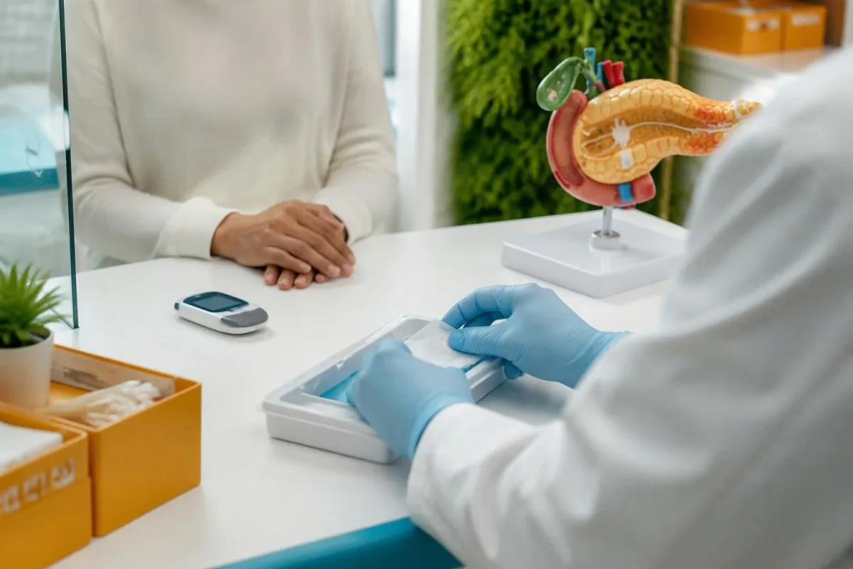
Whipple’s triad links symptoms to the low glucose rather than merely showing that both occurred on the same day. For example, shakiness with a laboratory glucose of 43 mg/dL followed by improvement after carbohydrate is more informative than shakiness with glucose of 91 mg/dL; our глюкозаның төмендеу себептері guide explains competing medication, illness and fasting explanations.
Continuous glucose monitors measure interstitial glucose, and their readings can lag behind circulating glucose during rapid changes. A nighttime sensor value of 48 mg/dL can also result from pressure on the sensor; discuss repeated alerts with a clinician, but do not deliberately avoid food to reproduce an unconfirmed episode or treat a sensor trace as biochemical proof.
Palpitations, sweating and tremor are autonomic symptoms, while confusion, impaired coordination and unusual behavior can reflect inadequate glucose delivery to the brain. Symptoms with glucose of 85 mg/dL need another explanation, potentially including causes covered in our жүрек қағуын бағалау нұсқаулығы; absence of classic sweating does not exclude hypoglycemia, especially after recurrent episodes.
Which results belong beside proinsulin during low glucose?
The critical sample includes plasma glucose, insulin, C-peptide, proinsulin, beta-hydroxybutyrate and hypoglycemic medication screening. When glucose is below 55 mg/dL, clinicians ask whether insulin-related markers are inadequately suppressed, rather than whether they exceed ordinary fasting reference ranges.
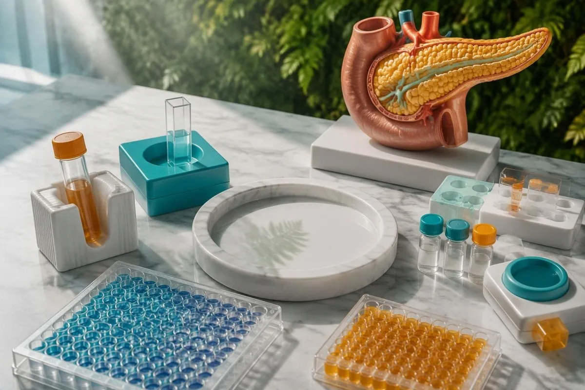
The Endocrine Society guideline describes insulin at least 3 µIU/mL, C-peptide at least 0.6 ng/mL and proinsulin at least 5 pmol/L during hypoglycemia as evidence supporting endogenous hyperinsulinism (Cryer et al., 2009). These values are context-dependent decision thresholds, not an independent checklist that diagnoses a pancreatic growth; clinical circumstances, kidney function and analytical reliability still matter.
Beta-hydroxybutyrate of 2.7 mmol/L or less supports an insulin-mediated mechanism because insulin restrains ketone production. A low ketone result is supportive rather than tumor-specific: injected insulin and insulin-releasing medications can also suppress ketones, while our бета-гидроксибутиратты интерпретациялау бойынша нұсқаулық explains why nutritional ketosis and ketoacidosis represent different clinical questions.
Samples should be collected during the same hypoglycemic episode, ideally before carbohydrate or intravenous glucose when that does not delay treatment. Pairing proinsulin from 09:00 with glucose from 11:00 can create a misleading pattern; in a supervised setting, a glucose increase of at least 25 mg/dL after intravenous glucagon provides additional evidence of preserved hepatic glycogen in an insulin-mediated episode.
| Деңгей | Аралық | Мағынасы |
|---|---|---|
| Low plasma glucose | <55 mg/dL (<3.0 mmol/L) | Common diagnostic context for adult hypoglycemia assessment; symptoms and sampling reliability remain necessary. |
| Insulin not suppressed | ≥3 µIU/mL | Supports inappropriate insulin activity during hypoglycemia; not an ordinary high-insulin definition. |
| C-peptide not suppressed | ≥0.6 ng/mL (≥0.2 nmol/L) | Supports endogenous secretion; insulin-releasing medications and kidney impairment must be considered. |
| Proinsulin not suppressed | ≥5 pmol/L | Guideline evidence supporting endogenous hyperinsulinism, not an insulinoma-specific cutoff. |
| Ketones suppressed | Beta-hydroxybutyrate ≤2.7 mmol/L | Supports insulin or insulin-like activity; does not distinguish a growth from medication effects. |
| Medication screen | Relevant secretagogues assessed | A detected sulfonylurea or meglitinide can explain an otherwise insulinoma-like pattern. |
Why is 5 pmol/L not a universal insulinoma cutoff?
Proinsulin of 5 pmol/L is a hypoglycemia decision threshold, not a universal normal-range ceiling. Its specificity for insulinoma is limited when used alone; higher end-of-fast thresholds have performed better in selected studies, but they cannot be transferred uncritically between assays and populations.

Guettier et al. examined this problem in the Journal of Clinical Endocrinology & Metabolism in 2013 and found that an end-of-fast proinsulin threshold around 22 pmol/L discriminated insulinoma cases from controls better than 5 pmol/L in their study population. That finding explains why a specialist may treat 6 pmol/L differently from 60 pmol/L, without declaring either value diagnostic in isolation.
A laboratory reference interval describes results expected under that laboratory’s specified conditions; it does not necessarily describe appropriate hormone suppression during hypoglycemia. In a clearly hypothetical laboratory with a fasting interval of 1–8 pmol/L, a result of 7 pmol/L would not be flagged high, yet it could still be inadequately suppressed if simultaneous plasma glucose were 41 mg/dL.
There is no defensible universal set of mild, moderate and critical proinsulin bands for diagnosing insulinoma. Comparing 12 pmol/L with 20 pmol/L requires the glucose context, method and episode timing—not just percentage change; our guide to зертханалық тексерулер арасындағы айырмашылықтар explains why analytical and biological variation can complicate trends.
How can medications mimic proinsulin and insulinoma patterns?
Sulfonylureas and meglitinides can reproduce the biochemical pattern of insulinoma by stimulating pancreatic insulin release. During glucose of 45 mg/dL, high insulin, C-peptide and proinsulin cannot distinguish a growth from these medications without an appropriately timed and sufficiently broad drug screen.
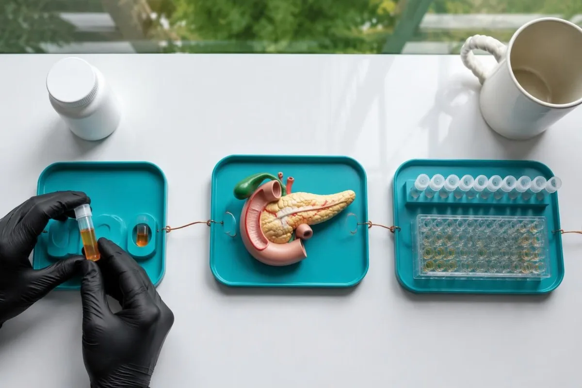
Examples include gliclazide, glimepiride, glipizide, repaglinide and nateglinide; a laboratory panel may not include every agent used internationally. A negative result from a sample collected 24 hours after an episode may be less informative than a simultaneous critical sample, depending on the drug and assay sensitivity; ask which medications were actually tested rather than accepting the word negative without qualification.
Injected insulin typically produces suppressed C-peptide and proinsulin during hypoglycemia, but insulin assays differ in how well they recognize insulin analogues. Thus, glucose of 39 mg/dL with unexpectedly low measured insulin does not always exclude insulin exposure; the same contextual issue is discussed in our low insulin interpretation guide, and specialist laboratories may need a different analytical method.
Medication reconciliation should include prescribed products, accidental mix-ups and access to another household member’s diabetes treatment, without assuming deliberate exposure. Older adults can have prolonged episodes after a single dose when clearance is impaired; our егде жастағы адамдарда дәрі-дәрмектердің әсері туралы нұсқаулығымыз guide supports this review, while insulin antibodies may be investigated when results and episode timing suggest an autoimmune mechanism.
Can kidney impairment or sample errors raise proinsulin?
Reduced kidney clearance can elevate proinsulin and C-peptide, while sample problems can distort the surrounding glucose or insulin results. An eGFR of 24 mL/min/1.73 m² makes straightforward application of standard hypoglycemia thresholds less reliable than it would be with preserved kidney function.
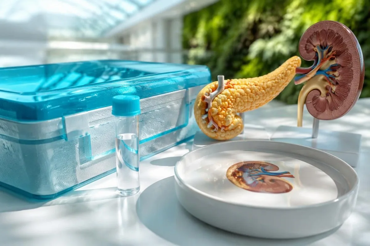
Kidney disease affects both marker clearance and susceptibility to hypoglycemia through medication accumulation, nutrition and altered glucose production. A C-peptide result of 2.1 ng/mL with glucose of 48 mg/dL therefore needs more cautious interpretation in advanced kidney impairment; our low eGFR assessment guide explains why kidney context changes the urgency and meaning of laboratory abnormalities.
Delayed sample processing can create artifactual low glucose because cellular elements continue consuming glucose after collection, while hemolysis can lower measured insulin in susceptible circumstances. A recorded glucose of 44 mg/dL without compatible symptoms may prompt review of processing and a repeat properly handled sample; our kidney chemistry interpretation guide also explains why one chemistry value should not stand in for overall clearance.
Kantesti can help organize a proinsulin result of 16 pmol/L alongside kidney markers, but our explanation cannot determine whether an assay suffered interference or a sample was mishandled. Ask the laboratory about intact versus total proinsulin, insulin cross-reactivity, relevant supplement interference and specimen quality; discordant results sometimes require repeat measurement using another method rather than more aggressive imaging.
What do combined proinsulin results look like in practice?
The same proinsulin value can mean different things at different glucose concentrations. The following 3 hypothetical examples illustrate clinical reasoning, not diagnostic predictions; each assumes a correctly processed sample, and none substitutes for a specialist assessment of the complete episode.
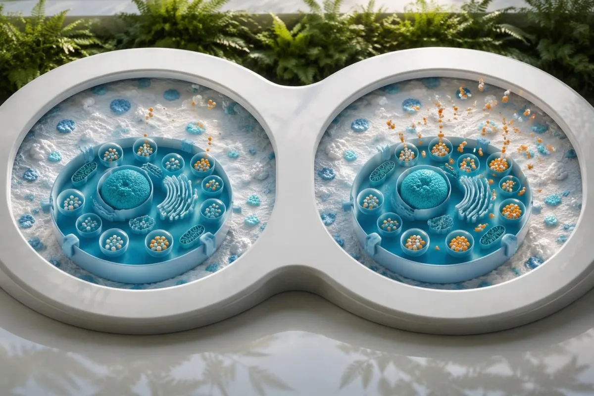
Example 1: an adult has glucose of 106 mg/dL, insulin of 24 µIU/mL, C-peptide of 4.2 ng/mL and proinsulin of 19 pmol/L during routine fasting testing. This pattern can fit increased secretory demand, including insulin resistance; without documented hypoglycemia, it does not establish an insulinoma pathway, even if proinsulin exceeds the laboratory’s reference interval.
Example 2: during confusion, plasma glucose is 42 mg/dL, insulin 8 µIU/mL, C-peptide 1.6 ng/mL, proinsulin 34 pmol/L and beta-hydroxybutyrate 0.4 mmol/L. This pattern strongly supports insulin-mediated endogenous secretion, but a detected sulfonylurea would change the explanation; a negative comprehensive screen and clinical evaluation would make autonomous pancreatic secretion a more serious consideration.
Example 3: plasma glucose is 46 mg/dL, insulin 1.4 µIU/mL, C-peptide 0.9 ng/mL and proinsulin 58 pmol/L during a supervised assessment. Unusually prominent proinsulin secretion can make the insulin result deceptively unimpressive, so one below-threshold marker should not automatically dismiss the pattern; our қан талдауын жүргізу бойынша нұсқаулық explains why same-episode alignment remains necessary.
How is this different from insulin-resistance testing?
Insulin-resistance testing evaluates metabolic demand, not failure to suppress insulin during hypoglycemia. A routine overnight fasting panel with glucose of 98 mg/dL is therefore not equivalent to a critical sample collected at glucose of 43 mg/dL, even when both include insulin and proinsulin.

HOMA-IR estimates insulin resistance from fasting insulin and glucose; the conventional formula using mg/dL is insulin multiplied by glucose, divided by 405. For example, insulin of 15 µIU/mL and glucose of 100 mg/dL produce about 3.7, but there is no universal diagnostic cutoff across assays and populations; our borderline fasting insulin guide explains those limitations.
High proinsulin levels and an increased proinsulin-to-insulin ratio have been studied as markers of beta-cell processing stress, but these measures are not standard stand-alone tests for insulin resistance or insulinoma. Ratios can become unstable when insulin approaches the assay’s detection limit, and mixing pmol/L with µIU/mL creates an invalid calculation; 2 apparently comparable ratios may therefore reflect different methods rather than different biology.
Кантести - бұл AI биомаркерлерін түсіндіру платформасы that explains why glucose of 100 mg/dL with elevated insulin belongs to a different reasoning pathway from symptomatic glucose of 43 mg/dL. In conditions such as PCOS, established metabolic assessment remains more useful than isolated precursor testing; our PCOS diagnostic criteria guide separates reproductive diagnosis from metabolic risk assessment.
When is a supervised fast appropriate for suspected insulinoma?
A supervised fast is appropriate when fasting hypoglycemia is convincingly suspected but a spontaneous critical sample cannot be obtained. The assessment may continue for up to 72 hours, with monitored sampling and predefined stopping rules; an isolated high proinsulin result is not sufficient reason to perform it.

An endocrinology team considers the episode pattern, medication history and quality of previous glucose documentation before arranging a fast. Symptoms before breakfast, during prolonged gaps between meals or after exercise may justify this pathway, whereas a single mildly elevated proinsulin value with glucose of 96 mg/dL usually calls for clarification rather than immediate admission for prolonged fasting.
During the fast, staff monitor symptoms and obtain repeated laboratory samples, increasing sampling frequency as glucose falls; the final critical sample is collected according to the protocol before treatment when safe. The 72-hour duration is a maximum, not a target, and many diagnostic episodes occur earlier; the Endocrine Society protocol explicitly places biochemical assessment and supervised stopping criteria ahead of simply completing a clock interval (Cryer et al., 2009).
Never attempt a 72-hour diagnostic fast at home, including while wearing a glucose sensor. Ordinary meal-timing practices discussed in our үзік-үзік ашығу кезіндегі зертханалық талдаулар жөніндегі нұсқаулығымыз are not substitutes for an endocrine investigation; suspected hypoglycemia may make intentional fasting unsafe, and changes to insulin or other diabetes medicines require a clinician-directed plan.
What if symptoms occur after meals rather than fasting?
Predominantly meal-related hypoglycemia may require a supervised mixed-meal test rather than a prolonged fast. Monitoring can extend to 5 hours after a representative meal, with glucose and hormone samples obtained during symptoms; the meal pattern alone neither proves nor excludes insulinoma.
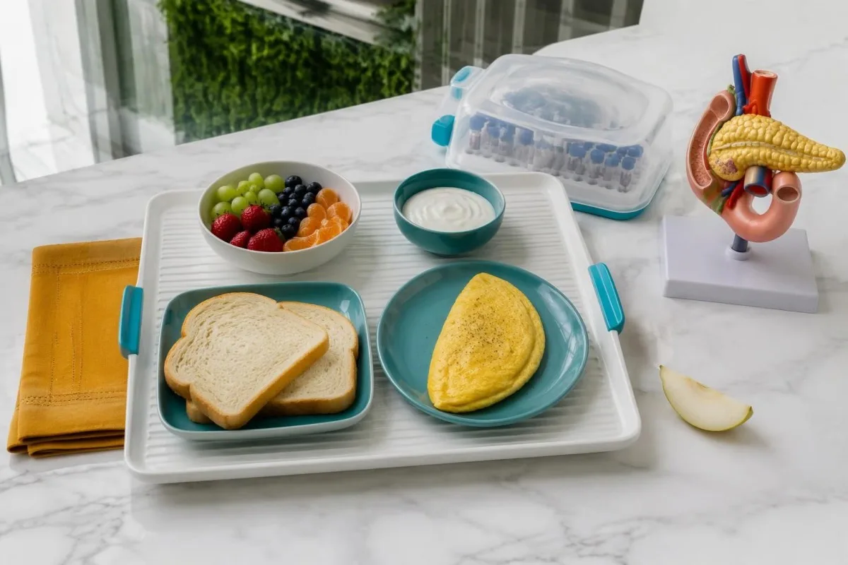
Hypoglycemia after upper gastrointestinal surgery can reflect altered nutrient delivery and exaggerated insulin release, rather than a pancreatic growth. An episode 2–3 hours after eating therefore requires the surgical history as well as the hormone pattern; endogenous hyperinsulinism is a biochemical description, not a promise that imaging will identify a solitary insulinoma.
An oral glucose tolerance test is generally not the preferred investigation for suspected postprandial hypoglycemia because a glucose drink can create results that do not reproduce everyday meals. A supervised mixed meal better matches the clinical trigger, although meal composition and interpretation are less standardized than a fasting protocol; uncertainty should be discussed rather than hidden behind a single post-meal proinsulin cutoff.
Sugar cravings, fatigue and irritability 1–2 hours after lunch do not establish biochemical hypoglycemia. Record meal timing, symptoms and available glucose readings, then seek an appropriate assessment rather than self-ordering repeated proinsulin panels; our sugar cravings testing guide explains why these symptoms alone are insufficient evidence of an insulin-secreting disorder.
When do scans enter the insulinoma diagnostic pathway?
Pancreatic imaging generally follows biochemical confirmation of endogenous hyperinsulinemic hypoglycemia and exclusion of relevant medications. Glucose of 42 mg/dL with an appropriate hormone pattern provides a reason to investigate localization; proinsulin of 20 pmol/L with normal glucose does not justify the same assumption.
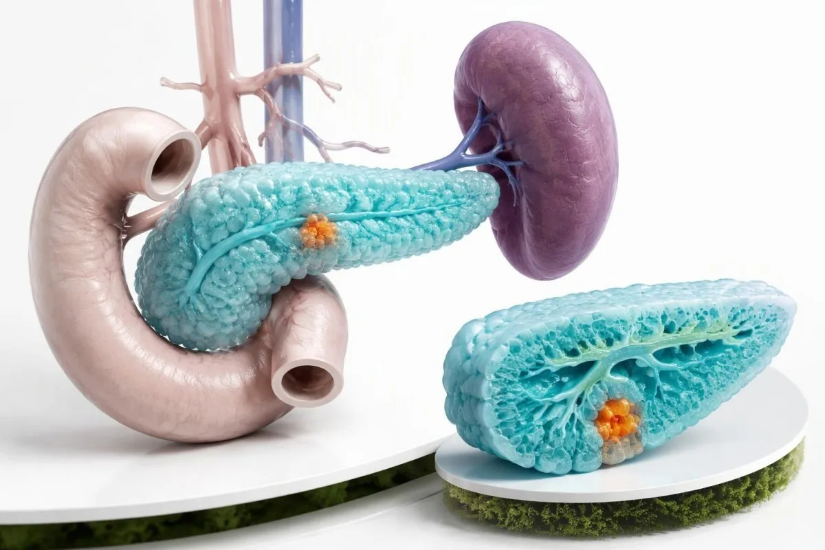
CT, MRI and endoscopic ultrasound can identify or characterize a pancreatic source, but small insulinomas may evade an initial scan. A negative first study does not cancel convincing biochemistry, while an incidental pancreatic finding does not prove that it caused hypoglycemia; those 2 directions of error are why localization and hormone diagnosis must remain separate steps.
When standard imaging is unrevealing, specialist centers may consider receptor-based imaging or selective arterial calcium stimulation with hepatic venous sampling. These are localization tools for a selected patient, not routine responses to a proinsulin result of 11 pmol/L; the choice depends on previous studies, local expertise, availability and whether the clinical pattern supports focal or more diffuse insulin secretion.
Multiple endocrine neoplasia type 1 may become relevant when pancreatic findings coexist with personal or family evidence of other endocrine disorders. A raised gastrin result—for example 300 pg/mL while taking a proton-pump inhibitor—does not independently establish that syndrome; our high gastrin interpretation guide explains why medication effects and clinical context are also necessary here.
What should you do with a high proinsulin report now?
Your next step depends on documented glucose and symptoms, not the proinsulin flag alone. Severe confusion, seizure or inability to swallow requires emergency help; an alert person who can swallow can take 15–20 g of fast-acting carbohydrate and recheck glucose after 15 minutes.
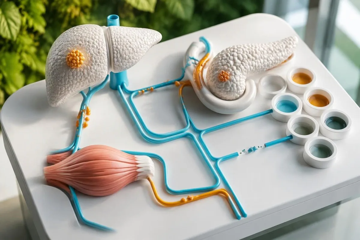
Do not give food or drink to someone who cannot swallow safely; use prescribed rescue glucagon if available and follow the emergency plan. Recurrent confirmed readings below 54 mg/dL, or episodes that require another person’s help, warrant prompt clinical review rather than waiting for another proinsulin result; avoid driving during symptoms and obtain advice about driving safety after recurrent episodes.
Кантести - бұл AI қан талдауы нәтижесін түсіндіру платформасы that can explain uploaded laboratory reports in about 60 seconds, but it cannot monitor a fast or certify that glucose of 43 mg/dL was measured during symptoms. Kantesti Ltd is a UK organization described on our company information page; our role is educational interpretation, not emergency monitoring or autonomous diagnosis.
Kantesti AI explanations should be checked against the report’s units, collection times, medication list and glucose context before any clinical decision. Our жауапты AI қағидаттарымыз explain the need for human oversight; the AI технологиясы бойынша нұсқаулық describes the interpretation workflow, which cannot replace the 6 components of a properly collected critical sample or a specialist-supervised assessment.
Which evidence supports proinsulin test interpretation?
The diagnostic framework is supported by clinical hypoglycemia guidance and studies evaluating insulin and proinsulin during supervised fasting. The 2 medical references below directly address those questions; the separate Figshare publications are general educational resources, not evidence validating an insulinoma cutoff.
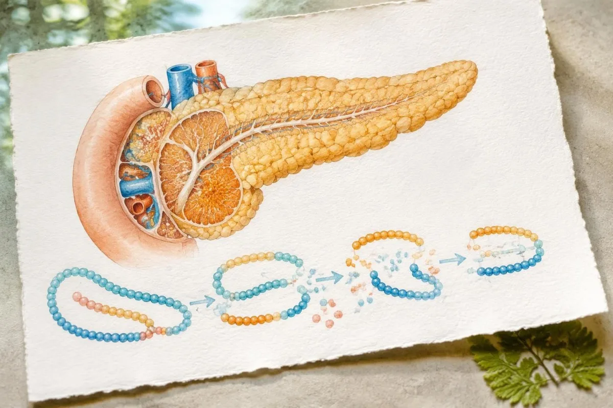
Cryer et al.’s 2009 Endocrine Society guideline provides the framework for Whipple’s triad, critical sampling and a supervised fast of up to 72 hours. Guettier et al.’s 2013 study adds a specific caution: the 5 pmol/L proinsulin decision point has limited specificity for insulinoma when treated as a stand-alone result, and higher study-derived cutoffs require assay-aware interpretation.
The 2 Figshare guides listed below cover hematology markers and digestive symptoms associated with fasting; neither establishes the sensitivity or specificity of a proinsulin test. Our ашығу және ас қорыту белгілері туралы нұсқаулығымыз addresses a different clinical question, and digestive discomfort is not a reason to attempt a prolonged insulinoma investigation without supervision.
Before an endocrine appointment, bring at least 1 complete report showing the glucose value, collection time, proinsulin method if available and any simultaneous insulin or C-peptide measurements. Add a medication list and a brief symptom timeline; our медициналық мазмұн стандарттары explain why we distinguish published clinical evidence, hypothetical examples and platform capabilities rather than presenting them as equivalent proof.
Жиі қойылатын сұрақтар
Проинсулин деңгейінің жоғары болуы инсулиноманы анықтай ала ма?
Проинсулин деңгейінің жоғары болуы глюкоза көрсеткіштері мен клиникалық мәнмәтінді ескермей инсулиноманы анықтауға мүмкіндік бермейді. Зертханалық жолмен расталған гипогликемия кезінде, әдетте 55 мг/дл-ден (3,0 ммоль/л) төмен болғанда, жеткілікті басылмаған проинсулин эндогендік инсулин секрециясын растауы мүмкін. Дәрігерлер инсулинді, C-пептидті, бета-гидроксибутиратты және тиісті дәрілік заттарға скрининг нәтижелерін де бағалайды. Эндогендік секрецияға сай келетін айқын көріністің өзін ұйқы безінің орналасуын анықтауға кіріспес бұрын инсулин бөлінуіне ықпал ететін дәрілерден және басқа себептерден ажырату қажет.
Қандай проинсулин деңгейі инсулиноманы меңзейді?
Гипогликемия кезінде кемінде 5 pmol/L болатын проинсулин эндогендік гиперинсулинизмді қолдайтын клиникалық нұсқаулықтағы шешім шегі болып табылады, бірақ инсулиномаға тән диагноз емес. Guettier және әріптестері 2013 жылы жүргізген зерттеуде зерттелген популяцияда ашығудың соңындағы шамамен 22 pmol/L шегі 5 pmol/L шегіне қарағанда ажырату қабілеті жоғары болғаны анықталды. Талдау әдістері, бүйрек функциясы және сынама алу жағдайлары бұл мәндердің нақты адамға қолданылуына әсер етеді. Глюкоза деңгейі қалыпты кезде алынған жоспарлы талдау нәтижесін гипогликемия кезіндегі ашығудың соңына арналған шек бойынша түсіндіруге болмайды.
Can proinsulin be high because of insulin resistance?
Proinsulin can rise with increased beta-cell secretory demand and altered precursor processing, including circumstances associated with insulin resistance. Proinsulin of 18 pmol/L with glucose of 105 mg/dL therefore does not establish insulinoma. Routine metabolic assessment may include glucose, HbA1c, lipids and selected insulin measurements, depending on the clinical question. Proinsulin and proinsulin-to-insulin ratios are not standard stand-alone diagnostic tests for insulin resistance.
Why are C-peptide and a medication screen needed?
C-peptide and medication screening help distinguish endogenous insulin secretion from injected insulin and insulin-releasing drugs. C-peptide of at least 0.6 ng/mL during hypoglycemia supports continued pancreatic secretion, but sulfonylureas and meglitinides can produce that same pattern. A negative medication screen is only as informative as its drug coverage, sensitivity and collection timing. Kidney impairment and insulin assay recognition of analogues can further complicate interpretation.
Do I need a 72-hour fast for a high proinsulin result?
An isolated high proinsulin result does not automatically justify a 72-hour fast. A supervised fast is considered when fasting hypoglycemia is convincingly suspected and a spontaneous critical sample cannot be obtained. The test has monitored sampling and stopping criteria, and it often ends before the maximum duration. Never perform a prolonged diagnostic fast at home, even with a continuous glucose monitor.
Can insulinoma be present when insulin is not high?
A low or ordinary-looking insulin result does not completely exclude insulinoma during hypoglycemia, particularly when proinsulin secretion is unusually prominent or assay problems are present. Insulin below 3 µIU/mL with markedly elevated proinsulin and inadequately suppressed C-peptide requires specialist interpretation rather than automatic dismissal. The glucose concentration, ketone response, medication screen and analytical method must be reviewed together. Insulin described as normal on a routine report may still be inappropriate at glucose of 42 mg/dL.
What should I do if my glucose is low while waiting for testing?
An alert person who can swallow can take 15–20 g of fast-acting carbohydrate and recheck glucose after 15 minutes, repeating treatment if glucose remains low according to their care plan. Severe confusion, seizure, unconsciousness or inability to swallow requires emergency help, and food or drink should not be given by mouth. Use prescribed rescue glucagon if available and follow the emergency instructions. Do not delay treatment to obtain a proinsulin sample; recurrent episodes need prompt medical review.
Қан анализінің нәтижесін түсіндіріп алыңыз
Зертханалық есепті жүктеңіз, сонда Kantesti AI әр биомаркерді бірнеше секунд ішінде қарапайым тілмен түсіндіреді. Әлем бойынша 2 миллионнан астам адам қолданады.
Сілтемелер
- Klein, T., Mitchell, S., & Weber, H. (2026). B теріс қан тобы, LDH қан талдауы және ретикулоциттер саны жөніндегі нұсқаулық. Kantesti AI медициналық зерттеулері. DOI
- Klein, T., Mitchell, S., & Weber, H. (2026). Ашыққаннан кейінгі диарея, нәжістегі қара дақтар және асқазан-ішек жолдары жөніндегі нұсқаулық, 2026. Kantesti AI медициналық зерттеулері. DOI
- Cryer PE et al. (2009). Evaluation and Management of Adult Hypoglycemic Disorders: An Endocrine Society Clinical Practice Guideline. Journal of Clinical Endocrinology & Metabolism. DOI PubMed
- Guettier JM et al. (2013). The Role of Proinsulin and Insulin in the Diagnosis of Insulinoma: A Critical Evaluation of the Endocrine Society Clinical Practice Guideline. Journal of Clinical Endocrinology & Metabolism. DOI PubMed
- 2 млн+Талданған тесттер
- 127+Елдер
- 100+Тілдер
Медициналық топ туралы


E-E-A-T сенім сигналдары
Тәжірибе
Дәрігер басқаратын зертханалық талдауды түсіндіру жұмыс ағындарын клиникалық шолу.
Сараптама
Биомаркерлердің клиникалық контексте қалай әрекет ететініне зертханалық медицина фокусы.
Билік
Доктор Томас Кляйн медициналық тұрғыдан тексерген, доктор Сара Митчелл мен профессор доктор Ханс Вебердің үлесімен.
Сенімділік
Дабыл қаупін азайту үшін айқын бақылау жолдарымен дәлелге негізделген түсіндіру.
Медициналық ескерту
Бұл мақала тек білім беру мақсатында ғана және медициналық кеңес болып табылмайды. Диагноз және ем туралы шешімдер үшін әрдайым білікті медицина қызметкеріне жүгініңіз.
Жарияланды · Редакция тобына хабарласыңыз
Англия мен Уэльсте тіркелген Kantesti Ltd басып шығарған, компания №. 17090423, Лондон, Біріккен Корольдік.
