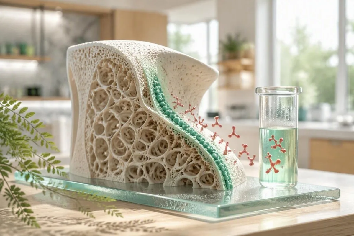
Օստեոկալցինի արդյունքը ոսկրի կառուցման ակտիվության լուսանկար է, ոչ թե ոսկրային խտության կորստի ինքնուրույն ախտորոշում։ Օգտակար հարցն այն է, թե արդյոք արժեքը համապատասխանում է ձեր տարիքին, երիկասարդական ֆունկցիային, գլյուկոզայի վիճակին, վերջին կոտրվածքին և բուժման պատմությանը։.


- Ոսկրի ձևավորման մարկեր. Օստեոկալցինն արտազատվում է օստեոբլաստների կողմից ոսկրի ձևավորման ժամանակ, բայց այն կարող է բարձրանալ նաև, երբ ոսկրի ընդհանուր շրջանառությունը բարձր է։.
- Համընդհանուր միջակայք չկա. Չափահասների օստեոկալցինի նորմալ արժեքները տարբերվում են վերլուծության մեջ; շատ N-MID վերլուծություններ ցույց են տալիս մոտավորապես 11-43 ng/մլ նախքան դաշտանադադարը, բայց լաբորատոր միջակայքը վերահսկվում է։.
- Բարձր արդյունք. Օստեոկալցինի բարձր մակարդակը կարող է առաջանալ աճի, դաշտանադադարի, կոտրվածքի վերականգնման, հիպերթիրեոզի, D վիտամինի անբավարարության հետևանքով երկրորդային հիպերպարաթիրեոզի կամ ոսկրի բարձր շրջանառությամբ հիվանդության դեպքում։.
- Ցածր արդյունք. Օստեոկալցինի ցածր մակարդակը կարող է արտացոլել ոսկրի շրջանառության ճնշումը հակա-ռեզորպտիվ բուժման, գլյուկոկորտիկոիդների, դիաբետի, ցածր էներգիայի մատչելիության կամ հիպոթիրեոզի պատճառով։.
- Ողջերի հնարք. eGFR-ի նվազումը կարող է բարձրացնել շրջանառվող օստեոկալցինը, քանի որ երիկամային մաքրումը նվազում է, ուստի երիկամների քրոնիկ հիվանդությունը փոխում է մեկնաբանությունը։.
- Լավագույն ուղեկիցներ P1NP, CTX, կալցիում, ֆոսֆատ, հանքային ֆոսֆատազ, 25-հիդրոքսիվիտամին D, PTH, կրեատինին/eGFR, TSH, և HbA1c-ն օստեոկալցինին հաղորդում են կլինիկական նշանակություն։.
- Բուժման մոնիտորինգ Ոսկրի դարձի մարկերները կարող են փոխվել թերապիայից 3-6 ամսվա ընթացքում, մինչդեռ DXA սկանը սովորաբար ավելի դանդաղ է փոխվում։.
- Կրկնել իմաստուն Հնարավորության դեպքում օգտագործեք նույն լաբորատորիան, անալիզը, հավաքագրման ժամանակը և ծոմապահության ռեժիմը օստեոկալցինի շարքային չափումների համար։.
Ինչ է իրականում չափում արյան մեջ օստեոկալցինի թեստը
Օստեոկալցինի արյան թեստը չափում է սպիտակուց, որն առաջանում է հիմնականում օստեոբլաստների կողմից, այն բջիջները, որոնք ստեղծում են նոր ոսկրային մատրից։. Բարձր արդյունքը սովորաբար նշանակում է, որ ոսկրի դարձը ակտիվ է, մինչդեռ ցածր արդյունքը հաճախ նշանակում է, որ ոսկրի ձևավորումը ճնշված է; ոչ մի արդյունք ինքնին չի ապացուցում, որ ոսկորը ուժեղ է կամ թույլ։.
Օստեոկալցինը ներառվում է ոսկրի մեջ վիտամին K-կախյալ կարբոքսիլացումից հետո, իսկ ավելի փոքր մասը մտնում է շրջանառության մեջ։ Լաբորատորիաները կարող են չափել ընդհանուր, անվնաս, N-տերմինալ միջին ֆրագմենտ կամ չկարբօքսիլացված օստեոկալցին; դրանք կապված չափումներ են, ոչ փոխարինելի թվեր։ Այդ պատճառով 25 նգ/մլ արժեքը կարող է սովորական լինել մեկ անալիզում և անհնար լինել ուղղակի համեմատել մեկ այլ անալիզի հետ։.
Իմ կլինիկական պրակտիկայում հիվանդները հաճախ ենթադրում են, որ օստեոկալցինը կալցիումի թեստ է։ Այն այդպիսին չէ. կալցիումը խստորեն կարգավորվում է շիճուկում, մինչդեռ օստեոկալցինը ոստեոբլաստների ակտիվության դինամիկ մարկեր է, որը կարող է նկատելիորեն շարժվել կոտրվածքից, նոր դեղորայքից կամ դաշտանադադարից հետո։.
Կանտեստին մի է AI արյան անալիզատոր որը կարդում է օստեոկալցինը հանքային, երիկամային, վահանագեղձի և նյութափոխանակության մարկերների կողքին, այլ ոչ թե ախտորոշումը կցում մեկ դրոշակված գծին։ Սա կարևոր է, քանի որ նորմալ կալցիումի արդյունքը չի բացառում արագացված կմախքային դարձը; մեր բիոմարկերների հղման ուղեցույց բացատրում է, թե ինչու են մարկերները պատասխանում տարբեր կենսաբանական հարցերի։.
Ինչու՞ օստեոկալցինը ոսկրային խտության մոնիտորինգի թեստ չէ
Օստեոկալցինը խորհուրդ չի տրվում որպես ոսկրային խտության մոնիտորինգի պոպուլյացիոն թեստ։ DXA ոսկրային խտության թեստավորումը և կոտրվածքների ռիսկի գնահատումը մնում են այն գործիքները, որոնք գնահատում են կոտրվածքների ռիսկը; օստեոկալցինը հիմնականում օգտակար է, երբ կլինիցիստը պահանջում է դարձի կամ բուժման պատասխանի բիոքիմիական պատկեր։.
Օստեոկալցինի նորմալ արժեքները կախված են վերլուծությունից և կյանքի փուլից
Օստեոկալցինի նորմալ արժեքները հատուկ են անալիզին և պետք է կարդացվեն ձեր հաշվետվության վրա տպված հղման միջակայքի համեմատ։. Շատ մեծահասակների N-MID օստեոկալցինի անալիզներն օգտագործում են մոտավորապես 11-43 նգ/մլ միջակայքեր նախադաշտանադադարային կանանց մոտ և 15-46 նգ/մլ դաշտանադադարից հետո, բայց այդ թվերը համընդհանուր սահմաններ չեն։.
Հղման միջակայքերը տարբերվում են, քանի որ անալիզները ճանաչում են տարբեր ֆրագմենտներ, և քանի որ օստեոկալցինը բավականաչափ անկայուն է, որ նմուշի մշակումը կարևոր է։ Որոշ եվրոպական լաբորատորիաներ զեկուցում են արդյունքները միկրոգրամ/լիտրով, որը թվային համարժեք է ng/mL-ին, մինչդեռ ուրիշները զեկուցում են pmol/L; միավորի փոխարկումը անվտանգ է միայն այն դեպքում, երբ հայտնի են ճշգրիտ անալիտը և կալիբրատորը։.
Երեխաների և դեռահասների մոտ հաճախ հանդիպում է մեծահասակներից զգալիորեն բարձր օստեոկալցին, քանի որ երկայնական աճը արագացնում է ոսկրի ձևավորումը։ 70 նգ/մլ-ից բարձր սեռական հասունության արժեքը կարող է ֆիզիոլոգիապես սպասելի լինել, մինչդեռ 68-ամյա մարդու մոտ նույն արդյունքը պահանջում է համատեքստ, ինչպիսին է վերջին կոտրվածքը, PTH մակարդակը և վահանագեղձի կարգավիճակը։.
Դոկտոր Թոմաս Քլայնը խորհուրդ է տալիս հիվանդներին պահել լաբորատորիայի անվանումը, անալիզի անվանումը, հավաքագրման ժամանակը և դաշտանադադարի կարգավիճակը յուրաքանչյուր արդյունքի հետ։ Արդյունքը, որը 20% տարբեր է, կարող է արտացոլել իրական դարձը, բայց այն կարող է նաև արտացոլել տարբեր անալիզ կամ նախավերլուծական մշակում; մեր ուղեցույցը արյան թեստերի իրական տարբերությունների մասին ընդգրկում է այդ տարբերակումը։.
Ինչ կարող է նշանակել օստեոկալցինի բարձր մակարդակը
A high osteocalcin level most often reflects increased bone turnover, not automatically bone gain. Menopause, adolescence, healing fractures, hyperthyroidism, hyperparathyroidism, osteomalacia, and some high-turnover bone disorders can all raise it.
The distinction between formation and turnover is the trap. When osteoclast-driven resorption and osteoblast-driven formation both accelerate, osteocalcin can be high while net bone density falls—particularly in untreated hyperthyroidism or early postmenopause.
A healing fracture can raise bone markers for months. I have seen a 59-year-old whose osteocalcin remained near twice her laboratory upper limit 10 weeks after wrist fixation; her calcium, PTH, and TSH were unremarkable, and the timing was far more informative than the isolated number.
A high osteocalcin paired with high alkaline phosphatase may support active skeletal remodeling, but liver disease can also raise total alkaline phosphatase. Review the pattern through our explanation of alkaline phosphatase after fracture before assuming the source is bone.
When high turnover deserves prompt review
Unexplained high osteocalcin with bone pain, recurrent low-trauma fractures, calcium above 10.5 mg/dL, phosphate abnormalities, or a suppressed TSH deserves clinician review. These combinations can point toward endocrine or mineral disorders that require targeted testing rather than supplements alone.
Ինչ կարող է նշանակել օստեոկալցինի ցածր մակարդակը
A low osteocalcin level usually indicates low bone formation activity or pharmacologic suppression of turnover. Common explanations include antiresorptive medicines, glucocorticoids, type 2 diabetes, hypothyroidism, low energy availability, and prolonged immobility.
Bisphosphonates and denosumab reduce bone resorption and, secondarily, formation markers; a lower osteocalcin after treatment may show the medicine is biologically active. It is not a reason to stop treatment unless the prescribing clinician sees a broader safety concern.
Type 2 diabetes is associated with lower bone-turnover markers despite a DXA result that can look normal or even high. This helps explain the so-called diabetic bone paradox: density does not capture all aspects of bone material quality and fall risk.
Severe calorie restriction, amenorrhea, and endurance training can suppress formation markers before a DXA scan changes. Athletes with fatigue, recurrent stress injury, or altered periods should consider a RED-S laboratory review rather than treating a low marker as an isolated nutrition problem.
Low is not always safer
Very low turnover is not automatically protective because bone needs ongoing remodeling to repair microdamage. The evidence does not support a single osteocalcin threshold for “over-suppression,” which is why symptoms, fracture history, duration of therapy, and DXA trends still lead clinical decisions.
Ինչու են տարիքը, սեռական հասունացումը և դաշտանադադարը փոխում օստեոկալցինի մակարդակը
Osteocalcin is highest during periods of rapid skeletal change, especially puberty and the first years after menopause. Estrogen decline removes a brake on bone remodeling, so formation and resorption markers often rise together after the final menstrual period.
Peak osteocalcin values often occur during adolescence, when growth plates and cortical expansion demand active matrix production. Adult reference ranges should never be applied to a 13-year-old without paediatric, age- and sex-specific intervals.
After menopause, marker changes can precede a measurable DXA decline. The 2020 Endocrine Society osteoporosis guideline recommends treatment decisions based on fracture risk and bone density rather than osteocalcin alone, while acknowledging that bone-turnover markers can help monitor adherence in selected patients (Eastell et al., 2019).
Menstrual timing has a smaller effect than menopause status, illness, and treatment, but consistency still helps when trending a result. For related hormone context, our article on estradiol results in menopause explains why one hormone measurement rarely dates the transition precisely.
Men receiving androgen deprivation therapy
Men receiving androgen deprivation therapy can develop rapid turnover and bone loss, often before symptoms arise. A baseline DXA, vitamin D measurement, calcium intake review, and fracture-risk assessment are more actionable than an osteocalcin value by itself.
Ողջերի ֆունկցիան կարող է օստեոկալցինը ավելի բարձր երևալ
Reduced kidney function can elevate osteocalcin because the kidneys contribute to clearance of circulating osteocalcin fragments. A high value in chronic kidney disease may therefore reflect retention, altered turnover, or both—not simply faster bone formation.
This is clinically significant once eGFR is persistently below 60 mL/min/1.73 m², and interpretation becomes especially unreliable in advanced CKD. The 2017 KDIGO CKD-MBD guideline favors serial PTH and bone-specific alkaline phosphatase to assess turnover because extremes of either marker help predict underlying turnover state (KDIGO CKD-MBD Update Work Group, 2017).
A high osteocalcin with eGFR 32 mL/min/1.73 m² and normal calcium is a different clinical problem from the same osteocalcin result with eGFR 95. Phosphate, corrected calcium, PTH, bicarbonate, 25-hydroxyvitamin D, and medication exposure often explain more than osteocalcin does in this setting.
Կանտեստին մի է ԱԻ լաբորատոր թեստի մեկնաբանման ծառայությունում that treats creatinine and eGFR as essential context before flagging a bone marker pattern. Patients can compare this with our practical CKD փուլերի ուղեցույցը and the kidney-focused BUN-to-creatinine review.
Շաքարախտը կարող է իջեցնել օստեոկալցինը, մինչդեռ կոտրվածքների ռիսկը բարձրանում է
Type 2 diabetes often lowers osteocalcin and other formation markers even when DXA bone density is not low. Chronic hyperglycaemia may impair osteoblast function and alter collagen cross-linking, contributing to fracture risk that density alone can underestimate.
HbA1c above 6.5% confirms diabetes in appropriate clinical circumstances, but the osteocalcin relationship is associative rather than a treatment target. An unexpectedly low osteocalcin in someone with HbA1c 8.4% should prompt a wider review of diabetes control, falls, kidney function, vitamin D, and medications—not an attempt to raise osteocalcin directly.
Advanced glycation end-products stiffen collagen and may reduce the bone's ability to absorb energy. That is one reason a person with diabetes and a “reassuring” DXA T-score can still warrant careful fracture-risk assessment after a low-trauma fracture.
When fasting insulin, triglycerides, and glucose move together, metabolic risk may be developing before diabetes is formally diagnosed. Our review of borderline fasting insulin shows which companion results make that pattern more convincing.
Undercarboxylated osteocalcin and glucose
Undercarboxylated osteocalcin has been studied as a possible metabolic signal, but results in humans remain inconsistent and assays are not standardized enough for diabetes management. I would not use it to diagnose insulin resistance or choose a glucose-lowering medicine.
Ոսկրային դեղամիջոցներն ու սովորական դեղերը կարող են փոխել օստեոկալցինը
Antiresorptive osteoporosis medicines generally lower osteocalcin, while anabolic therapies usually raise formation markers early in treatment. The direction and size of change depend on the medicine, baseline turnover, adherence, renal function, and assay.
Oral bisphosphonates commonly reduce formation and resorption markers by roughly 30-70% within 3-6 months in adherent patients, although there is wide individual variation. Denosumab can suppress resorption markers more rapidly, so timing relative to the injection matters when interpreting a panel.
Teriparatide and abaloparatide usually increase formation markers within 1-3 months; romosozumab produces an early formation rise followed by a return toward baseline. The IOF and IFCC recommend P1NP as the reference formation marker and serum CTX as the reference resorption marker for many osteoporosis studies, not osteocalcin, because standardization is better (Vasikaran et al., 2011).
Prednisone at doses of 5 mg daily or more for 3 months can suppress osteoblast activity and increase fracture risk. Do not self-adjust calcium, vitamin D, or osteoporosis medication based on one result; our medication trend safety guide explains what clinicians look for over time.
Other medications to disclose
Thyroid hormone excess, anticonvulsants, aromatase inhibitors, androgen deprivation therapy, proton-pump inhibitors in selected patients, and loop diuretics can affect bone risk through different mechanisms. Bring a complete medicines and supplement list, including injections and intermittent steroid courses, to the appointment.
Ուղեկցող թեստերը, որոնք օստեոկալցինին տալիս են կլինիկական համատեքստ
Osteocalcin is most useful when paired with P1NP or CTX, calcium, phosphate, alkaline phosphatase, 25-hydroxyvitamin D, PTH, creatinine/eGFR, TSH, and HbA1c. Each companion test answers a different question about formation, resorption, mineral supply, endocrine drive, or clearance.
P1NP reflects type I collagen formation and CTX reflects type I collagen breakdown; both are usually collected fasting in the morning for serial comparison, especially CTX because it has pronounced day-to-day and circadian variation. A raised CTX plus raised osteocalcin supports high turnover more clearly than osteocalcin alone.
Calcium should be interpreted with albumin or measured as ionized calcium when clinically necessary. Phosphate below 2.5 mg/dL, elevated PTH, and low 25-hydroxyvitamin D can suggest mineral stress or secondary hyperparathyroidism, while a suppressed TSH can uncover an overlooked driver of bone loss.
Kantesti AI interprets osteocalcin results by mapping these companion markers into a time-based pattern, not a simplistic high-or-low verdict. Our vitamin D retest schedule և ֆոսֆատի միջակայքի ուղեցույցում are useful preparation for that discussion.
Ինչպես պատրաստվել օստեոկալցինի թեստին և այն արդար պայմաններում կրկնել
Use the same laboratory and, when possible, collect osteocalcin at the same morning time for repeat testing. Fasting is not always mandatory for osteocalcin, but fasting morning collection improves comparability when CTX or other bone-turnover markers are measured at the same visit.
Avoid scheduling a trend test immediately after a major fracture, orthopedic procedure, acute illness, or an unusually hard endurance event unless the clinician specifically wants that information. These states can temporarily change turnover markers enough to obscure the baseline signal.
Tell the laboratory and clinician about biotin, because high-dose supplements can interfere with some immunoassays. Biotin doses of 5-10 mg daily, common in hair and nail products, are more relevant than the 30 micrograms found in a typical multivitamin; laboratory-specific instructions should guide any pause.
Record the date of your last denosumab injection, anabolic dose, menstruation changes, and steroid use. A useful baseline blood-test checklist can prevent the common problem of having numbers without the clinical timeline needed to read them.
Can exercise change the number?
A single workout has less predictable effect on osteocalcin than on CK or lactate, but strenuous training, low energy availability, and recent injury can shift turnover biology. For a planned serial result, keeping exercise ordinary for 24 hours beforehand is a reasonable pragmatic approach unless your own laboratory instructs otherwise.
Երբ բժիշկներն օգտագործում են օստեոկալցինը, և երբ նրանք սովորաբար չեն օգտագործում
Clinicians may order osteocalcin to investigate unusual bone turnover or monitor a selected treatment response, but they do not use it to diagnose osteoporosis by itself. A DXA scan, vertebral imaging when indicated, fracture history, and medication review remain more decisive.
I most often find bone markers helpful when a patient has an unexpected fracture, a strikingly abnormal PTH or TSH result, uncertain adherence to therapy, or a mismatch between symptoms and DXA. They can also help distinguish an actively remodeling state from a lower-turnover state, though bone biopsy remains the definitive test in rare difficult CKD cases.
Routine testing in a well adult without fracture risk factors rarely changes management. A 2026 panel with a modestly high osteocalcin but no symptoms, normal kidney function, normal calcium, normal TSH, and recent menopause generally calls for risk assessment and follow-up—not panic.
Dr. Thomas Klein has seen the greatest value when the request includes a specific clinical question: “Is turnover suppressed after six months of therapy?” is far better than “Is my bone marker normal?” Patients can prepare with our blood tests for women over 60 ակնարկ։.
DXA and FRAX remain central
DXA measures areal bone mineral density, whereas osteocalcin reflects current biology; one cannot substitute for the other. A low-trauma hip or vertebral fracture warrants medical assessment even if a turnover marker appears unremarkable.
Օստեոկալցինի արդյունքների չորս ձևերը, որոնք բժիշկները ճանաչում են
The most informative osteocalcin results are patterns: high with high CTX, high with reduced eGFR, low after antiresorptive therapy, or low with diabetes and low energy availability. The same numerical result can mean different things in each pattern.
High osteocalcin plus high CTX, normal eGFR, low 25-hydroxyvitamin D, and high PTH suggests a high-turnover, secondary hyperparathyroid pattern. In contrast, high osteocalcin plus eGFR 28 mL/min/1.73 m² should make renal retention and CKD-mineral bone disorder central considerations.
Low osteocalcin with a 50% fall in CTX after six months of alendronate may support adherence and expected antiresorptive effect. Low osteocalcin with HbA1c 9.1%, low activity, and no bone medication points in a very different direction: metabolic and mechanical bone-quality risk.
A borderline number should not be upgraded to disease merely because it is marked red. Our explanation of սահմաններից դուրս լաբորատոր «դրոշակների» is particularly relevant for assays with wide biological variation.
Why trends beat single values
A personal baseline is often more clinically useful than a population interval for a dynamic marker. A 35% sustained change on the same method, aligned with a medicine start or corrected vitamin D deficiency, is generally more persuasive than a one-time mild elevation.
Գործնական հաջորդ քայլերը բարձր կամ ցածր օստեոկալցինի դեպքում
A high or low osteocalcin result should lead to a focused review of fracture history, medicines, kidney function, calcium-phosphate balance, vitamin D, PTH, thyroid status, and DXA timing. Seek urgent care for acute severe bone pain with confusion, marked weakness, dehydration, or symptoms of significant calcium disturbance—not for osteocalcin alone.
Bring prior results rather than only the newest report. A clinician can interpret whether an osteocalcin shift followed a fracture, menopause, a 20 mg prednisone course, a denosumab injection, reduced eGFR, or a major change in training volume.
If results are mildly abnormal but you feel well, repeat testing is commonly planned after 3-6 months under comparable conditions, alongside whichever companion markers were abnormal. If you have a fragility fracture, new height loss, persistent bone pain, kidney disease, or calcium above the reference range, do not wait for a routine repeat.
Կանտեստին մի է AI-ով աշխատող արյան թեստի վերլուծության գործիք used by more than 2 million people across 127+ countries to organize longitudinal laboratory context for a clinician conversation. Our medical review standards are described in the կլինիկական վավերացման ակնարկ, և մեր Բժշկական խորհրդատվական խորհուրդ helps keep patient-facing interpretations appropriately cautious.
Questions to ask at your appointment
Ask which osteocalcin assay was used, whether a repeat should use the same laboratory, whether P1NP and CTX would change management, and whether you need DXA or vertebral fracture assessment. That short list usually produces a more useful consultation than asking whether the number is simply “good” or “bad.”
Օստեոկալցինի թեստավորման սահմանափակումները և այն, ինչն ապացուցված է
Osteocalcin has biological value, but evidence does not support a universal diagnostic cutoff for osteoporosis, diabetes, or “bone age.” Its strongest role is as a contextual turnover marker, while treatment and screening decisions still rest on validated fracture-risk tools.
The evidence for osteocalcin as a metabolic hormone in humans is honestly mixed. Associations with insulin sensitivity and fat mass are interesting, but they are not sufficient to make osteocalcin a diabetes screening or nutrition-targeting biomarker.
International work has focused on standardizing P1NP and CTX because their analytical and clinical use is more mature for osteoporosis trials. Osteocalcin remains useful in specific endocrine and research settings, but its fragment diversity makes comparison across laboratories difficult.
As of September 22, 2026, Dr. Thomas Klein's practical advice is simple: use osteocalcin to ask a better question about turnover, then verify the answer with the right companion tests and fracture-risk assessment. Readers who want to understand how our reasoning system handles such uncertainty can review the AI մեկնաբանության տեխնոլոգիայի ուղեցույցը.
Հաճախակի տրվող հարցեր
Ոսկրացողինի արյան թեստի նորմալ սահմանները որո՞նք են։
Osteocalcin normal values depend on the exact assay, age, sex, menopause status, and units used by the laboratory. Many N-MID osteocalcin assays report approximate adult intervals of 11-43 ng/mL before menopause and 15-46 ng/mL after menopause, but your report's interval takes priority. Children and teenagers can have values above 70 ng/mL during rapid growth without bone disease. A result should be compared only with previous tests performed by the same method whenever possible.
Ի՞նչ է նշանակում օստեոկալցինի բարձր մակարդակը։
A high osteocalcin level usually means bone turnover is increased, with osteoblasts actively producing bone matrix. Common causes include puberty, the first years after menopause, fracture healing, hyperthyroidism, vitamin D deficiency with raised PTH, and some high-turnover bone disorders. Chronic kidney disease can also raise osteocalcin because renal clearance falls, particularly when eGFR is below 60 mL/min/1.73 m². High osteocalcin alone does not diagnose osteoporosis or prove that bones are gaining density.
Ինչ է նշանակում ոսկրային ալցինի ցածր մակարդակը։
A low osteocalcin level usually reflects reduced bone formation activity or successful suppression of turnover by an antiresorptive medicine. Bisphosphonates and denosumab may reduce bone-turnover markers by roughly 30-70% within 3-6 months, depending on the marker and treatment. Low osteocalcin also occurs with type 2 diabetes, glucocorticoid exposure, hypothyroidism, immobility, and low energy availability. There is no validated single low cutoff that proves dangerous over-suppression of bone turnover.
Բարձր օստեոկալցինը կարող է երիկամային հիվանդությունների պատճառ դառնալ:
Yes, kidney disease can cause a high osteocalcin result because circulating osteocalcin fragments are partly cleared by the kidneys. When eGFR is persistently below 60 mL/min/1.73 m², the result becomes less specific for bone formation alone. In chronic kidney disease, clinicians usually interpret PTH, calcium, phosphate, vitamin D, and bone-specific alkaline phosphatase together. Advanced CKD may require nephrology or endocrine input when mineral markers are abnormal or fractures occur.
Պետք է արդյոք պահել սննդային ռեժիմ (չուտել) մինչև օստեոկալցինի արյան ստանդարտ թեստավորումը:
Fasting is not universally required for osteocalcin, but a fasting morning sample is sensible when osteocalcin is measured with CTX or used for serial monitoring. CTX has substantial circadian variation and is commonly collected in the morning after an overnight fast. Use the same collection time, laboratory, and assay for repeat tests to reduce noise. Avoid major training sessions and report recent fractures, acute illness, high-dose biotin, and osteoporosis medicine timing.
Օստեոկալցինն ավելի լավ է, քան DEXA սկանավորումը օստեոպորոզի համար:
No, osteocalcin is not better than DXA for diagnosing osteoporosis or estimating fracture risk. DXA measures bone mineral density, while osteocalcin measures current bone formation activity and can change without a meaningful density change. A DXA T-score of -2.5 or lower at accepted sites meets the densitometric definition of osteoporosis in appropriate adults, whereas no universal osteocalcin cutoff does. Osteocalcin can complement DXA when clinicians are assessing turnover, endocrine causes, or treatment response.
Ստացեք AI-ով աշխատող արյան անալիզի վերլուծություն այսօր
Միացեք ավելի քան 2 միլիոն օգտատերերի ամբողջ աշխարհում, ովքեր վստահում են Kantesti-ին՝ լաբորատոր թեստերի ակնթարթային, ճշգրիտ վերլուծության համար։ Վերբեռնեք ձեր արյան անալիզի արդյունքները և ստացեք 15,000+ բիոմարկերների համապարփակ մեկնաբանություն վայրկյանների ընթացքում։.
📚 Հղված հետազոտական հրապարակումներ
Klein, T., Mitchell, S., & Weber, H. (2026). Kantesti LTD. (2026). BUN/Creatinine հարաբերակցության բացատրություն. Երիկամների ֆունկցիայի թեստի ուղեցույց։ Zenodo։..։ Kantesti AI Medical Research.
Klein, T., Mitchell, S., & Weber, H. (2026). Kantesti LTD. (2026). Ուրոբիլինոգենը շեղումի թեստում՝ 2026-ի ամբողջական շեղումի ուղեցույցը։ Zenodo..։ Kantesti AI Medical Research.
📖 Արտաքին բժշկական հղումներ
⚕️ Բժշկական հրաժարում
Այս հոդվածը միայն կրթական նպատակների համար է և չի հանդիսանում բժշկական խորհրդատվություն։ Ախտորոշման և բուժման որոշումների համար միշտ խորհրդակցեք որակավորված բուժաշխատողի հետ։.
E-E-A-T վստահության ազդանշաններ
Փորձառություն
Բժշկի կողմից ղեկավարվող կլինիկական վերանայում՝ լաբորատոր մեկնաբանության աշխատանքային հոսքերի համար։.
Մասնագիտություն
Լաբորատոր բժշկության կենտրոնացում՝ այն բանի վրա, թե ինչպես են բիոմարկերները դրսևորվում կլինիկական համատեքստում։.
Հեղինակություն
Գրված է դոկտոր Թոմաս Քլայնի կողմից՝ դոկտոր Սառա Միթչելի և պրոֆեսոր դոկտոր Հանս Վեբերի կողմից վերանայմամբ։.
Հուսալիություն
Ապացույցների վրա հիմնված մեկնաբանություն՝ հստակ հետագա քայլերի ուղիներով՝ ահազանգերը նվազեցնելու համար։.