
Creatine kinase can reveal muscle-cell injury, but pain may also come from medicines, electrolyte shifts, thyroid disease, inflammation, nerves, or joints.

Əsas nəticələr
- Kreatin kinaza rises after muscle-cell injury; a result above 5 times the laboratory upper limit is commonly used as a rhabdomyolysis warning, although no single cutoff fits every patient.
- Elektrolitlər matter because potassium, calcium, magnesium, phosphate and sodium abnormalities can cause cramps, weakness or abnormal muscle contraction.
- Qalxanabənzər vəz testi usually begins with TSH and free T4; hypothyroidism can cause aching and sometimes a raised CK, especially when combined with a statin.
- İlkinci testlər such as CRP, ESR, ANA and myositis antibodies can support an inflammatory muscle diagnosis, but normal results do not exclude every myopathy.
- Timing changes results: strenuous exercise can raise CK for several days, while a late test may miss a short-lived electrolyte disturbance or early muscle injury.
- Blood tests are not enough when pain follows a nerve pattern, affects one joint, causes persistent weakness, or continues despite normal laboratory results; examination, imaging or nerve testing may be needed.
Which blood tests should come first for muscle pain?
A blood test for muscle pain usually starts with creatine kinase, a comprehensive metabolic panel, kidney function, urinalysis, TSH and selected inflammation or deficiency tests. Severe weakness, rapidly worsening pain, reduced urine, confusion, chest symptoms or dark tea-coloured urine need urgent medical assessment rather than routine laboratory interpretation.

The first-line panel usually includes CK, creatinine, estimated glomerular filtration rate, potassium, calcium, magnesium, phosphate, sodium, bicarbonate, AST and ALT. A CK result above 5 times the local upper limit of normal is a commonly used signal for substantial muscle breakdown, but the clinical picture determines urgency. Our biomarker bələdçimiz explains why related results should be read as patterns rather than isolated flags.
A normal CK does not prove that muscle is normal. CK may remain normal in nerve-related weakness, mild medication symptoms, some endocrine disorders, early inflammatory disease, or pain originating in a tendon or joint. Conversely, a CK of 400 U/L after an unfamiliar strength workout may be less concerning than a CK of 300 U/L with weakness, kidney injury and rising potassium.
Kantesti is an AI blood test analyzer that organizes CK, kidney, electrolyte, thyroid and liver-related results into a symptom-focused pattern; it does not replace an examination or diagnose an emergency. For example, a hypothetical 45-year-old with new thigh pain, CK 2,400 U/L, creatinine rising from 80 to 135 µmol/L and potassium 5.8 mmol/L needs same-day clinical assessment, not reassurance from one normal sodium result.
| Səviyyə | Diapazon | Nə deməkdir |
|---|---|---|
| İlkin skrininq | CK within local range; creatinine stable; potassium 3.5-5.1 mmol/L | Makes major muscle breakdown or a major electrolyte disturbance less likely, but does not exclude nerve, tendon or inflammatory causes. |
| Possible muscle stress | CK about 1-3 times the local upper limit | May follow exercise, injections, minor injury, medicines or early disease; interpret with weakness, timing and repeat testing. |
| Substantial injury | CK about 3-5 times the local upper limit | Needs prompt review, especially with weakness, dehydration, abnormal urine or kidney-function changes. |
| Possible rhabdomyolysis | CK above 5 times the local upper limit or rapidly rising | Requires urgent assessment when paired with dark urine, reduced urine, electrolyte changes, severe weakness or kidney injury. |
How does the symptom pattern choose the lab panel?
The location, timing and type of pain determine which blood tests are useful: exertional soreness points toward CK and electrolytes, diffuse aching suggests medication or endocrine causes, and weakness with rash or swallowing difficulty raises concern for inflammatory muscle disease.
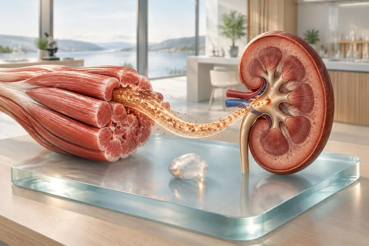
Muscle pain beginning within 24 to 72 hours after unfamiliar exercise often reflects delayed-onset soreness, which may produce a modest CK rise. Pain that is focal, worsens when a tendon is loaded, or follows a joint movement may need examination and imaging rather than broad blood testing. A sciatica symptom guide shows why burning, electric or one-sided pain may be neurological rather than muscular.
Persistent proximal weakness is different from soreness: difficulty rising from a chair, lifting the arms above the head or climbing stairs deserves CK, thyroid tests, electrolytes, medication review and often a neurological examination. Weakness that progresses over 2 to 6 weeks is more informative than a pain score alone because it can indicate a myopathy even when CK is only mildly raised.
A practical history changes interpretation by more than a decimal place. Record exercise, falls, fever, diarrhoea, fasting, alcohol, new medicines, dose changes and supplements during the preceding 7 days; the same CK value has different meaning after a marathon, a new statin, prolonged immobility or no clear trigger.
What does a creatine kinase test show?
The creatine kinase test detects enzyme released when muscle fibres are stressed or damaged, but CK is not specific for one disease. Reference intervals vary by laboratory, sex, age, ancestry, muscle mass and recent exercise, so a result should be compared with the report’s own upper limit.
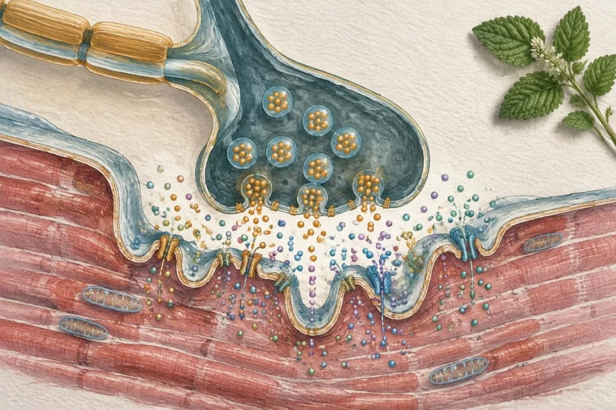
Many adult laboratories report approximate CK upper limits near 150 to 200 U/L for women and 250 to 320 U/L for men, but these are illustrative rather than universal. A CK of 600 U/L may be three times the upper limit in one laboratory and less than twice the upper limit in another. The high aldolase guide explains why a second muscle enzyme can help when CK and symptoms disagree.
CK can rise after resistance training, long-distance running, seizures, intramuscular procedures, falls or prolonged pressure on a limb. In a hypothetical 52-year-old marathon runner with AST 89 IU/L and CK 720 U/L the likely explanation may be recent muscle exertion, whereas the same CK with progressive weakness and no exercise requires a different work-up.
The CK-MB fraction is not the routine test for ordinary muscle pain; high-sensitivity cardiac troponin is preferred when heart injury is suspected. This distinction matters because skeletal muscle injury can confuse older CK-MB interpretation, while chest pressure, breathlessness, fainting or palpitations require urgent assessment regardless of total CK.
| Səviyyə | Diapazon | Nə deməkdir |
|---|---|---|
| Yerli diapazonda | Often approximately 30-200 U/L, depending on the laboratory and person | Major ongoing muscle-cell injury is less likely, but mild or non-muscle causes remain possible. |
| Yüngül dərəcədə yüksəlmiş | About 1-3 times the laboratory upper limit | Common after exercise, minor injury or medication exposure; review timing and consider a repeat. |
| Orta dərəcədə yüksək | About 3-5 times the laboratory upper limit | Prompt review is sensible, particularly if weakness, dehydration or abnormal kidney results are present. |
| Əhəmiyyətli dərəcədə yüksək | Above 5 times the laboratory upper limit | Possible rhabdomyolysis; urgent evaluation depends on symptoms, trend, urine findings, kidney function and electrolytes. |
Which electrolytes can cause cramps or muscle pain?
Low potassium, calcium or magnesium and, less often, abnormal sodium or phosphate can cause cramps, twitching, weakness or diffuse muscle discomfort. Kidney-function tests are paired with electrolytes because muscle breakdown can release potassium and threaten renal clearance.
Typical adult reference ranges are sodium 135-145 mmol/L, potassium 3.5-5.1 mmol/L, total calcium 2.15-2.55 mmol/L, magnesium 0.70-1.00 mmol/L and phosphate 0.8-1.5 mmol/L, although laboratories differ. Potassium below 3.0 mmol/L can produce marked weakness or arrhythmia risk, while potassium above 6.0 mmol/L is potentially dangerous and needs urgent confirmation and treatment.
Low calcium can cause tingling, spasms and carpopedal cramps, but total calcium can look low when albumin is low; ionized calcium may clarify the physiology. Low magnesium below roughly 0.5 mmol/L can cause tremor, cramps and refractory low potassium, so replacing potassium alone may fail until magnesium is addressed. A urine potassium explanation shows how urine testing can distinguish loss through the kidneys from loss elsewhere.
Kidney injury changes the risk calculation. A hypothetical patient with CK 8,000 U/L, creatinine 180 µmol/L, potassium 6.2 mmol/L and bicarbonate 18 mmol/L has a potentially dangerous rhabdomyolysis pattern even if pain is improving; the reason is that released muscle contents and reduced filtration can combine to disturb heart rhythm and acid-base balance.
Can medicines cause muscle pain or a high CK?
Medicines can cause muscle symptoms with a normal CK, a modest CK rise or severe muscle breakdown; statins are well known, but interactions, dose changes and kidney impairment often determine risk. Never stop a prescribed medicine abruptly without medical advice, especially a steroid, anticonvulsant or cardiovascular medicine.
Statin-associated muscle symptoms commonly occur without major CK elevation, while CK above 10 times the upper limit is unusual and warrants prompt clinician review. The European Atherosclerosis Society consensus emphasizes symptom timing, CK, dechallenge and rechallenge rather than assuming every ache is caused by a statin (Stroes et al., 2015). A SLCO1B1 statin-risk guide explains why genetics can contribute without proving that a symptom is genetic.
Other possible contributors include fibrates, colchicine, corticosteroids, antimalarials, some antivirals, antipsychotics and drug combinations that raise exposure. Supplements are not automatically harmless: excessive vitamin A, niacin, red-yeast-rice products and stimulant mixtures may complicate the picture, while undocumented ingredients make causality difficult to assess.
Write down the start date, dose, manufacturer, recent antibiotics, grapefruit intake and supplement changes for the preceding 4 weeks. For example, new aching 10 days after adding a strong enzyme-inhibiting medicine to a stable statin is a stronger interaction clue than pain that began 2 years after an unchanged prescription.
How can thyroid and endocrine tests explain muscle pain?
Thyroid muscle pain is usually evaluated with TSH and free T4, with CK added when weakness or marked aching is present. Hypothyroidism can produce stiffness, cramps and proximal weakness, while hyperthyroidism more often causes muscle wasting or weakness with a normal CK.

A common adult TSH reference interval is approximately 0.4-4.0 mIU/L, but pregnancy, age, assay method and laboratory policy change interpretation. A high TSH with low free T4 supports primary hypothyroidism; a low TSH with high free T4 supports thyrotoxicosis. The T4 and thyroid guide explains why free hormone values should be read with TSH rather than alone.
Hypothyroidism can raise CK, sometimes to several hundred or several thousand U/L, and untreated disease may increase susceptibility to statin-related muscle symptoms. The evidence is less tidy for borderline TSH elevations: clinicians differ on treatment thresholds, and a TSH of 5.5 mIU/L does not by itself prove that muscle pain is thyroid-driven.
Other endocrine clues include morning cortisol when adrenal disease is suspected, glucose and HbA1c when metabolic symptoms coexist, and calcium or phosphate testing for parathyroid disorders. A hypothetical person with TSH 18 mIU/L, low free T4, CK 950 U/L and slow reflexes has a coherent endocrine pattern; CK 950 U/L with normal thyroid tests after heavy exercise points elsewhere.
Which blood tests look for inflammatory muscle disease?
Inflammatory muscle disease is investigated with CK, aldolase, AST, ALT, LDH, CRP or ESR, ANA and selected myositis antibodies, but no single blood test confirms or excludes it. Progressive symmetrical weakness, difficulty swallowing, breathlessness or a characteristic rash is more diagnostically useful than pain alone.
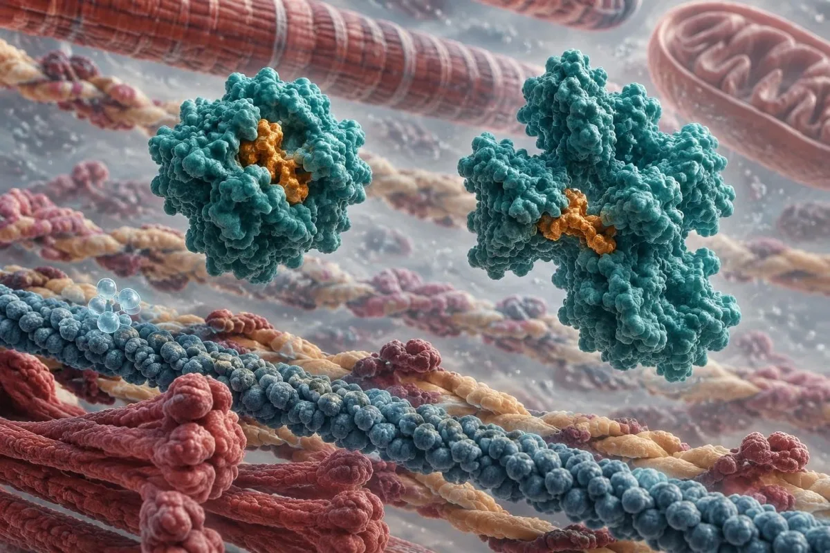
CK is often raised in polymyositis, immune-mediated necrotizing myopathy and dermatomyositis, but levels can be normal in some inclusion body myositis cases or late disease. CRP may be normal despite active muscle disease, so a CRP below 5 mg/L cannot independently rule it out. The 2017 EULAR/ACR classification criteria use clinical, laboratory and biopsy-related features together rather than a single antibody (Lundberg et al., 2017).
ANA is a screening clue, not a muscle-specific diagnosis; low-positive results occur in healthy people and in several autoimmune conditions. Myositis-specific antibodies such as anti-Jo-1, Mi-2, SRP or HMGCR can refine the differential, but a negative panel does not exclude disease because assays differ and not every relevant antibody is included. Our ANA interpretation guide covers titre, pattern and pre-test probability.
The combination of symptoms matters: CK 4,000 U/L plus new shoulder and hip weakness is more concerning than CK 400 U/L plus isolated tenderness after exercise. A clinician may add pulmonary testing, MRI, electromyography or tissue examination when weakness persists, because antibody results cannot show whether the affected muscle is actively inflamed or whether another process is responsible.
Can vitamin or systemic illness blood tests find the cause?
Vitamin D, B12, iron studies, glucose, liver tests and selected infection testing may identify contributors to aching or fatigue, but deficiency results rarely explain severe focal weakness by themselves. Testing should follow symptoms, diet, bleeding risk, medication exposure and recent illness rather than an indiscriminate supplement panel.
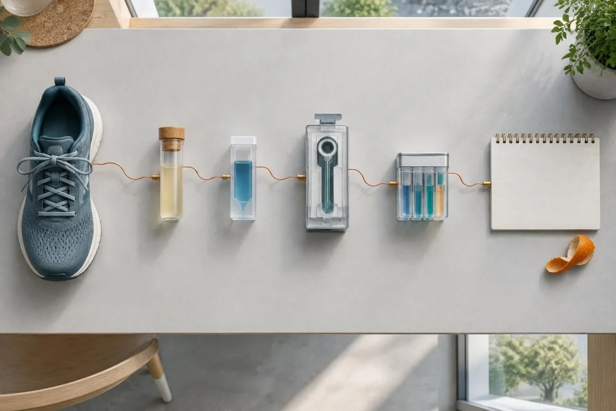
The NIH considers 25-hydroxyvitamin D below 12 ng/mL deficient for most people, 12-20 ng/mL potentially inadequate and 20 ng/mL or higher generally adequate for bone health, although target debates continue. Vitamin D deficiency can contribute to proximal weakness and bone or muscle discomfort, but a level of 18 ng/mL does not prove it is the sole cause. See our guide to vitamin D retesting before starting high-dose replacement.
B12 deficiency may cause numbness, balance problems and weakness through nerve injury rather than direct muscle-cell damage; methylmalonic acid can help when B12 is borderline. Ferritin below approximately 15-30 ng/mL often indicates depleted iron stores, but iron deficiency more commonly causes fatigue or exercise intolerance than isolated muscle pain. A ferritin guide for athletes explains why inflammation and training can distort interpretation.
Viral illness, diarrhoea, prolonged fasting and fever can cause muscle pain through dehydration, immune signalling or electrolyte shifts. Creatine kinase, kidney function and electrolytes are more useful than a broad viral antibody panel when acute muscle injury is suspected; antibodies may remain positive long after the triggering illness has ended.
What do AST, ALT, LDH and aldolase mean with muscle pain?
AST, ALT, LDH and aldolase can rise from muscle as well as liver or other tissues, so they cannot identify the source alone. CK, GGT, bilirubin and the symptom timeline help separate a muscle enzyme pattern from primary liver disease.
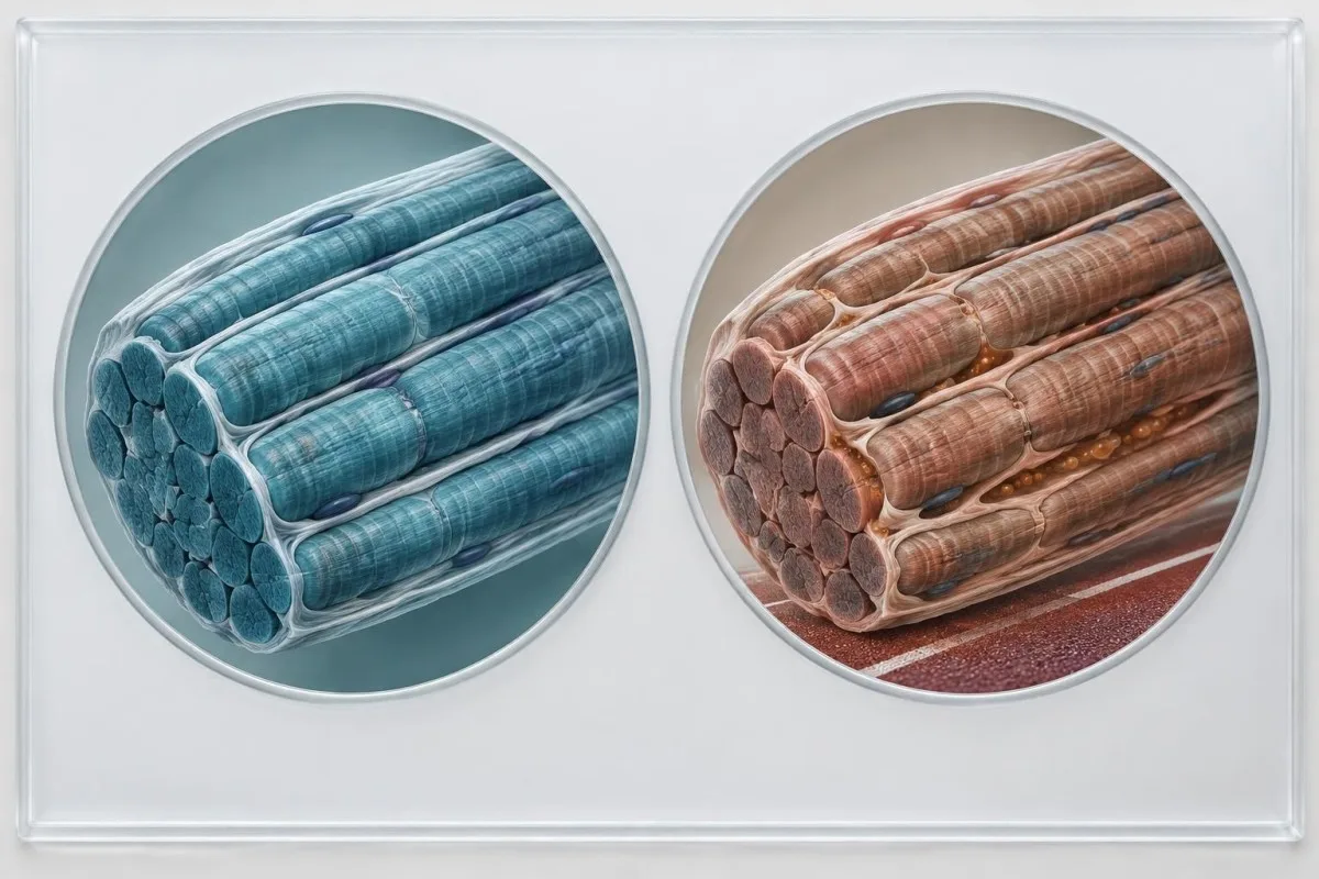
AST is found in skeletal muscle and may rise after exercise or muscle injury; ALT is more liver-enriched but can also increase with substantial muscle damage. A high AST with CK 2,000 U/L, normal bilirubin and normal GGT suggests muscle contribution more strongly than isolated AST with high bilirubin. Our guide to yüksək AST nəticələri explains why the ratio and accompanying tests matter.
LDH is distributed across muscle, liver, red cells and other tissues, so a raised result is a signal rather than a diagnosis. Haemolysis during collection can falsely increase LDH and AST, while intense exercise can produce a real increase; the laboratory comment about sample quality may be as useful as the number.
Aldolase can support a muscle source when CK is normal or only slightly elevated, but it is not perfectly specific. In a hypothetical patient with proximal weakness, CK 110 U/L, aldolase 14 U/L, AST 76 IU/L and normal GGT, repeating enzymes and arranging a neuromuscular assessment may be more informative than labelling the result as liver disease.
When should a muscle-pain blood test be repeated?
Repeat testing depends on the suspected trigger: CK is often repeated after several days without strenuous exercise, while urgent electrolyte or kidney abnormalities are rechecked sooner. The correct interval is determined by severity, trend and symptoms rather than by a fixed waiting period.
Strenuous exercise can keep CK elevated for 3 to 7 days, sometimes longer after unfamiliar eccentric exercise. If a well person has CK 450 U/L after heavy lifting, a clinician may repeat it after roughly 5 to 7 quiet days; this is not appropriate when weakness, dark urine or kidney abnormalities are present. The qan analizi zaman xəritəsi bələdçimiz explains why timing is part of interpretation.
Fasting is usually not required for CK, electrolytes or TSH, but follow the laboratory instructions if lipids, glucose or other tests are included. Avoiding unaccustomed exercise and recording hydration can make a repeat more interpretable, while deliberately drinking excessive water can distort sodium and does not treat rhabdomyolysis safely.
Trend often beats one value. A CK falling from 6,000 to 2,000 U/L over 48 hours may indicate improvement, whereas a rise from 600 to 3,000 U/L after a new medicine is a different signal even though both results are called elevated.
When will blood tests miss the cause of muscle pain?
Blood tests may miss nerve compression, tendon injury, osteoarthritis, fibromyalgia, compartment problems and some neuromuscular disorders because these conditions do not reliably release abnormal blood markers. Persistent symptoms with normal CK require a better examination, not simply an ever-larger laboratory panel.

Burning pain, pins and needles, electric shocks or weakness confined to one nerve territory suggest a neurological source; CK may remain normal. Electromyography and nerve-conduction studies can assess electrical function, while MRI or ultrasound can examine tendons, joints and selected muscle changes. Blood tests cannot show a meniscus tear, rotator-cuff problem or spinal nerve compression.
Fibromyalgia and central pain syndromes usually have normal CK, CRP and thyroid results, although clinicians first exclude conditions suggested by the history. Pain that is widespread for more than 3 months with sleep disturbance is a different diagnostic pattern from CK-positive muscle injury. A focused sciatica and nerve-sign guide illustrates how distribution can be more revealing than intensity.
One-sided swelling, severe pain after a cast or tight equipment, pain out of proportion, or worsening pain with passive stretch needs urgent examination because a pressure-related muscle emergency may evolve before routine blood results become dramatic. A normal CK at the first hour does not safely exclude every early process.
Which muscle-pain results need urgent care?
Urgent care is warranted for severe or rapidly increasing muscle pain with weakness, dark urine, reduced urine, fainting, confusion, fever, chest symptoms or abnormal potassium. Rhabdomyolysis is commonly defined in practice by CK above 5 times the upper limit of normal, but kidney function, electrolytes and the clinical course determine risk.
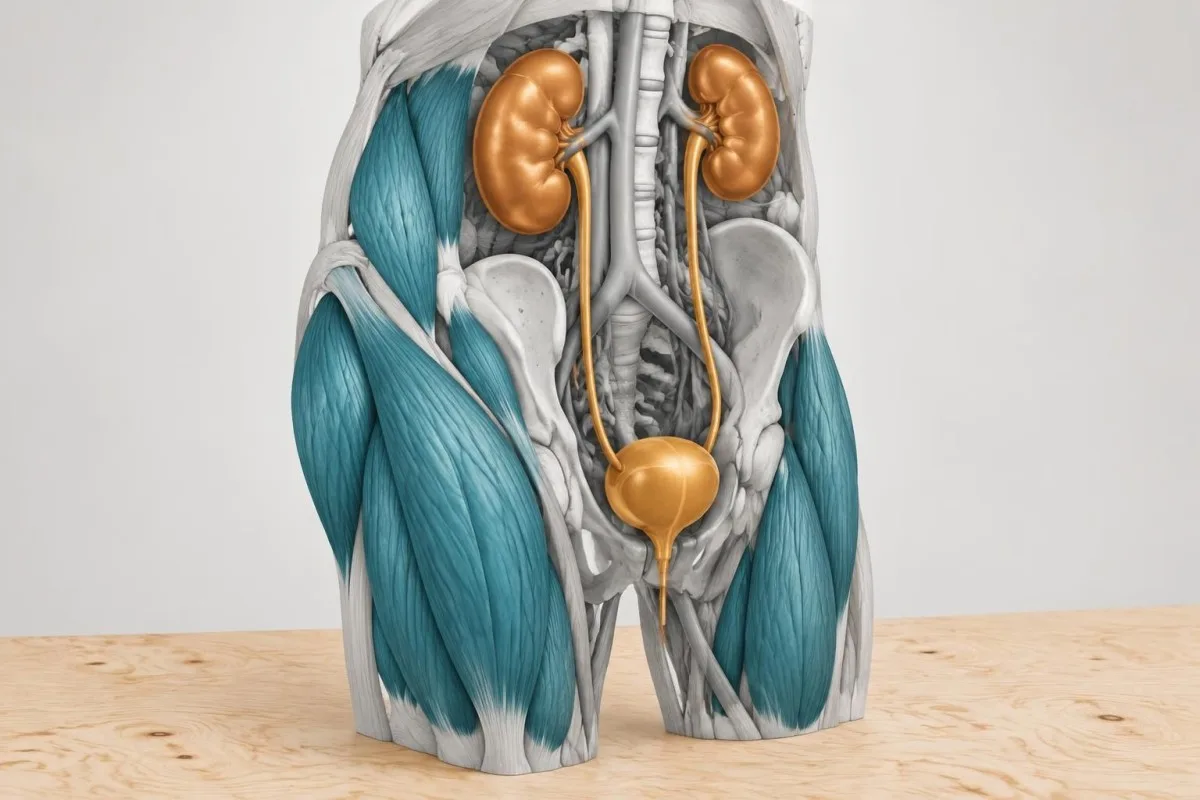
The classic laboratory pattern includes markedly raised CK, rising creatinine, high potassium or phosphate, low calcium early in some cases, and urine positive for blood with few or no red cells because of myoglobin. Myoglobin can disappear quickly, so a negative urine result does not exclude a later presentation. Bosch, Poch and Grau describe rhabdomyolysis as a syndrome with varied causes, including exertion, medicines, seizures and prolonged immobilisation (Bosch et al., 2009).
CK above 5,000 U/L is often treated as a clinically significant level, but it is not a universal admission threshold. A hypothetical 30-year-old with CK 12,000 U/L, normal creatinine, normal potassium and reliable follow-up differs from an older adult with CK 3,000 U/L, potassium 6.1 mmol/L and reduced urine; the second pattern is immediately more dangerous.
Do not attempt to manage a concerning result by force-drinking water or taking unprescribed diuretics. Emergency clinicians may repeat CK and electrolytes, monitor urine output and heart rhythm, review medicines, and provide carefully measured fluids when appropriate. A qaranlıq sidik xəbərdarlıq bələdçisi explains why colour change is a triage clue rather than a diagnosis.
How should you read a muscle-pain lab report?
Read a muscle-pain report as a timeline and a cluster: compare CK with its local upper limit, kidney function, potassium, AST, ALT, urine findings and symptoms on the same date. A single flagged value rarely identifies the cause without medication, exercise and illness context.
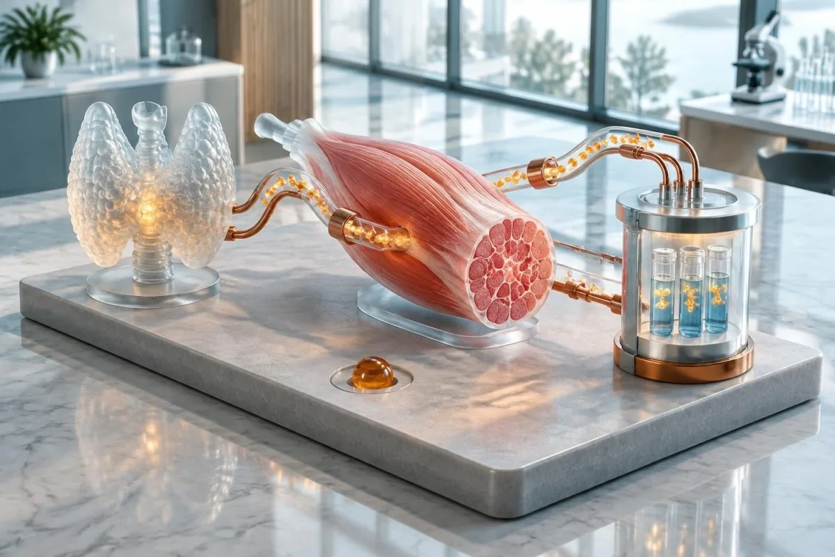
Kantesti is an AI-powered blood test analysis tool that can compare current and previous results, highlight related changes and explain units such as U/L, mmol/L and µmol/L. Our platform can help a reader prepare focused questions, but it cannot examine strength, assess urine output or decide whether a high potassium result is real; those limits matter most when a result is urgent.
Check whether the report uses a sex-specific CK interval, whether the sample was haemolysed, and whether creatinine is being compared with a personal baseline. A creatinine of 110 µmol/L may be normal for one muscular adult but a meaningful rise for someone whose baseline is 70 µmol/L. Trends are also affected by hydration, recent exercise and laboratory method changes.
For a hypothetical report with CK 1,100 U/L, AST 140 IU/L, ALT 48 IU/L, GGT 22 U/L, normal bilirubin and a hard workout 36 hours earlier, muscle release is plausible but not proven. Uploading a complete report to our AI with the collection date and symptom timeline gives more useful context than entering CK alone.
What should you do after abnormal muscle-pain labs?
After abnormal muscle-pain labs, contact the ordering clinician with the result, symptoms, exercise history and full medicine list; seek urgent care for red flags or dangerous electrolyte and kidney changes. The next step may be a repeat panel, medication review, thyroid treatment, imaging, neurological testing or specialist referral.

Ask four concrete questions: How far is CK above my laboratory limit? Are potassium, creatinine, bicarbonate and urine findings safe? Could a medicine, supplement, thyroid problem or recent exercise explain this? What result or symptom should trigger same-day review? These questions reduce the risk of treating a number without identifying the mechanism.
Do not self-prescribe high-dose vitamin D, magnesium or potassium from a single flagged result. Kidney disease, pregnancy, heart rhythm conditions and medicine interactions can make supplementation harmful, and a low total calcium may reflect low albumin rather than true calcium deficiency.
Kantesti is an AI blood test interpretation platform that can help turn a multi-page report into a structured list of biomarkers, trends and follow-up questions; clinical decisions still belong with a qualified healthcare professional. For information about how our medical content and review standards are developed, see the tibbi validasiya səhifəsi.
What information improves the next muscle-pain assessment?
The most useful information is a dated symptom timeline, exact medicines and supplements, recent exercise or immobility, urine changes, weakness distribution and previous laboratory results. These details help clinicians decide whether to repeat CK, investigate thyroid or electrolytes, assess nerves and joints, or refer for specialist testing.
Bring the collection date, symptom-onset date and the last 7 days of activity, including heavy lifting, running, seizures, falls, fever, vomiting or diarrhoea. Also record whether pain is symmetrical, whether weakness affects stairs or overhead lifting, and whether symptoms began after a dose change within the last 30 days.
Ask whether CK should be repeated after 5 to 7 days without strenuous exercise, whether TSH and free T4 are needed, and whether urine, aldolase, CRP, ESR, ANA or myositis antibodies fit the presentation. Testing should be selective: a larger panel can produce incidental positives that create anxiety without explaining the pain.
If symptoms persist despite normal blood tests, ask what non-laboratory assessment is appropriate. Depending on the pattern, that may include joint examination, ultrasound, MRI, electromyography, nerve-conduction studies or physiotherapy assessment rather than another round of nonspecific blood markers.
Tez-tez verilən suallar
Əzələ ağrısı üçün ən yaxşı qan analizi hansıdır?
Əzələ ağrısı zamanı ilk növbədə adətən kreatinkinaza üçün qan testi aparılır, çünki bu test gərginliyə məruz qalmış və ya zədələnmiş əzələ liflərindən ferment ifrazını aşkar edir. İlkin müayinə panelinə çox vaxt kreatinin, kalium, kalsium, maqnezium, fosfat, natrium, bikarbonat, AST, ALT və TSH də daxil edilir. CK-nın laboratoriyanın yuxarı həddindən 5 dəfədən çox olması əzələlərin əhəmiyyətli dərəcədə parçalanması ehtimalını artırır, lakin CK-nın normal olması sinir, vətər, oynaq, endokrin və ya dərmanlarla əlaqəli yüngül səbəbləri istisna etmir.
Elektrolit balansının pozulması əzələ ağrısına səbəb ola bilərmi?
Bəli, kalium, kalsium və ya maqneziumun aşağı olması, yaxud natrium və ya fosfat səviyyələrinin normadan kənara çıxması qıcolmalara, əzələ seğirmələrinə, zəifliyə və ya yayılmış əzələ narahatlığına səbəb ola bilər. Böyüklər üçün tipik diapazonlar kalium 3.5–5.1 mmol/L, natrium 135–145 mmol/L, ümumi kalsium 2.15–2.55 mmol/L və maqnezium 0.70–1.00 mmol/L-dir, lakin laboratoriyalara görə dəyişə bilər. Kaliumun 3.0 mmol/L-dən aşağı və ya 6.0 mmol/L-dən yuxarı olması, xüsusilə zəiflik, ürəkdöyünmə və ya böyrək funksiyasının pozulması olduqda, təhlükəli ola bilər.
Qalxanabənzər vəz problemləri əzələ ağrılarına və CK səviyyəsinin yüksəlməsinə səbəb ola bilərmi?
Hipotireoz əzələ ağrılarına, sərtliyə, qıcolmalara, proksimal zəifliyə və CK səviyyəsinin yüksəlməsinə səbəb ola bilər. Hipertireoz isə daha çox əzələ itkisinə və ya CK normal olduğu halda zəifliyə səbəb olur. Qalxanabənzər vəzinin qiymətləndirilməsinə adətən TSH və sərbəst T4 göstəricilərinin yoxlanılması ilə başlanır; böyüklər üçün TSH-nin ümumi normal intervalı təxminən 0.4-4.0 mIU/L-dir. TSH-nin yüksək, sərbəst T4-ün aşağı olması və CK səviyyəsinin yüksəlməsi qalxanabənzər vəzlə əlaqəli uyğun bir mənzərədir, lakin TSH-nin təkcə cüzi yüksəlməsi ağrıya qalxanabənzər vəz xəstəliyinin səbəb olduğunu sübut etmir.
What CK level is dangerous with muscle pain?
A CK level above 5 times the laboratory upper limit is commonly used as a rhabdomyolysis warning, and CK above 5,000 U/L often prompts close clinical assessment. There is no universal dangerous number because potassium, creatinine, urine output, hydration, symptoms and CK trend change the risk. Severe weakness, dark urine, reduced urine, confusion, fainting or potassium above about 6.0 mmol/L requires urgent medical evaluation regardless of the CK value.
Can a blood test rule out inflammatory myositis?
No, blood tests cannot rule out every inflammatory myopathy. CK is often raised, but some people with inflammatory muscle disease have normal or only mildly elevated CK, and CRP below 5 mg/L does not exclude active muscle disease. Clinicians combine weakness pattern, examination, CK or aldolase, ANA, myositis antibodies and sometimes MRI, electromyography or tissue examination.
What if my blood tests are normal but my muscles still hurt?
Normal blood tests make major muscle-cell breakdown, severe electrolyte disturbance and some inflammatory conditions less likely, but they do not rule out tendon injury, joint disease, nerve compression, fibromyalgia or mild medication symptoms. Burning or electric pain, one-sided symptoms or numbness may require neurological assessment rather than more CK tests. Persistent weakness, symptoms lasting more than 2 to 6 weeks, or worsening function should be reviewed even when CK is normal.
Qan analizinin nəticələrini necə oxumaq
Laboratoriya hesabatınızı yükləyin, Kantesti-nin AI-si isə saniyələr ərzində hər biomarkeri sadə dildə izah etsin. Dünyada 2 milyondan çox insan istifadə edir.
İstinadlar
- Klein, T., Mitchell, S., & Weber, H. (2026). Klinik Təsdiqləmə Çərçivəsi v2.0 (Tibbi Təsdiqləmə Səhifəsi). Kantesti AI tibbi araşdırmaları. DOI
- Klein, T., Mitchell, S., & Weber, H. (2026). AI Qan Analizi Aparatı: 2,5M Test Təhlil Edildi | Qlobal Sağlamlıq Hesabatı 2026. Kantesti AI tibbi araşdırmaları. DOI
- Stroes ES et al. (2015). Statin-associated muscle symptoms: impact on statin therapy—European Atherosclerosis Society Consensus Panel Statement. European Heart Journal. DOI PubMed
- Lundberg IE et al. (2017). 2017 European League Against Rheumatism/American College of Rheumatology classification criteria for adult and juvenile idiopathic inflammatory myopathies and their major subgroups. Annals of the Rheumatic Diseases. DOI PubMed
- Bosch X et al. (2009). Rhabdomyolysis and acute kidney injury. The Lancet.
- 2M+Təhlil edilmiş testlər
- 127+ölkələr
- 100+Dillər
Tibbi komanda haqqında


E-E-A-T Etibar Siqnalları
Təcrübə
Həkim rəhbərliyi ilə laborator şərh iş axınlarının klinik icmalı.
Ekspertiza
Biomarkerlərin klinik kontekstdə necə davrandığına dair laboratoriya təbabəti diqqəti.
Səlahiyyətlilik
Dr. Thomas Klein tərəfindən tibbi baxımdan nəzərdən keçirilib; Dr. Sarah Mitchell və Prof. Dr. Hans Weber töhfə veriblər.
Etibarlılıq
Aydın izləmə yolları ilə həyəcanı azaltmağa yönəlmiş sübuta əsaslanan şərh.
Tibbi məsuliyyətdən imtina bildirişi
Bu məqalə yalnız təhsil məqsədlidir və tibbi məsləhət təşkil etmir. Diaqnoz və müalicə qərarları üçün həmişə ixtisaslı səhiyyə işçisi ilə məsləhətləşin.
Dərc edilmişdir · Redaksiya komandası ilə əlaqə saxlayın
İngiltərə və Uelsdə qeydiyyatdan keçmiş Kantesti Ltd tərəfindən nəşr olunub, şirkət №. 17090423, London, Birləşmiş Krallıq.