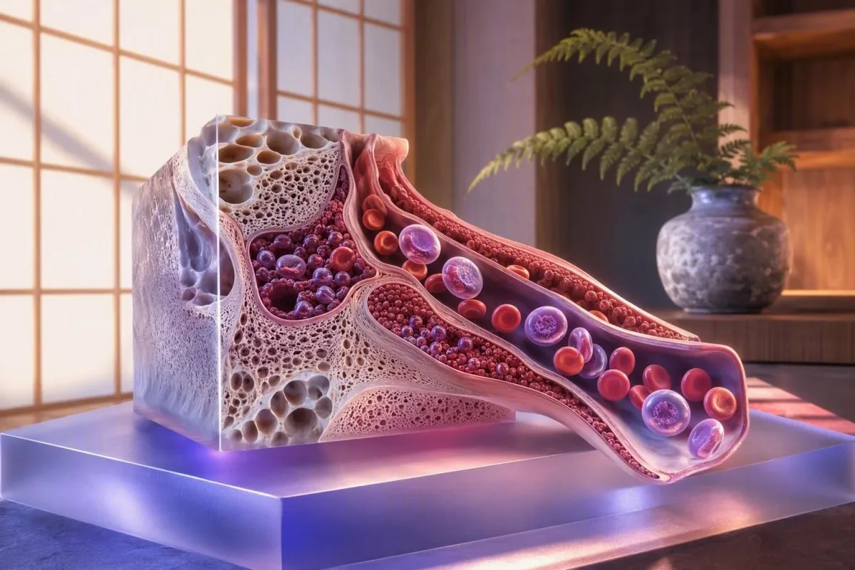
Цусны алдагдал, улаан эсийн устгал, эсвэл цус багадалтын эмчилгээнээс эдгэрч байгаа үед ясны ясны хариу урвалыг илтгэдэг залуу улаан эсүүд болох өндөр ретикулоцитууд нь ховорхон шинж тэмдэг үүсгэдэг. Чухал ач холбогдолтой шинж тэмдэг нь амьсгаадах, ядарч сульдах, шарлах, шээс харлах, эсвэл цус алдалтын шинж тэмдэг бөгөөд эргэн тойрон дахь CBC нь аль тайлбар нь тохирч байгааг тодорхойлдог.


- High reticulocytes ихэвчлэн шинж тэмдэг үүсгэдэггүй; тэдгээр нь улаан эсийн үйлдвэрлэл эсвэл эргэлтийг нэмэгдүүлж байгааг илтгэнэ.
- Насанд хүрэгчдийн ретикулоцитийн хувь нь ихэвчлэн 0.5-2.5% байдаг боловч цусны ретикулоцитийн абсолют тоо нь эмнэлзүйн хувьд илүү ашигтай байдаг.
- 100 × 10⁹/Л-аас дээш цусны ретикулоцитийн абсолют тоо цус багадалтын үед ясны ясны хариу урвалыг илтгэдэг бөгөөд ихэвчлэн цус алдалт эсвэл гемолизээс үүдэлтэй байдаг.
- Цус багадалтын үед залруулсан ретикулоцитийн хувь 2%-аас дээш байх нь ихэвчлэн ясны бүтэлгүйтлээс илүүтэйгээр ясны ясны зохих хариу урвалыг илтгэнэ.
- Шарлалт, хар цай өнгөтэй шээс, нуруу эсвэл хэвлий өвдөх цус багадалтын үед эмнэлзүйн хувьд чухал ач холбогдолтой гемолизыг илтгэж болно.
- Сүү шиг хар өтгөн, шулуун гэдсээс улаан цус алдах, эрчтэй сарын тэмдэг ирэх, эсвэл ухаан алдах ретикулоцит их байсан ч шуурхай үнэлгээ хийх шаардлагатай.
- Төмөр, витамин B12, эсвэл фолийн хүчлээр эмчилснээс хойш 3-7 хоногийн дараа ретикулоцит нэмэгдэж болно гемоглобин бүрэн сайжрахаас өмнө.
- Гемоглобин бага, шууд бус билирубин их, LDH их хамтад нь авч үзвэл гемолиз (цусны улаан эсийн задрал) батална.
- Анеми (цуцралт) үед ретикулоцитийн хувь нь төөрөгдүүлж болно учир нь улаан эсийн тоо бага байх нь хувийн тоог хиймээр өндөр харагдуулдаг.
Бодит амьдрал дээр өндөр ретикулоцитууд ямар мэдрэмж төрүүлдэг вэ
Өндөр ретикулоцитийн шинж тэмдгүүд нь ихэвчлэн улаан эсийн эргэлтийг удирдаж буй нөхцөл байдлын шинж тэмдгүүд байдаг бөгөөд ретикулоцитээс үүдэлтэй шинж тэмдгүүд биш юм. Орон нутгийн лавлах хязгаараас дээш гарсан утга нь ядаргаа, дасгал хийх үед амьсгаадах, зүрх дэл Алдах, шарлалт, эсвэл ямар ч шинж тэмдэггүй байх зэрэгтэй хамт байж болно.
Ретикулоцит бол эргэлтэнд ойролцоогоор 1-2 хоногийн дотор боловсордог боловсордоггүй улаан эсийн элементүүд юм. Тэдний РНХ-ийн үлдэгдэл нь өвдөлт, халуурах, эсвэл ядрах шалтгаан болохгүй. Би үр дүнг шалгаж байхдаа ретикулоцитын тоо өндөр эхлээд гемоглобин бага эсэхийг асуудаг: хэвийн гемоглобинтой үед өндөр тоо нь зүгээр л нөхөн төлбөртэй үйл явц байж болох бөгөөд анеми нь ихэнх шинж тэмдгүүдийг тайлбарладаг.
Төмрийн эмчилгээ хийлгэсний дараа 3.0% ретикулоцитийн хувьтай байж, эрүүл саруул мэт санагдаж болно. 3.0%-тай өөр нэг өвчтөн гемоглобин нь 78 г/л (7.8 г/дл) байсан тул бүүртийж магадгүй, ретикулоцитээс болоод биш. Энэ ялгаа нь портал үр дүнгүүд дээр ихэвчлэн анзаарагддаггүй; манай бүрэн CBC гарын авлага тоог гемоглобин болон индексийн хажууд байрлуулахад тусалдаг.
2026 оны 8-р сарын 21-ний байдлаар, ноён Томас Кляйны практик дүрэм нь энгийн: ретикулоцитийн хэмжээ нэмэгдсэн нь гемоглобин буурч, билирубин нэмэгдэж, шинэ цус алдалт, эсвэл тухайн хүний өмнөх суурь түвшнээс ихээхэн өөрчлөлттэй байвал клиник ач холбогдолтой болно. Гадаа гарсан хувийн тоо нь ховорхон оношлогдоно.
Яагаад хувийн тоо нь төөрөгдүүлж болох вэ?
31% -ийн ретикулоцитын хувь нь ясны чөмөгний эрчимтэй хариу урвал биш юм. Гемоглобин 24% -тай хүний хувьд 3 × 24/45 гэсэн томъёогоор бодоход залруулсан ретикулоцитын хувь нь ойролцоогоор 1.6% байх бөгөөд энэ нь цус багадалтын зэрэгт хангалтгүй байж болно. Тийм учраас гематологууд абсолют тоог, хүнд хэлбэрийн цус багадалтын үед ретикулоцитын үйлдвэрлэлтийн индексийг илүүд үздэг.
Ретикулоцитийн үр дүнг хэтрүүлэлгүйгээр хэрхэн унших вэ
Ясны чөмөгний хариу урвалыг хамгийн түрүүнд үнэлэхэд абсолют ретикулоцитын тоо хамгийн сайн үзүүлэлт болдог. Олон лабораториуд насанд хүрэгчдэд зориулсан лавлагааны хязгаарыг 25-100 × 10⁹/л гэж тэмдэглэдэг боловч төхөөрөмжийн арга болон орон нутгийн хүн ам харилцан адилгүй байдаг.
Ретикулоцитын хувь нь эргэлдэж буй бүх улаан эсээс хамаарч тооцогддог бол абсолют ретикулоцитын тоо нэг литр тутам дахь ретикулоцитын бодит тоог тооцоолдог. Насанд хүрэгчдийн 120 × 10⁹/л гэсэн үр дүн ихэвчлэн өндөр байдаг боловч гемоглобин 70 г/л байхад 90 × 10⁹/л гэсэн үр дүн хангалтгүй бага байж болно. Тоо нь хуваарь болон эмнэлзүйн түүхтэй байх ёстой.
Боловсорч гүйцээгүй ретикулоцитын фракц эсвэл IRF нь ясны чөмөг хүүхдийн эсийг эрт гаргадаг үед нэмэгддэг. Энэ нь хими эмчилгээний дараа, цочмог цус алдалтын дараа, цус задралын дараа, эритропоэтин эмчилгээ хийсний дараа, эсвэл төмрийн нөхөн төлбөрийн дараа нэмэгдэж болно. Манай тайлан дээр нэмэлт параметр гарч ирвэл манай тайлбарыг уншина уу. боловсроогүй ретикулоцитын фракц хэмээх нэмэлт параметр таны тайланд гарч ирвэл манай тайлбарыг уншина уу.
Кантести бол AI цусны шинжилгээний анализатор нь гемоглобин, MCV, RDW, билирубин, LDH, төмрийн шинжилгээ, өмнөх үр дүнгүүдтэй харьцуулан ретикулоцитын үр дүнг уншдаг бөгөөд зөвхөн нэг өндөр үзүүлэлтэд утга өгдөггүй. Энэ нь чухал учир нь эмчилгээний дараах 2.8% -ийн үр дүн нь тайвшруулах боловч хурдан буурч буй гемоглобин хажууд ижил үр дүн нь тийм биш юм.
Өндөр ретикулоцитууд нь цус алдалтыг зааж өгөх үед
Цочмог эсвэл үргэлжилж буй цус алдалт нь ясны чөмөг хангалттай төмөртэй, ажиллаж байгаа тохиолдолд ойролцоогоор 3-5 хоногийн дараа ретикулоцитыг нэмэгдүүлж болно. Шинж тэмдэг нь сулрах, босоход толгой эргэх, түргэн хэмнэлтэй цохилох, ер бусын хүнд хэлбэрийн сарын тэмдэг, хар өтгөн, эсвэл шууд гэдэсний цус алдалт зэргийг агуулж болно.
Цусны эргэлтийн хэмжээг гэнэт алдсаны дараа ясны чөмөгний нөхөн авах хугацаа шаардагддаг тул ретикулоцитын тоо хэвийн хэвээр байж болно. Мөн цусны шингэнүүд тэнцвэржихээс өмнө гемоглобин хуурамчаар тайвшруулах боломжтой. 4-7 хоногийн дараа зохих хариу урвал нь ихэвчлэн ретикулоцитоз, зураг дээр полихромази, дараа нь RDW-ийн өсөлт хэлбэрээр илэрдэг.
Сарын тэмдэгийн цус алдалтыг тодорхой асуулт тавьж, тодорхой бус баталгаа өгөхгүйгээр асуух хэрэгтэй. 2.5 см-ийн зоос том бөөгнөрөл гарах, 1-2 цаг тутамд хамгаалалтаа солих, эсвэл 7 хоногоос дээш хугацаагаар цус алдах нь ретикулоцит эхэндээ нэмэгдэж байсан ч төмрийн дутагдалд хүргэж болно. Эрэгтэйчүүд манай хэлэлцүүлгийг олж магадгүй сарын тэмдэгийн үед гемоглобины өөрчлөлт цаг хугацааг ойлгоход тустай.
Мeлена гэгддэг хар наалдамхай өтгөн нь ходоодны дээд хэсгийн цус алдалтыг илтгэж болох ч төмрийн эм, висмут нь цус алдалгүйгээр өтгөнийг хар болгож болно. Гэдэсний шинжилгээний эерэг хариу нь эх үүсвэрийг тодорхойлохгүй; манай FOBT үр дүн нь удирдамж нь эмийн түүх болон дараачийн шинжилгээнүүд яагаад чухал болохыг тайлбарлаж байна. Ухаан алдах, амралт чөлөөтэй байхдаа амьсгаадах, эсвэл хар өтгөн тогтмол гарах нь тухайн өдөр эмнэлгийн тусламж авах шаардлагатай.
Зургийг өөрчилж буй гемолитик цус багадалтын шинж тэмдэг
Ясны чөмөг нөхөн төлж чадахгүйгээр улаан эсүүд устдаг үед цус задралын цус багадалтын шинж тэмдэг илэрдэг. Нийтлэг шинж тэмдгүүд нь ядрах, цайх, шарлах, шээс харлах, хэвлийгээр өвдөх, цус багадалт хүндрэх үед амьсгаадах зэрэг орно.
Цус задрал нь нэгэн зүйл биш, харин шинжилгээний үр дүнгээс хамаарсан байдал юм. Шууд бус билирубиныг ихэсгэх, ЛДГ-ийг ихэсгэх, хаптоглобиныг багасгах, ретикулоцитозыг ихэсгэх нь нэг шинжилгээний үр дүнгээс илүү хүчтэй батлагдана. Барчеллини ба Фаттиццо (2015) нь шинжилгээ бүрд төөрөгдөл учруулж болох зүйлс байдгийг онцолсон: хаптоглобин үрэвслийн улмаас хэвийн хэмжээнд байж болно, харин ЛДГ нь цусны бус олон өвчинд ихэсдэг.
Цус задрал дахь шарлалт нь холбогдоогүй билирубины улмаас нүд, арьс шарлах шинж тэмдэг бөгөөд ялангуяа гемоглобин шээсэнд ялгарч байвал шээс нь бараан өнгөтэй байж болно. Өнгөгүй өт, шээсэнд билирубин илэрч байвал элэг, цөсний замын өөр өвчнийг илтгэнэ. Энэ ялгаа нь практик бөгөөд бид шууд ба шууд бус билирубиний харьцаа үүнийг нарийвчлан авч үзсэн.
Миний туршлагаас үзэхэд, G6PD дутмагшилтай хүнд шинэ эм, халуурах, эсвэл тодорхой хоол хүнс хэрэглэсний дараа гэнэт бараан шээс гарах нь яаралтай эмчид хандах шаардлагатай. Энэ тохиолдолд цус задрал нь гайхалтай хурдан явагдаж болно. Манай бага G6PD идэвхжил -ийн шинжилгээний хязгаарлалт болон аюулгүй байдлын талаарх мэдээллийг үзнэ үү.
Яагаад цус багадалтын үед ретикулоцитууд нэмэгддэг вэ
Өндөр ретикулоцитын тоо нь төмөр, В12 витамин, фолийн хүчил, эсвэл эритропоэтин эмчилгээнээс 3-7 хоногийн дараа ихэссэн тохиолдолд эмчилгээ үр дүнтэй байгааг илтгэнэ. Энэ үед ихэнх өвчтөнүүд гемоглобин нөхөн сэргэхэд 2-4 долоо хоногийн хугацаанд амьсгаадах нь аажмаар багасна.
Эрт үеийн ретикулоцитын өсөлтийг заримдаа ретикулоцитын хариу урвал гэж нэрлэдэг. Хангалттай В12 витамин эмчилгээ хийсний дараа энэ нь ихэвчлэн 5-8 хоногт оргилдоо хүрдэг; төмрийн хувьд шимэгдэлт, тун, үрэвсэл, үргэлжилсэн алдагдал зэргээс шалтгаалан хугацаа нь тогтворгүй байдаг. Эмчилгээ, онош зөв бол гемоглобин ихэвчлэн 2-3 долоо хоногийн хугацаанд ойролцоогоор 10 г/л-ээр нэмэгддэг.
Нэгэн ашигтай боловч дутуу үнэлэгдсэн зүйл бол RDW. Нөхөн сэргэлтийн үед хуучин жижиг эсвэл том улаан эсүүд шинээр үйлдвэрлэгдсэн эсүүдтэй хамт оршдог тул RDW нь хэвийн болохоос өмнө нэмэгдэж болно. Энэ түр зуурын өсөлтийг нөхцөл байдлыг дордуулах шинж тэмдэг гэж андуурах ёсгүй; төмрийн дараа RDW яагаад нэмэгдэж болох вэ -ийг үзнэ үү.
Кантести бол AI-аар ажилладаг цусны шинжилгээний үр дүнгийн шинжилгээний хэрэгсэл нь одоогийн болон өмнөх CBC-г харьцуулж, ретикулоцит нь гемоглобин сайжрахаас өмнө нэмэгдсэн эсэхийг илрүүлж болно - энэ нь хариу урвалыг илтгэх байдал юм. Гэхдээ энэ нь төмрийн тун хангалттай гэдгийг баталж чадахгүй; ферритин, трансферрины ханалт, эмчилгээг дагаж мөрдөх байдал, дутмагшил үүсэх шалтгаан зэргийг эмнэлзүйн талаас нь авч үзэх шаардлагатай.
Яагаад төмрийн дутагдал нь ретикулоцитийн хариу урвалыг хязгаарлаж болох вэ
Төмрийн дутагдал нь цус алдалт эсвэл цус задрал байгаа үед ч гэсэн хүлээгдэж буй ретикулоцитын өсөлтийг саатуулж болно. Ферритин 15 мкг/л-ээс доогуур байх нь эрүүл насанд хүрсэн хүмүүст төмрийн нөөц байхгүйг өндөр нарийвчлалтайгаар илтгэнэ, харин үрэвсэл нь ферритинийг хуурамчаар хэвийн эсвэл өндөр харагдуулж болно.
Ретикулоцитуудад гемоглобин нийлэгжүүлэхэд төмөр шаардлагатай. Давтагдсан хүнд цус алдалттай байгаа хүнд өндөр хэвийн ретикулоцитын хувь байж болох ч ясны чөмөгний төмөр шавхагдсан тул хариу урвал муу байж болно. Трансферрины ханалт 20%-ээс доогуур байх нь төмрийн нийлүүлэлт хязгаарлагдмал байгааг дэмжиж болно, ялангуяа ферритин, CRP, MCV, болон эмнэлзүйн нөхцөл байдалтай хамт авч үзвэл.
Ферритин нь цочмог фазын урвалд ордог; идэвхтэй үрэвсэлт өвчний үед 60 мкг/л-ийн утга нь төмрийн дутагдлыг найдвартай үгүйсгэж чадахгүй. Уусамтгай трансферрины рецептор нь стандарт төмрийн судалгаанууд зөрчилдөх үед ашигтай байж болно. Манай трансферрины ханалт бага гарын авлага нь зөвхөн ийлдсэнд байгаа төмрийг хэрхэн буруу шийдвэр гаргахад хүргэдэг болохыг тайлбарлана.
2021 оны Их Британийн хоол боловсруулахын нийгэмлэгийн Snook болон бусад гишүүдийн гаргасан удирдамжид шалгалтын өмнө төмрийн дутагдлыг баталгаажуулах, шалтгаангүй төмрийн дутагдлын цус багадалттай тохирох насанд хүрэгчдэд хоол боловсруулах тогтолцооны үнэлгээ хийхийг зөвлөсөн. Энэ нь төмрийн алдагдаж буй шалтгааныг үл тоомсорлож, зөвхөн төмрийн тоон үзүүлэлтийг эмчлэх алдаанаас хамгаалдаг.
Өндөр улаан эсийн эргэлтийг дэмжих цусан дахь дэлгэцийн түлхүүрүүд
Автомат тоолох арга нь чадахгүй тохиолдолд захын цусны шүүлтүүр нь ретикулоцитозын ард байгаа механизмыг харуулж чадна. Полихромази нь залуу улаан эсийн ялгаралтыг нэмэгдүүлж байгааг дэмждэг бол сфероцитууд, шизоцитууд, хазуулсан эсүүд, эсвэл бөөгнөрөл нь тодорхой шалтгааныг илтгэж болно.
Поли хромази нь будагдсан хальсан дээрх цэнхэр-саарал том эсүүдийг дүрсэлдэг бөгөөд энэ нь ретикулоцитууд дахь РНХ-ийн үлдэгдлийг илэрхийлдэг. Энэ нь ретикулоцитын тоотой ерөнхийдөө нийцэж байх ёстой; Анализатортой харьцах, саяхан цус сэлбэх, эсвэл ер бусын эсийн морфологи нь зөрчил үүсгэж болно. Клиникийн зураг болон машины үр дүн хоорондоо зөрчилдөх үед гар аргаар хийсэн хальсны хяналт нь онцгой ач холбогдолтой.
Шизоцитууд нь улаан эсийн хэлтэрхий бөгөөд тэдгээрийг анхааралтай морфологийн хяналт шаарддаг, энгийн харагдлаар хангалтгүй. Насанд хүрэгчдэд, зөв бэлтгэгдсэн үрэвслийн үед шизоцитын тоо 1%-аас дээш байвал тромботик бичил судасны эмгэгийг тохирох нөхцөлд, ялангуяа тромбоцитопени болон эрхтний гэмтэлтэй байвал дэмжиж болно. Унших шизоцитууд яаралтай байх үед лабораторийн тайлбараас өөрийгөө оношлохыг оролдохын оронд.
Сфероцитууд нь дархлааны гемолиз эсвэл удамшлын сфероцитоз үед тохиолдож болно, харин хазуулсан эсүүд нь исэлдлийн гэмтлийн боломжийг нэмэгдүүлдэг. Ягер болон бусад (2020) автоиммун гемолиз сэжиглэгдэж байвал шууд антиглобулины шинжилгээ, эсвэл DAT хийхийг зөвлөдөг - гэхдээ DAT сөрөг байвал бүх тохиолдлыг үгүйсгэхгүй. Гематологийн тайлбар заримдаа нарийн төвөгтэй байдаг.
Гемолизээс гадна өндөр ретикулоцитийн нийтлэг шалтгаанууд
Ретикулоцитын өндөр түвшний шалтгаанууд нь саяхан цус алдах, гемолиз, цус багадалтын эмчилгээ амжилттай болох, ясны ургамлын дарангуйлал дараах эдгэрэлт, өндөрлөг газар дасан зохицох, заримдаа улаан эсийн удамшлын өвчнүүд орно. Хэвийн гемоглобин нь үр дүнгийн яаралтай байдлыг өөрчилдөг боловч шинээр тогтвортой өндөр байдлыг тайлбарлах шаардлагагүй болгодоггүй.
Өндөрлөг газар шилжсэний дараа эритропоэтиний өдөөлт нь гемоглобин өсөхөөс өмнө ретикулоцитыг нэмэгдүүлж болно. Үр нөлөө нь өндөрлөг, хугацаа, төмрийн статус, хувь хүний физиологиос хамаарна; богино цасны амралт ховор тохиолдолд мэдэгдэхүйц эсвэл тогтвортой үр дүнг тайлбарладаг. Тамирчид нь мөн цусны сийвэнгийн эзэлхүүний өөрчлөлтийг харгалзан үзэх ёстой бөгөөд энэ нь харагдах CBC-ийн концентрацийг өөрчилж болно.
Саяхан хими эмчилгээ, вирусын ясны ургамлын дарангуйлал, эсвэл хүнд ширүүн тэжээлийн дутагдал нь ясны ургамлын үйл ажиллагаа эргэж ирэхэд эдгэрлийн ретикулоцитоз үүсгэж болно. Энэ нь ерөнхийдөө урам зориг өгдөг боловч цаг нь чухал: шинээр халуурах, цус алдах, эсвэл цөөхөн нейтрофил бүхий өндөр тоо нь энгийн эдгэрлийн түүх биш юм. Бидний нийтлэл цөөхөн ретикулоцит ба сул ясны ургамал эсрэг хэв маягийг тайлбарладаг.
Лабораторийн асуудал нь холбогдох тайлбарыг заримдаа гажуудуулж болно. In-vitro дээжний гемолиз нь бие махбодод гемолиз нотлохгүйгээр кали, LDH болон бусад химийн утгыг нөлөөлж болно. Лабораторийн тайлбарыг шалгаж, шаардлагатай үед дахин шинжлэхийг анхаарч үзээрэй; манай гемолизийн дахин авах (redraw) гарын авлага нь зохистой дараагийн алхмуудыг тоймлон харуулсан болно.
Жирэмслэлт, нярай болон хүүхдэд байгаа ретикулоцитийн үзүүлэлт
Ретикулоцитын лавлах интервал нь наснаас хамааралтай байдаг бөгөөд нярайн утгууд нь насанд хүрэгчдийн утгуудаас хамаагүй өндөр байдаг. Нярайн ретикулоцитын хувь нь ихэвчлэн 3-7% хүрдэг тул насанд хүрэгчдийн 0.5-2.5% интервалыг хэрэглэх нь шаардлагагүй айдсыг бий болгож болно.
Жирэмсний үед цусны сийвэнгийн эзэлхүүн нэмэгдэж, ялангуяа хоёр дахь гурван сард гемоглобины хэмжээг бууруулдаг тул анемийн босго болон харагдах ретикулоцитын хувь хоёулаа контекст шаарддаг. Бага гемоглобинтой, сул дугаарлагдсан ретикулоцитын хариу урвал нь төмрийн дутагдал, фолийн хүчлийн дутагдал, эсвэл бусад үйлдвэрлэлийн асуудлыг сэжиглэх хэвээр байна. Клиникийн асуулт нь зүгээр л утга нь тэмдэглэгдсэн эсэх тухай биш юм.
Нярайн үед өндөр ретикулоцит нь физиологийн шинж чанартай байж болно, гэхдээ тэдгээрийг гемолитик өвчин, цусны бүлгийн нийцгүй байдал, эсвэл бусад нялхсын өвчинтэй хамт авч болно. Төрснөөс хойш 24 цагийн дотор шарлалт үргэлж түргэн мэргэжлийн үнэлгээ шаарддаг; гэрээр тайлбарлах нь энд аюулгүй биш юм. Манай нялх хүүхдийн цусны шинжилгээний гарын авлага нь хүүхдийн интервалууд яагаад жижиг насанд хүрэгчдийн интервалууд байдаггүйг тайлбарладаг.
Хүүхдүүд арьс өнгөө алдах, идэвхгүй болох, амьсгаадах, шээс харлах, эсвэл давтагдах шарлалт зэрэг шинж тэмдгүүдтэй байвал ялангуяа гэр бүлийн анамнез, цөсний чулуу, эсвэл спленэктоми зэрэг түүхтэй байвал яаралтай үнэлгээ өгөх ёстой. Ретикулоцитын үр дүн нь идэвхтэй нөхөн төлжилтийг илрүүлж болох боловч зорилтот судалгаагүйгээр удамшлын оношийг тогтоож чадахгүй.
Эргэлтийг өдөөж болох эм, халдвар, өртөх байдал
Зарим эм, халдвар нь гемолиз үүсгэж эсвэл улаан эсийн далд эмзэг байдлыг илрүүлж, хэд хоногийн дараа өндөр ретикулоцитыг үүсгэж болно. Эмийн цаг хугацаа - антибиотик, халуун буцалтай ус, дапсоне, болон нэмэлтүүд - нь тооны өөрөөсөө адил мэдээлэл өгч болно.
Эмийн улмаас үүдэлтэй цус задрал нь ховор тохиолддог боловч эмнэлзүйн хувьд чухал ач холбогдолтой, учир нь сэжиглэж буй эмийн үйлдлийг түргэн зогсоох шаардлагатай байдаг. G6PD дутагдлын үед тодорхой эм, вакцин, халдварын улмаас үүссэн исэлдэлтийн стресс нь нэг эпизодыг өдөөж болно; цаг хугацаа нь хэдэн цагаас хэдэн хоног хүртэл үргэлжилж болно. Хэрэв яаралтай тусламжийн эмч нар заагаагүй бол эмчийн зөвлөгөөгүйгээр жороор олгосон эмийг бүү зогсоо.
Шууд антиглобулины тест нь дархлааны урвалын улмаас үүдэлтэй цус задралыг дархлааны бус механизмаас ялгахад тусалж болох боловч энэ нь тодорхойгүй ядаргааг илрүүлэх скрининг тест биш юм. Халуурах, хоолой өвдөх, ханиалгах, аялал эсвэл шинэ тууралт гарах нь алга болсон мэдээллийг өгч болно. Аялалаас хойш шалтгаан нь тодорхойгүй халууралттай үед манай Малярын шинжилгээний цаг хугацааны гарын авлага нь ялангуяа хамаатай.
Доктор Томас Клайн өвчтөнүүдийг эмийн шошгыг зураг аваад, эхэлсэн огноо, тун, ургамлын гаралтай бүтээгдэхүүн, ойрын инфузийг томилолтын үеэр оруулж өгөхийг зөвлөж байна. Харанхуй шээс гарахаас 48 цагийн өмнө эхэлсэн 500 мг антибиотик нь 'саяхан уусан нэг шахмал' гэсэн ой санамжаас илүүтэй эмчлэгчдэд ашигтай байдаг.'
Өндөр ретикулоцитууд хэзээ яаралтай эмнэлгийн тусламж шаарддаг вэ
Цусны улаан эсийн хэмжээ ихсэх нь хүнд шинж тэмдэг, идэвхтэй цус алдалт, цээж өвдөх, ухаан алдах, ойлголтоо алдах, шарлалт хурдан муудах, эсвэл кола шиг өнгөтэй шээс зэрэг шинж тэмдгүүд илэрвэл яаралтай үнэлгээ шаардлагатай. Яаралтай байдал нь цусны улаан эсийн хэмжээний хязгаараас хамааралтай биш, харин эмнэлзүйн тогтвортой байдал, гемоглобины хандлагад тулгуурладаг.
Цээжээр хүндрэх, унах, амьсгалахад хүндрэлтэй болох, ойлголтоо алдах, эсвэл хүйтэн, чийглэг арьс, босож чадахгүй байх зэрэг шокийн шинж тэмдгүүд илэрвэл яаралтай тусламжийн үйлчилгээнд хандаарай. Олон насанд хүрэгчдэд гемоглобины хэмжээ 70 г/л-ээс доош байх нь эмнэлэгт хэвтэж буй тогтвортой өвчтөнүүдэд цусаар солих нийтлэг хязгаарлалт боловч шинж тэмдэг, идэвхтэй цус алдалт, зүрхний өвчин, орон нутгийн протокол нь шийдвэрийг өөрчилнө.
Шарлалт, хар өтгөн, хүнд, үргэлжилж буй сарын тэмдэг, эсвэл саяхан мэдэгдэж байсан хэмжээнээс 20 г/л-ээс их гемоглобины бууралт зэрэг шинж тэмдгүүд илэрвэл тухайн өдөр нь үнэлгээ хийлгэх нь зүйтэй. Цусны улаан эсийн хэмжээ ихсэх нь тухайн хүн одоо ч гэсэн цусны улаан эсээ нөхөн төлөх хурдаас хурдан алдаж эсвэл устгаж байгаа үед итгэл төрүүлж болно. Симптом-холбоотой тунгалаглах байдлын тухай манай практикийг үзнэ үү амьсгал давчдах шинж тэмдэгтэй цусны шинжилгээний гарын авлага шинж тэмдэгтэй холбоотой эмчилгээг яаралтай хийхэд.
Цусны улаан эсийн хэмжээ ихсэхийг эмчилгээг хойшлуулах шалтгаан болгон бүү ашигла. Энэ нь жижиг гэдэс нь хичээж байгааг илтгэнэ, хариу урвал хангалттай гэдгийг баталж чадахгүй. Миний харж буй хамгийн санаа зовоосон хослол бол хүнд шинж тэмдэг, буурч буй гемоглобин, тоон үзүүлэлтээр өндөр боловч цус багадалтын залруулгын дараа хангалтгүй байдал юм.
Эмч нар шалтгааныг тодорхойлоход ашигладаг шинжилгээ
Эмч нар ихэвчлэн цусны улаан эсийн хэмжээг давтан CBC, цусны улаан эсийн тоо, цусны бүлэг, билирубины фракц, LDH, гаптоглобин, DAT, төмрөний судалгаа, шинж тэмдгүүд дээр үндэслэн зорилтот шинжилгээ хийж шалгадаг. Цус алдалтыг цус задралаас ялгах нэг панель байхгүй.
Эхний асуулт бол жижиг гэдэсний хариу урвал тохиромжтой эсэх явдал юм. Гемоглобин, гематокрит, MCV, RDW, цусны улаан эсийн тоо, цусны улаан эсийн үйлдвэрлэлийн индекс нь зөвхөн хувиас илүү сайн хариуг өгдөг. Дараа нь бүлэг ба билирубины хэв маяг нь цус алдалт, цус задрал, эмчилгээнээс нөхөн сэргэлтийг ялгахад тусалдаг.
Цус алдалт үүсэх боломжтой үед төмрийн шинжилгээ нь ялангуяа ашигтай байдаг. Ферритин, трансферрин ханалт, CRP, заримдаа ууссан трансферрин рецептор нь жижиг гэдэс нь нөхөн төлөх субстраттай эсэхийг харуулж чадна; манай төмрийн шинжилгээний лавлах гарын авлага TIBC ба ханалтын хоорондын харилцан уялдааг тайлбарласан.
Кантести AI лабораторийн шинжилгээний тайлбар үйлчилгээ Эдгээр үр дүнгүүдийг хэв маягт ангилж, чиг хандлагын контекстийг хадгалдаг боловч DAT, гемоглобины электрофорез, өтгөн шинжилгээ, хэт авиан эсвэл мэргэжилтэн рүү илгээх шаардлагатай эсэхийг эмч шийддэг. Манай эмнэлгийн хяналт, арга зүй хэрхэн зохицуулагддаг талаар ил тод байдлыг хангахад манай эмнэлзүйн баталгаажуулалтын стандартуудтай.
Ретикулоцит ба гемоглобиныг цаг хугацаатай хэрхэн хянах вэ
Цусны улаан эсийн чиг хандлагыг ижил өдөр гемоглобинтой харьцуулж, дараа нь 1-3 долоо хоногийн дараа харьцуулах үед хамгийн их ашигтай байдаг. Нөхөн сэргэлтийн үед цусны улаан эсийн хэмжээ ихэвчлэн түрүүлж, гемоглобин дагалддаг; үргэлжилж буй алдалт эсвэл устгалын үед гемоглобин өндөр хэвээр байсан ч буурч болно.
Огноо, шинж тэмдэг, холбогдох сарын тэмдэг, эмийн өөрчлөлт, халдвар, өндөрлөгт өртөх, цусны хандив, цусан сэлбэлт, эмчилгээ эхэлсэн огноог тэмдэглэнэ. Энэхүү богино хугацааны цаг хугацаа нь ойлгомжгүй зөрчлийг ихэвчлэн шийддэг. Инфуз эсвэл хурц өвчний дараа хоёр өдрийн дараа цуглуулсан үр дүнг тогтвортой суурь гэж үзэх ёсгүй.
Давтан шалгах хугацаа нь эмнэлзүйн асуултад нийцэх ёстой. Унтраагүй амаар уух төмрийг эхэлсний дараа 2-4 долоо хоногийн дараа CBC ба цусны улаан эсийн хэмжээг үнэлэх нь ихэвчлэн ашигтай байдаг бол идэвхтэй цус задрал эсвэл цус алдалт сэжиглэгдэж байгаа тохиолдолд тэр өдөр эсвэл эмнэлэгт хэвтэж байгаа үед цуврал хэмжилт хийхийг шаардаж болно. Ашиглах зэрэгцээ үр дүнгийн харьцуулалт тус тусдаа тугийг харьцуулахаас зайлсхийх.
Kantesti нь лабораторийн PDF эсвэл зургийн үр дүнг олон удаагийн үзлэгийн дараалсан хэлбэрт оруулах боломжтой бөгөөд энэ нь өөр өөр лабораториуд өөр өөр нэгж эсвэл хэвийн хязгаарыг ашигладаг тохиолдолд онцгой ашигтай юм. Гэсэн хэдий ч шинэ шинж тэмдэг нь цэвэрхэн график байдлаас илүү чухал юм. Тоо нь зөвлөмж болохоос биш, баталгаа биш.
Өндөр үр дүн гарсаны дараа эмчтэйгээ асуух асуултууд
Өндөр ретикулоцитын дараа хамгийн сайн асуулт бол “Миний ясны хэмжээ миний гемоглобины түвшинд тохирсон эсэх, үүнийг юу шалтгаан болж байна вэ?” Ийм байдлаар асуух нь зөвхөн тэмдэглэгдсэн зүйлээс илүүтэйгээр цус алдалт, цус задрал, төмрийн бэлэн байдал, нөхөн сэргэлт рүү чиглэсэн хэлэлцүүлгийг удирддаг.
Дурьдсан үр дүн нь абсолют тоо, хувь, залруулсан хувь, эсвэл ретикулоцитын үйлдвэрлэлийн индекс эсэхийг асуу. Цусны кино нь полихромази, хэлтэрхий, сфероцит эсвэл бусад морфологийг харуулсан эсэхийг асуу. Эдгээр дэлгэрэнгүй мэдээллүүд нь гематокрит 24%-тай хүнтэй, гематокрит 45%-тай хүний 3% хувийг ижил гэж үзэн эмчлэх нийтлэг алдаанаас урьдчилан сэргийлэх боломжтой.
Цус алдалтын шинж тэмдэг, эм, гэр бүлийн цус багадалтын түүх, саяхан болсон өвчин, аялал, хоолны хөдөлгөгч хүчин зүйлс, өмнөх CBC-ийн жагсаалтыг авч ир. Хэрэв төмрийн дутагдал илэрвэл түүний шалтгааныг болон хэзээ дахин ферритин, гемоглобиныг шалгахыг асуу. Манай гарын авлага төмрийн эмчилгээ ба дахин шалгалт танд бэлтгэхэд тусалж чадна.
Kantesti нь нарийн төвөгтэй үр дүнг ойлгомжтой болгоход чиглэсэн олон талт байгууллагаас бүтээгдсэн боловч өндөр эргэлттэй загварууд нь хүний клиникийн шүүлт хийх шаардлагатай байдаг. Манай эмч нар болон гадны хянагчид хувьсан өөрчлөгдөж буй стандартыг дагаж мөрддөг; Та энэ хяналтын талаар тэдний хийсэн ажлыг манай Эмнэлгийн зөвлөх зөвлөл.
Өндөр байдаг ретикулоцитийн тооллогын практик дүгнэлт
Өндөр ретикулоцитын тоо нь ихэвчлэн ясны хэмжээ хариу үйлдэл үзүүлж байгааг, өвчний эх үүсвэр биш гэдгийг нотлох баримт юм. Дараагийн алхам бол энэ хариу үйлдэл нь нөхөн сэргэлт, цус алдалт, цус задрал эсвэл өөр шалтгааныг тусгаж байгаа эсэхийг, мөн цус багадалтын зэргээс хангалттай эсэхийг тогтоох явдал юм.
Өндөр тоо ба эмчилгээ хийсний дараа тогтвортой эсвэл нэмэгдэж буй гемоглобин нь ихэвчлэн сайн мэдээ юм. Өндөр тоо ба улаан гемоглобин, шарлалт, харанхуй шээс, эсвэл цус алдалт нь шуурхай үнэлгээ хийх шаардлагатай. Энэ бол тоо нь үнэ цэнэтэй боловч тоон утга нь илүү чухал байдаг нэг талбар юм.
Ихэнх насанд хүрэгчид зөвхөн ретикулоцит өндөр байгаагаас хоолны дэглэмээ өөрчлөх эсвэл нэмэлт тэжээл хэрэглэх шаардлагагүй. Төмөр, фолийн хүчил, B12 нь дутагдал эсвэл эмчээр батлагдсан заалт байвал хэрэглэх ёстой; төмрийг буруу хэрэглэх нь оношлогооны мөрийг бүдгэрүүлж болно. Дутагдлыг баталгаажуулсны дараа хоолны дэглэмийн талаархи мэдээллийг хянана уу гем болон гемгүй тосны хоол.
Хэрэв та шинэ CBC-ийг тайлбарлаж байгаа бол зөвхөн гаж нөлөөний тэмдэглэгээг биш, бүрэн тайланг хадгал. Kantesti-ийн дээр боломжтой. ретикулоцитын үр дүнг тусад нь уншихтай харьцуулахад хамааралтай тайлбар хийх нь илүү аюулгүй байдгийг тайлбарлаж байна.
Байнга асуудаг асуултууд
Ретикулоцитын түвшин өндөр байх нь таныг ядрахад хүргэж болох уу?
Ретикулоцитын тоо ихсэх нь ерөнхийдөө шууд ядаргаа үүсгэдэггүй. Учир нь тэдгээр нь дөнгөж ялгарсан улаан эсүүд юм. Харин суурь шалтгаан нь цус багадалт, цус алдалт, гемолиз, халдвар эсвэл өөр системийн эмгэг үүсгэсэн үед ядаргаа илэрдэг. Ретикулоцитын тоо 100 × 10⁹/л-ээс их, гемоглобины хэмжээ лабораторийн хэвийн хязгаараас доогуур байвал ясны чөмөг идэвхтэйгээр нөхөн төлжүүлж байгааг示ж болох боловч эрсдэлийг гемоглобины түвшин болон шинж тэмдгийн хүндийн зэрэг тодорхойлно. Тайван үед шинээр амьсгал давчдах, ухаан алдах, цээжээр өвдөх эсвэл идэвхтэй цус алдах тохиолдолд яаралтай эмнэлгийн үзлэгт орно уу.
Ретроцитийн түвшин хэр их байвал өндөр гэж үзэх вэ?
Ихэнх насанд хүрэгчдийн лабораториуд 0.5-2.5% орчим ретикулоцитын хувь болон 25-100 × 10⁹/L орчим ретикулоцитын тоог ашигладаг боловч орон нутгийн интервал харилцан адилгүй байдаг. 2.5%-аас дээш хувь эсвэл 100 × 10⁹/L-аас дээш тоо нь ихэвчлэн өндөр гэж тэмдэглэгддэг. Цус багадалтад ретикулоцитын хувь дангаас илүүтэйгээр ретикулоцитын тоо болон залруулсан ретикулоцитын хувь нь илүү мэдээлэлтэй байдаг, учир нь цус багадалт нь хувийг хиймлаар нэмэгдүүлж болзошгүй юм. Эмч үр дүнг гемоглобин, гематокрит, MCV, билирубин, LDH болон хаптоглобинтой хамт тайлж унших ёстой.
Өндөр ретикулоцит үргэлж цус задрах шинжтэй анеми байдаг уу?
Үгүй, ретикулоцитын өндөр түвшин нь үргэлж цус задралын улмаас үүссэн цус багадалтыг илтгэдэггүй. Тэд цус алдсанаас хойш 3-7 хоногийн дараа, төмөр, фолийн хүчил, витамин B12 эмчилгээний дараах сэргэлтийн үед, ясны чөмөг дарангуйлагдахаас эхлээд, өндөрлөгт дасан зохицох үед нэмэгдэж болно. Цус задралын улмаас үүссэн цус багадалтыг шууд шингэлэх нь өндөр ретикулоцит, шууд бус билирубин, LDH-ийн түвшин өндөр, хаптоглобин бага, шарлалт эсвэл шээс бараантах зэрэг шинж тэмдгүүдтэй хамт тохиолдоход илүү өндөр магадлалтай болдог. Механизмыг тодорхойлохын тулд цусны зураг, шууд антиглобулины шинжилгээ хийх шаардлагатай байж болно.
Цус багадалт нь ретикулоцитын түвшинг ихэсгэж чадах уу?
Төмөр дутагдлын улмаас улаан эсийн үйлдвэрлэл буурдаг тул, тогтмол илэрдэг төмрийн дутагдал нь ретикулоцитийн хариу урвалыг хангалтгүй болгодог ба хэмжээг ихэсгэдэггүй. Үр дүнтэй төмөр нөхөн хийсний дараа ретикулоцит нэмэгдэж болно, энэ нь ихэвчлэн 2-4 долоо хоногийн дараа гемоглобин сайжирхаас өмнө ажиглагддаг. 15 мкг/л-ээс доош ферритин нь бусад талаар эрүүл насанд хүрэгчдэд төмрийн нөөц багассаныг баттай харуулдаг, харин үрэвсэл нь ферритинийг нэмэгдүүлж, дутагдлыг нууж болно. Цус алдалт нь өндөр ретикулоцит ба төмөр хомсдолтой зэрэгцэн оршиж болно, тийм учраас хоёр процессыг хоёуланг нь үнэлэх шаардлагатай.
Хэмоглобин хэвийн байхад ретикулоцит өндөр байвал санаа зовох хэрэгтэй юу?
Эритроцит байнга өндөр, гемоглобин хэвийн байгаа нь шууд аюултай гэсэн үг биш ч шинээр, тасралтгүй ийм үр дүн гарч байвал шалтгааныг нь тодруулах нь зүйтэй. Энэ нь саяхан болсон бага зэргийн цус алдалтын дараах нөхөн төлжилт, нөхөн төлжих шинж чанартай цус задрал, өндөрлөгт амьдрах, эсвэл лабораторийн хязгаарын ойролцоох хэвийн биологийн хувьсамж зэргийг илтгэж болно. Гемоглобин хэвийн байдал нь нөхөн төлжих шинж чанартай цус задралыг бүрэн үгүйсгэхгүй, ялангуяа билирубин ихэссэн, эсвэл шарлалт, шээс харлах, эсвэл улаан эсийн эмгэгтэй удамшлын түүхтэй бол. Учир нь тайлагдаагүй тохиолдолд цусны бүх шинжилгээ (CBC), абсолют ретикулоцит, цус задралын маркеруудыг давтан хийлгэх нь ихэвчлэн зүйтэй байдаг.
Цус багадалт сайжирсны дараа ретикулоцит хэр хурдан буурах ёстой вэ?
Улаан эсийн эх эсийн тоо цусны гемоглобин нөхөн сэргэхэд эсвэл ясны чөмөгний өдөөлт тогтворжих үед лабораторийн хэвийн хэмжээнд буурах хандлагатай байдаг боловч энэ хугацаа нь шалтгаанаас хамаарч өөр өөр байдаг. Төмөр эсвэл В12 витаминаар эмчилгээ хийсний дараа улаан эсийн эх эсийн тоо ихэвчлэн 3-8 хоногийн дотор оргилдоо хүрч, гемоглобин нэмэгдэхийн хэрээр сүүлийн хэдэн долоо хоногт хэвийн болж болно. 4-8 долоо хоногоос хэтэрсэн үргэлжилсэн өндөр түвшин нь цус алдалт, цус задрал, эмчилгээнд бүрэн хариу өгөхгүй байх, өндөрлөг газар байх, эсвэл удамшлын өвчин байгаа эсэхийг шалгах шаардлагатай. Хамгийн үр дүнтэй харьцуулалт бол зөвхөн нэг удаагийн үр дүн биш, гемоглобин болон шинж тэмдгүүдтэй хавсарсан улаан эсийн эх эсийн тооны чиг хандлага юм.
Ретикулоцитын цусны шинжилгээ өгөхийн өмнө мацаг барих шаардлагатай юу?
Зөвхөн ретикулоцитын тооллого хийхэд ихэвчлэн мацаг барих шаардлагагүй тул та хэвийн хооллож, ууж болно. Хэрэв тухайн үзлэгээр өөр шинжилгээнүүдийг мөн авах бол лабораторийн бэлтгэл хийх зааврыг дагана уу.
Өнөөдөр AI-аар дэмжигдсэн цусны шинжилгээний тайлал аваарай
Лабораторийн шинжилгээний агшин зуур, үнэн зөв тайлалд итгэдэг дэлхийн 2 сая гаруй хэрэглэгчтэй нэгдээрэй. Цусны шинжилгээний хариугаа байршуулж, 15,000+ биомаркеруудын цогц тайллыг хэдхэн секундын дотор аваарай.
📚 Иш татсан судалгааны нийтлэлүүд
Клейн, Т., Митчелл, С., ба Вебер, Х. (2026). Төмрийн судалгааны гарын авлага: TIBC, Төмрийн ханалт ба холбох чадвар. Kantesti AI Медикл Рисерч.
Клейн, Т., Митчелл, С., ба Вебер, Х. (2026). aPTT-ийн хэвийн хэмжээ: D-Dimer, Уураг C цусны бүлэгнэлтийн гарын авлага. Kantesti AI Медикл Рисерч.
📖 Гадаад эмнэлгийн лавлагаа
⚕️ Эмнэлгийн мэдэгдэл
Энэ нийтлэл зөвхөн боловсролын зорилготой бөгөөд эмнэлгийн зөвлөгөө биш болно. Оношилгоо, эмчилгээний шийдвэр гаргахдаа мэргэшсэн эрүүл мэндийн үйлчилгээ үзүүлэгчтэй заавал зөвлөлдөнө.
E-E-A-T итгэлийн дохио
Туршлага
Эмчийн удирдлагатай лабораторийн тайлалын ажлын урсгалын клиник хяналт.
Мэргэшсэн байдал
Биомаркерууд клиникийн нөхцөлд хэрхэн ажиллаж/өөрчлөгдөж байгаад чиглэсэн лабораторийн анагаах ухаан.
Эрх мэдэл
Доктор Томас Клейн бичсэн, Доктор Сара Митчелл, Проф. Доктор Ханс Вебер нар хянан тохиолдуулсан.
Найдвартай байдал
Сэрэмжлүүлгийг бууруулахын тулд тодорхой дараагийн алхамтай, нотолгоонд суурилсан тайлал.