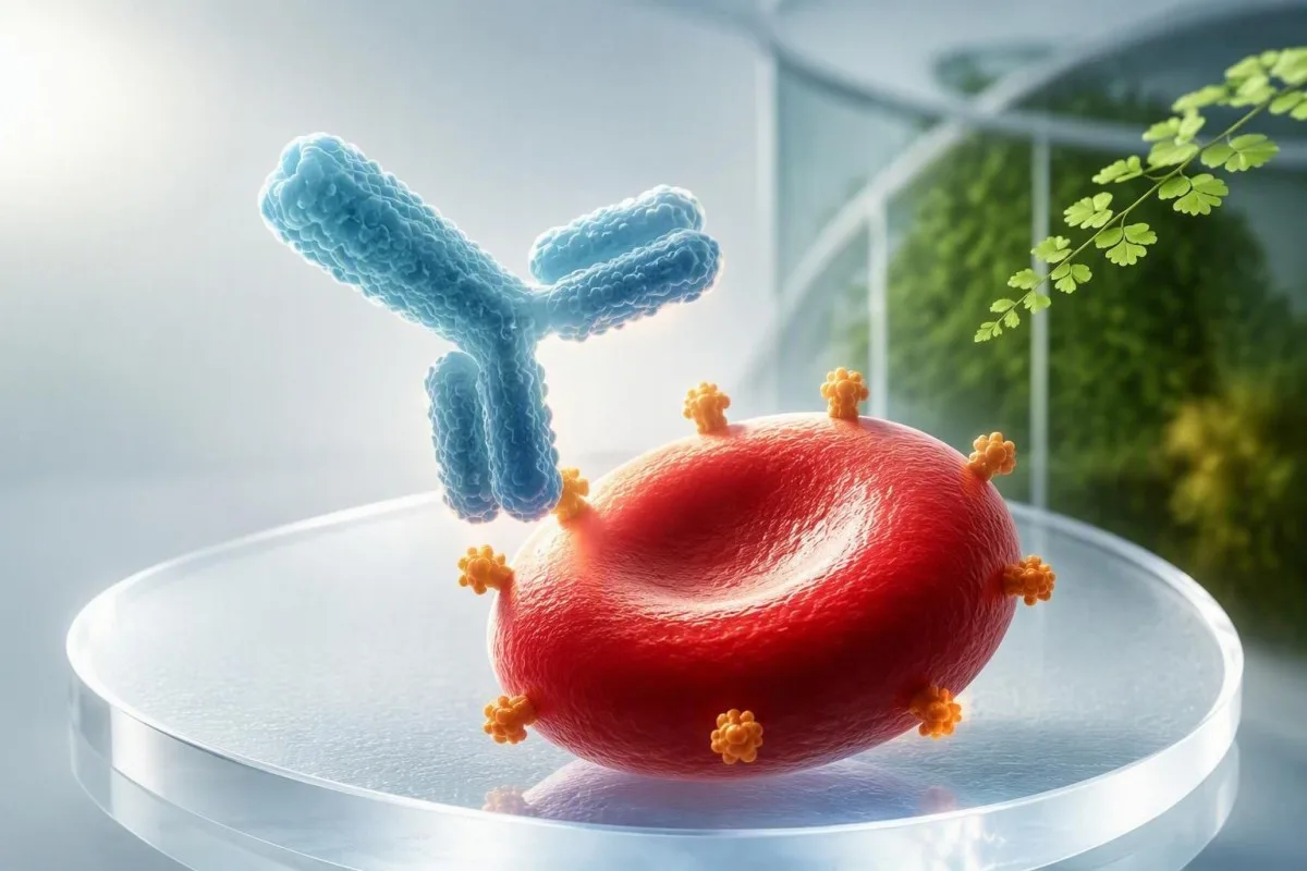
Кызыл кан клеткаларына каршы антителонун оң жыйынтыгы сиздин төрөт боюнча командаңызга антителону аныктоону жана анын маанилүүлүгүн баалоону билдирет. Көптөгөн жыйынтыктар балага эч качан зыян келтирбейт, бирок аз сандагы учурларда адистердин кылдат көзөмөлү талап кылынат.


- Оң скрининг бул күтүлбөгөн кызыл кан клеткаларына каршы антитело аныкталганын билдирет; бул өзүнчө түйүлдүктүн анемиясын диагноз койбойт.
- Anti-D, anti-c жана anti-K Улуу Британиянын кош бойлуулук боюнча көрсөтмөлөрүндө түйүлдүктүн катуу анемиясы менен эң ырааттуу байланышкан антителолор болуп саналат.
- 1:16 же 1:32 титри көпчүлүк антителолор үчүн түйүлдүк-медициналык талкууга себеп болот, бирок ар бир лаборатория өзүнүн критикалык чегин белгилейт.
- 4 IU/mL жогору Anti-D же RCOG көрсөтмөсү боюнча 7.5 IU/mL жогору anti-c адистердин баалоосуна татыктуу.
- Anti-K кош бойлуулук учурунда дароо медициналык жардам талап кылат себеби ал тургай төмөн титрде да катуу түйүлдүктүн аз кандуулугу пайда болушу мүмкүн.
- MCA Doppler 1,5 MoM жогору орточо же катуу түйүлдүктүн аз кандуулугун текшерүү үчүн колдонулган инвазивдик эмес чектөө болуп саналат.
- Пассивдүү анти-D Rh иммуноглобулиндин үзгүлтүксүз колдонуусунан болжол менен 8–12 жумага чейин байкалып турушу мүмкүн жана иммундук анти-D менен бирдей эмес.
- Атасынын антигенин же түйүлдүктүн ДНКсын текшерүү түйүлдүк тийиштүү эритроцит антигенин алып жүрөөр-жүрбөсүн аныктай алат.
- Симптомдорду күтпөңүз: түйүлдүктүн аз кандуулугу, адатта, кош бойлуу аял үчүн байкалбайт жана пландаштырылган байкоо аркылуу аныкталат.
Кош бойлуулук учурунда антителонун оң анализи чындыгында эмнени билдирет
A кош бойлуулук учурунда антителонун скринингинин оң болушу сиздин плазмаңызда сиздин ээ болбогон эритроцит антигенине реакция кылуучу антитело бар экендигин билдирет; бул сиздин балаңыз жабыркаганын далилдебейт. Кийинки кадам - антителону идентификациялоо, андан кийин анын деңгээли, тенденциясы жана түйүлдүктүн мураскорлук антигени байкоо жүргүзүүнү талап кылабы же жокпу чечүү.
Тест көбүнчө индиректтик антиглобулин тест же индиректтик Кумбс тест кош бойлуулук скрининг деп аталат. Ал эненин плазмасындагы ABO системасынан тышкары антителолорду издейт, ал эми түз антиглобулин тест эритроциттерге мурунтан эле тиркелген антителолорду издейт. Скрининг, адатта, катталуу учурунда жана Улуу Британияда болжол менен 28 жумада кайрадан жүргүзүлөт; экинчи үлгү маанилүү, анткени клиникалык жактан маанилүү антитело эрте терс натыйжадан кийин пайда болушу мүмкүн.
Менин практикамда “оң” деген сөз эч ким антителонун атын түшүндүрө электе эле түшүнүктүү тынчсызданууну жаратат. Төмөн деңгээлдеги anti-Lea же anti-P1 натыйжасы көбүнчө түйүлдүк үчүн аз өзгөрөт, ал эми anti-D, anti-c, же anti-K дароо жолду өзгөртөт. Позитивдүү антителонун натыйжалары эмнени билдирет кененирээк жалпы түшүндүрмө болуп саналат, бирок кош бойлуулук чечимдери эритроциттердин так спецификалык мүнөзүнө жараша болот.
Кантести - бул AI кан анализи анализатору бул кош бойлуулук лабораториялык отчетун жөнөкөй тилге которо алат, бирок ал түйүлдүктүн абалын аныктай албайт же кан куюу лабораториясын жана акушердик командасын алмаштыра албайт. 2026-жылдын 18-сентябрына карата, мен бейтаптарга баштапкы отчетту, алардын кан тобун, мурунку кан куюу тарыхын жана ар бир анти-D инъекциясынын датасын сактап коюуну сунуштайм; бул төрт деталь көбүнчө көрүнгөн карама-каршылыктарды чечет.
Кош бойлуулук учурунда скрининг эмне үчүн өзгөрүшү мүмкүн
Эритроцит аллоантителолору мурунку кош бойлуулук, бойдон алдыруу, жатын ичиндеги кош бойлуулук, кан куюу же кээде байкалбаган түйүлдүк-эненин канынын агымынан кийин пайда болушу мүмкүн. Антителонун концентрациясы жылдар бою туруктуу болушу мүмкүн, аныктоо үчүн азайтып, андан кийин антигендин жаңы экспозициясынан кийин тез көбөйүшү мүмкүн.
Кайсы кызыл кан клеткаларына каршы антителолор түйүлдүккө таасир этиши мүмкүн?
Anti-D, anti-c, жана anti-K эритроцит антителолору болуп саналат, кош бойлуулук топтору эң жогорку артыкчылыктуу деп эсептешет, анткени алар түйүлдүктүн аз кандуулугун жана жаңы төрөлгөн ымыркайдын сары оорусун пайда кылышы мүмкүн. Anti-E, anti-C, anti-e, anti-Fya, anti-Jka, жана anti-S да гемолитикалык ооруну пайда кылышы мүмкүн, бирок орточо тобокелдик, адатта, төмөн жана аз болжолдуу.
Antibodies that cross the placenta are usually IgG менен коштоп келет; large IgM antibodies generally do not cross in clinically meaningful amounts. Anti-Lea, anti-Leb, anti-M when naturally occurring IgM, and anti-P1 are therefore often reassuring findings, though the transfusion laboratory still records them because they can complicate selecting compatible blood for the mother.
Anti-K behaves differently from anti-D. It can suppress fetal red-cell production as well as shorten circulating red-cell survival, so a modest titre may still matter. RCOG’s Green-top Guideline No. 65 recommends referral once anti-K is detected, rather than waiting for a numerical threshold (RCOG, 2014).
The practical question is never simply “Is an antibody present?” It is “Does the fetus have the target antigen, and is the antibody behaving in a way that could lower fetal hemoglobin?” For related red-cell patterns, our guide to ретикулоциттерди текшерүү боюнча түшүндүрмөсү explains why young red-cell production is central to anemia assessment.
Лабораториялар оң антитело анализин кантип тастыктайт
A pregnancy antibody screen positive result is confirmed by antibody identification and, when relevant, quantitative measurement or titration in the same specialist laboratory. One isolated screen result is not enough to estimate fetal danger because assay method and antibody strength both influence the report.
The laboratory tests maternal plasma against a panel of reagent red cells with known antigen profiles. The pattern of reactions identifies anti-D, anti-c, anti-K, or another specificity; it can also expose multiple antibodies. If the screen is weak or inconsistent, the laboratory may repeat it using a different enhancement medium, because cold-reacting antibodies and nonspecific reactivity can muddy the first result.
For most non-D antibodies, a titre is reported as a doubling dilution such as 1:2, 1:8, 1:16, or 1:32. A rise from 1:4 to 1:16 represents a fourfold increase, not a small change. Results should be compared in the same laboratory where possible, since a 1:16 from one method cannot safely be treated as interchangeable with 1:16 from another.
A prior transfusion can be the missing clue, particularly after surgery or postpartum hemorrhage. I ask specifically about transfusions received abroad, because patients may not realize that a unit given 10 years earlier is relevant. Blood type and reticulocyte markers provide useful context, but the antibody identification card from the blood bank is the document to keep permanently.
Титри, IU/mL жана багыттоого алып келген сандар
Titre thresholds help select monitoring intensity, but they do not directly measure fetal hemoglobin. A critical titre of 1:16 or 1:32 is commonly used for many antibodies in US practice, while UK services quantify anti-D and anti-c in IU/mL and use antibody-specific thresholds.
For anti-D, RCOG advises fetal-medicine referral when the level exceeds 4 IU/мл and identifies levels above 15 IU/mL as associated with a high risk of severe hemolytic disease. For anti-c, referral begins above 7.5 IU/mL, with risk rising substantially above 20 IU/mL. These cutoffs describe population risk; a prior severely affected pregnancy outweighs a seemingly modest current number.
Many maternity units repeat anti-D and anti-c testing every 4 weeks until 28 weeks, then every 2 weeks until delivery. For other antibodies, a repeat at 28 weeks may be enough if there is no history of hemolytic disease and the fetus is not known to be antigen-positive. The ACOG Practice Bulletin on alloimmunization emphasizes that laboratory trends and obstetric history must be read together (ACOG, 2018).
Dr. Thomas Klein’s practical advice is to ask for the actual number and unit, not merely “high” or “low.” A laboratory flag is not a fetal diagnosis, and a falling titre does not guarantee safety once Doppler surveillance has begun. Our article on сапаттык жана сандык тесттер explains why those result formats answer different questions.
Инъекциядан алынган пассивдүү Anti-D болушу мүмкүнбү?
Ооба—passive anti-D after Rh immunoglobulin can make the antibody screen positive for roughly 8 to 12 weeks, sometimes longer with sensitive assays. Passive anti-D protects against sensitization; immune anti-D means sensitization has already occurred and needs a different plan.
Routine Rh immunoglobulin is often given at 28 weeks in the UK, sometimes as two doses earlier in the third trimester, and after a potentially sensitizing event such as vaginal bleeding, abdominal trauma, or an invasive procedure. A standard UK dose is commonly 1,500 IU at 28–30 weeks, though local schedules vary. The laboratory needs the injection date, dose, and product record before it can interpret anti-D correctly.
No single laboratory test perfectly separates passive anti-D from early immune anti-D. Timing, reaction strength, previous negative screens, and whether the result persists or rises guide the interpretation. In a patient screened 6 days after prophylaxis with weak anti-D and a previously negative booking sample, passive antibody is much more likely; a rising level over several weeks is more concerning.
Do not decline indicated Rh immunoglobulin just because a report says “anti-D detected” unless your maternity unit has confirmed immune anti-D. If there is uncertainty, the transfusion laboratory and fetal-medicine clinician should decide together. Pregnancy test timing and false results is a separate issue, but it is a reminder that test timing can completely change interpretation.
Эмне үчүн кээ бир антителолордон кийин өнөктөштү текшерүү сунушталат
Partner antigen testing estimates whether the fetus can inherit the red-cell antigen targeted by the maternal antibody. If the biological father lacks that antigen, the fetus cannot be antigen-positive for it and antibody-related fetal anemia from that specificity is not expected.
A father who is homozygous for the relevant antigen passes it to every fetus; a heterozygous father passes it about 50% of the time. For example, paternal K-negative status makes anti-K unable to affect that fetus, while K-positive status needs zygosity or fetal genotyping to refine the risk. This is a risk calculation, not a test of paternity.
Partner testing should be offered sensitively and never assumed to be straightforward. Some patients prefer direct fetal testing, use donor conception, or cannot access partner testing safely. In those situations, treating the fetus as potentially antigen-positive until non-invasive fetal genotyping is available is clinically sensible.
A positive antibody screen can also affect maternal care if an emergency transfusion becomes necessary. The maternity team should notify the blood bank early so antigen-negative units can be sourced; this is particularly relevant with multiple antibodies. Read our гемоглобин жана гематокрит жетектейт for why pregnancy anemia still deserves separate attention.
Инвазивдик эмес түйүлдүк генотипирлөө: Ал сизге эмнени айтып бере алат
Cell-free fetal DNA testing from maternal plasma can determine fetal D, C, c, E, e, and K antigen status without sampling fetal fluid in many specialist services. A fetus reported antigen-negative for the relevant antibody usually does not need surveillance for anemia caused by that antibody.
Fetal genotyping is commonly performed from around 16 жума for most Rh antigens and may be feasible from around 20 жумадан кийин for K because of assay performance and local protocol. A result may be inconclusive when fetal DNA fraction is low, especially early in pregnancy or with higher maternal body weight. An inconclusive result is not reassuring; clinicians usually repeat it or monitor as if the fetus is antigen-positive.
Кантести - бул AI лабораториялык тест чечмелөө кызматы аркылуу жарыяланат that can help patients file serial maternal antibody results by date and unit, but fetal genotyping results require clinician review because test availability and confidence intervals differ between laboratories. Our медициналык валидация стандарттары explain why laboratory source, method, and clinical oversight remain essential.
Invasive testing with amniocentesis or fetal blood sampling is now rarely used solely to establish antigen status when cell-free DNA is available. It still has a role when results conflict, when fetal anemia needs confirmation, or when another diagnostic question justifies the procedure. The shift toward non-invasive testing has reduced procedure-related risk, which most patients find genuinely reassuring.
MCA Допплер мониторинги түйүлдүктүн анемиясын кантип аныктайт
Middle cerebral artery peak systolic velocity (MCA-PSV) Doppler is the main non-invasive test for moderate or severe fetal anemia in an antigen-positive at-risk fetus. A value above 1.5 multiples of the median (MoM) prompts urgent fetal-medicine assessment, often including consideration of fetal blood sampling.
Anemic fetal blood is less viscous, so it moves faster through the middle cerebral artery. The sonographer measures the vessel near its origin with the Doppler beam aligned as closely as possible to flow; a poor angle can falsely increase the number. This is why a Doppler scan should be performed in an experienced fetal-medicine unit rather than interpreted from a single still image.
Monitoring commonly starts around 18 to 20 weeks when the fetus is antigen-positive and maternal antibody levels or history justify it. Scans may be weekly or every 1–2 weeks, depending on the antibody, concentration, and prior pregnancy outcome. MCA-PSV is less reliable after about 35 weeks, when delivery may become safer than invasive confirmation in some cases.
The scan is watching a physiological consequence, not the antibody itself. Hydrops—fluid accumulation in at least two fetal compartments—is a late and serious sign, so clinicians aim to intervene long before it appears. For another pregnancy-specific laboratory emergency pattern, see our explanation of HELLP syndrome results.
Түйүлдүктүн анемиясы шектелсе эмне болот
Suspected significant fetal anemia leads to urgent fetal-medicine review, not automatic treatment. The specialist team weighs gestational age, MCA-PSV, ultrasound findings, and previous pregnancy history before recommending fetal blood sampling, intrauterine transfusion, early delivery, or close repeat assessment.
If fetal blood sampling confirms significant anemia, an intrauterine transfusion may be given through the umbilical cord under continuous ultrasound guidance. Donor cells are selected to be antigen-negative for the maternal antibody, CMV-safe according to local policy, irradiated, and crossmatched against maternal plasma. These procedures are concentrated in expert centers because timing and technique matter enormously.
The first transfusion is often performed after 18 weeks, although individual circumstances vary. Repeat transfusions may be needed every 2–4 weeks because donor cells gradually age and the fetus continues to grow. A successful transfusion does not cure maternal alloimmunization; it buys time for safer fetal maturation.
I have seen families focus on the procedure and miss a practical detail: delivery location. A baby at risk of hemolytic disease should be delivered where neonatal bilirubin testing, compatible blood, and intensive care are available if needed. High bilirubin warning patterns explain why newborn jaundice is monitored so closely after birth.
Эмне үчүн мурунку жабыркаган кош бойлуулук планын өзгөртөт
A prior baby with anemia, neonatal exchange transfusion, severe jaundice, or an intrauterine transfusion predicts risk better than a single current antibody titre. In a subsequent antigen-positive pregnancy, fetal-medicine teams may begin surveillance earlier even when the first measured antibody level is low.
Immune memory can produce a sharp antibody rise after fetal antigen exposure, sometimes before the routine 28-week screen. Anti-D and anti-c concentrations can lag behind fetal risk, particularly when there was severe disease in a prior pregnancy. This is one reason clinicians ask about phototherapy, neonatal anemia, and transfusions—not just whether a previous pregnancy was “normal.”
Records from a prior delivery can be more useful than a verbal memory of the antibody name. Ask for the newborn discharge letter, direct antiglobulin test, bilirubin peak, hemoglobin nadir, and any transfusion details. A bilirubin level of 20 mg/dL (342 µmol/L) has a very different implication in a well newborn at 4 days than in a rapidly jaundiced infant at 18 hours.
Kantesti AI can preserve a dated trend view for maternal lab reports, yet historic neonatal records should sit alongside it rather than be reduced to a graph. Our узунунан (лонгитюддик) натыйжа боюнча колдонмо offers practical ideas for organizing those records before the first fetal-medicine appointment.
Эмне үчүн сиздин трансфузия планыңыз да маанилүү
A clinically significant maternal red-cell antibody can delay the supply of compatible blood for the pregnant person, even when the fetus is antigen-negative. Antigen-negative, crossmatch-compatible units may need to be sourced from a regional center, especially with anti-K, anti-Jka, or multiple antibodies.
This issue matters most if there is placenta previa, suspected placenta accreta spectrum, severe anemia, a planned caesarean birth, or any elevated hemorrhage risk. The obstetric and transfusion teams can arrange a current group-and-screen sample and reserve appropriate units. In most uncomplicated pregnancies, no blood is pre-booked, but the antibody record remains visible.
Tell your care team about every known antibody, even if a newer screen is negative. Some antibodies fall below detection yet return after transfusion exposure, and electronic records do not always follow patients across countries. A wallet antibody card or a clear phone photo of the formal laboratory report is surprisingly useful in an emergency.
Maternal hemoglobin affects resilience if blood loss occurs, but it does not tell us whether the fetus is anemic. Iron deficiency can coexist with alloimmunization, which is why кош бойлуулук учурундагы ферритиндин диапазону deserve a separate, early conversation.
Оң жыйынтыктар жөнүндө жалпы туура эмес түшүнүктөр
A positive antibody screen does эмес mean you have an autoimmune disease, an infection, or that your baby has the same blood group as you. It means an antibody was detected against a red-cell antigen, and only some antibodies can cross the placenta and affect an antigen-positive fetus.
The phrase “Coombs positive” is often used loosely, which creates confusion. The indirect Coombs test in pregnancy finds free antibodies in plasma; a direct Coombs test on a newborn’s cells may support hemolytic disease after birth, but neither test alone measures anemia severity. These tests answer different clinical questions at different times.
Another misconception is that dietary changes can lower an alloantibody titre. They cannot. Good nutrition supports maternal health, but there is no food, supplement, or detox that removes anti-D, anti-c, or anti-K; be wary of anyone offering one. For safe pregnancy supplement decisions, our lab-guided supplement guide keeps the focus on deficiencies that can actually be corrected.
As Dr. Thomas Klein, I also remind patients that anxiety can make them read every antibody result as a countdown to harm. Most positive screens do not lead to intrauterine treatment, and planned monitoring is what turns a rare serious risk into a managed clinical pathway.
Кийинки төрөт боюнча жолугушууда кандай суроолорду берүү керек
Сураңыз exact antibody name, current level or titre, previous result, and whether the fetus is known or assumed to be antigen-positive. Those four answers determine nearly every next step after a positive antibody screen in pregnancy.
Useful questions include: Is this passive or immune anti-D? Was my result measured in IU/mL or titre? Do I need paternal testing or fetal genotyping? When is my next sample, and what specific result would trigger MCA Doppler? Write the answers down; the distinction between anti-D and anti-G, for instance, can change whether Rh immunoglobulin remains appropriate.
Ask who will coordinate results after hours and where you should deliver if the fetus becomes at risk. You should also ask whether the blood bank has been notified if you have a planned caesarean birth or a history of postpartum bleeding. Preparing for a useful baseline draw includes simple habits that prevent missing key report details.
Кантести - бул AI биомаркер чечмелөө платформасы designed to make laboratory language easier to follow across 75+ languages, but a pregnancy antibody screen needs obstetric-led decisions and a transfusion-laboratory safety net. Readers can review how our clinical team works on the Медициналык кеңеш; in this setting, responsible interpretation means knowing exactly when AI should hand the decision back to the specialist.
Качан шашылыш жардамга кайрылуу керек жана төрөттөн кийин эмне болот
A positive red-cell antibody screen alone is not usually an emergency, but reduced fetal movements, vaginal bleeding, abdominal trauma, or labour symptoms require prompt maternity assessment regardless of antibody level. After birth, an at-risk newborn may have cord blood testing for blood group, direct antiglobulin test, hemoglobin, and bilirubin.
Fetal anemia itself rarely causes a symptom the pregnant person can feel, which is why keeping Doppler appointments matters. Contact your maternity triage unit the same day for reduced movements; do not wait until the next antibody blood test. Sudden swelling, severe headache, visual changes, or upper abdominal pain have other pregnancy causes and need urgent evaluation too.
Newborn bilirubin can rise quickly in immune hemolysis, sometimes within the first 24 hours. Treatment may include frequent feeding support, phototherapy, intravenous immunoglobulin in selected cases, or exchange transfusion for severe hyperbilirubinemia; the neonatal team uses age-specific bilirubin thresholds rather than one universal number. Late anemia can occur at 2–6 жума, so follow-up hemoglobin checks may be arranged even after a seemingly calm discharge.
The best outcome is often quiet and uneventful: an identified low-risk antibody, an antigen-negative fetus, or normal serial Dopplers followed by routine neonatal observation. If you are comparing laboratory reports, our AI technology guide describes the limits of automated interpretation; no app should reassure or alarm you instead of the maternity team when fetal monitoring is due.
Көп берилүүчү суроолор
Кош бойлуулук учурунда оң антитело скринингинин жыштыгы кандай?
Положительный скрининг эритроцитарных антител встречается примерно у 1% - 2% беременностей во многих группах населения с высоким уровнем доходов, проходящих скрининг, хотя частота зависит от происхождения, истории переливаний крови и местного использования Rh-иммуноглобулина. Только часть этих антител может вызывать гемолитическую болезнь плода и новорожденного. Анти-D, анти-c и анти-K подвергаются наиболее пристальному наблюдению, поскольку они наиболее четко связаны с клинически значимой анемией плода. Лаборатория должна определить специфическое антитело, прежде чем можно будет оценить индивидуальный риск.
Кош бойлуулук учурунда кайсы антителонун титри коркунучтуу?
Көпчүлүк D эмес эритроциттик антителолор үчүн, 1:16 же 1:32 титри феталдык медицина боюнча жолдомо алуунун критикалык чеги катары каралат, бирок чектөө лабораторияга жана антителого жараша болот. Улуу Британиянын жетекчилиги кээ бир антителолор үчүн санды аныкталган деңгээлдерди колдонот: 4 IU/млден жогору анти-D жана 7.5 IU/млден жогору анти-c жолдомо талап кылат, ал эми биринчи жолу аныкталганда анти-K жолдомо талап кылат. Буга чейинки кош бойлуулуктагы феталдык анемия учурдагы ишенимдүү титрди жокко чыгарат. Титрлер тобокелдикти баалайт; MCA доплери феталдык анемия чындап өнүгүп жаткандыгын баалайт.
Анти-D инъекциясынан улам антитело скрининги оң болушу мүмкүнбү?
Ооба, Rh иммуноглобулинди сайгандан кийин болжол менен 8-12 жума бою антителонун скринингинде пассивдүү anti-D пайда болушу мүмкүн. Профилактикадан кийин пассивдүү anti-D күтүлөт жана бул адамдын сезимтал болуп калганын билдирбейт. Кан куюу лабораториясы сайылган күнүн, мурунку скринингдерди, реакциянын күчүн жана деңгээли сакталып калабы же көбөйөбү дегенди колдонуп, пассивдүү жана иммундук anti-Dны айырмалайт. Эч качан сунушталган Rh иммуноглобулинди anti-D отчетто төрөт тобунун ырастамасысыз эле пайда болду деген шылтоо менен өткөрүп жибербеңиз.
Эгер өнөктөшүм антиген-терс болсо, балам көзөмөлгө муктажбы?
Эгер биологиялык атасы кызыл кан клеткасынын белгилүү антигенине терс деп тастыкталса, түйүлдүк ал антигенди тукум кууп өтө албайт жана ошол эненин антителасынан коркунучта болбойт. Мисалы, K-терс атасы эненин anti-K кош бойлуулукта түйүлдүктүн анемиясына себеп боло албайт дегенди билдирет. Өнөктөштү сыноо милдеттүү эмес жана үй-бүлөлүк жагдайлар ар кандай болгондуктан, кылдаттык менен жүргүзүлүшү керек. Жука клеткалуу түйүлдүк ДНК тестирлөө антигенге жана жергиликтүү кызматка жараша, болжол менен 16-20 жумадан баштап, көбүнчө түз инвазивдик эмес жоопту бере алат.
Кызыл кан клеткасынын антителолору бойдон түшүүгө себеп болобу?
Кызыл кан клеткаларына каршы аллоантителолор, адатта, эрте бойдон түшүүгө алып келбейт; алардын негизги таасири плацента IgG антителолорун өткөргөндөн кийин эненин канында анемия пайда болушу, көбүнчө кош бойлуулуктун кийинки этаптарында. Гидропс же түйүлдүктүн жоголушу өтө оор дарыланбаган аллоиммунизацияга алып келиши мүмкүн, бирок антигендерди аныктоо, MCA Доплер жана жатын ичиндеги кан куюу бул абалды бир топ жакшыртты. Бирок, бойдон түшүү же ectopic кош бойлуулук антителолордун пайда болушуна алып келүүчү сезгичтик окуясы болушу мүмкүн. RhD-терс адамдарга, эгерде алар буга чейин эле иммундук анти-D позитивдүү болбосо, кош бойлуулуктун айрым эрте окуяларынан кийин Rh иммуноглобулини сунушталышы мүмкүн.
Менин антитело скринингим оң болсо, төрөттөн кийин менин балам дарылоого муктаж болобу?
Ымыркайга төрөттөн кийин дарылоо керек, эгерде ал тиешелүү антигенди мурастап алса жана гемолиз, анемия же билирубиндин көбөйгөндүгүн көрсөтсө. Киндік канды текшерүү кан тобун, түз антиглобулин тестти, гемоглобинди жана билирубинди камтышы мүмкүн, андан кийин 24 сааттын ичинде билирубинди кайталап өлчөө керек, эгерде коркунуч болсо. Фототерапия жалпы жана инвазивдик эмес; интенсивдүү дарылоо оор учурлар үчүн сакталат. Ымыркайлар 2-6 жумада кеч анемияны да иштеп чыгышы мүмкүн, андыктан кээ бирлерине амбулатордук гемоглобинди көзөмөлдөө керек.
Бүгүн AI менен күчөтүлгөн кан анализин талдоону алыңыз
Дүйнө жүзү боюнча 2 миллиондон ашык колдонуучу Kantestiти заматта, так лабораториялык анализ талдоосу үчүн ишенет. Кан анализиңизди жүктөп, бир нече секунд ичинде 15,000+ биомаркерлеринин комплекстүү чечмелөөсүн алыңыз.
📚 Шилтемеленген изилдөө басылмалары
Кляйн, Т., Митчелл, С., & Вебер, Х. (2026). Kantesti Изилдөө тобу. (2026). Iron Studies Guide: TIBC, Iron Saturation & Binding Capacity. Zenodo.. Kantesti AI медициналык изилдөө.
Кляйн, Т., Митчелл, С., & Вебер, Х. (2026). Kantesti Изилдөө тобу. (2026). aPTT Normal Range: D-Dimer, Protein C Blood Clotting Guide. Zenodo.. Kantesti AI медициналык изилдөө.
📖 Тышкы медициналык шилтемелер
Royal College of Obstetricians and Gynaecologists (2014). The Management of Women with Red Cell Antibodies during Pregnancy: Green-top Guideline No. 65. RCOG Green-top Guideline.
⚕️ Медициналык эскертүү
Бул макала билим берүү максатында гана жана медициналык кеңеш болуп саналбайт. Диагноз коюу жана дарылоо чечимдери үчүн дайыма квалификациялуу саламаттыкты сактоо адисине кайрылыңыз.
E-E-A-T ишеним сигналдары
Тажрыйба
Дарыгер жетектеген лабораториялык чечмелөө иш процесстерин клиникалык кароо.
Экспертиза
Биомаркерлер клиникалык контекстте кандай жүрөрүнө лабораториялык медицина багыты.
Авторитеттүүлүк
Доктор Томас Клейн жазган, доктор Сара Митчелл жана профессор доктор Ханс Вебер тарабынан каралган.
Ишенимдүүлүк
Тынчсызданууну азайтуу үчүн так кийинки кадамдар менен далилге негизделген чечмелөө.