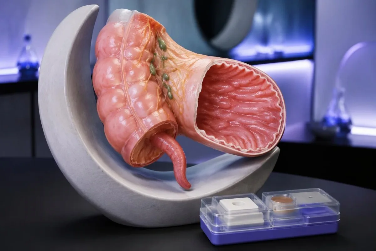
A positive stool result usually identifies a bacterial intestinal infection; mild cases often need fluids rather than antibiotics. Severe right-sided abdominal pain, dehydration, or signs of sepsis require urgent assessment—even when the test appears to explain your symptoms.

Pokok-pokok penting
- Positive stool PCR means Yersinia genetic material was detected; it does not measure bacterial viability or prove that every symptom has the same cause.
- Incubation is typically 4–7 days, although symptoms can begin 1–14 days after exposure.
- Appendicitis mimic occurs because Yersinia can affect the terminal ileum and nearby lymph nodes; a positive stool test cannot exclude actual appendicitis.
- Supportive care is usually sufficient for uncomplicated illness in an otherwise healthy person who can drink and is improving.
- Oral rehydration replaces water and salts; adults without fluid restrictions can often start with approximately 200–250 mL after each loose stool.
- Antibiotik are considered for severe disease, bloodstream infection, or selected high-risk patients—not automatically for every positive result.
- Higher-risk situations include iron overload, deferoxamine treatment, significant immunosuppression, and serious underlying illness.
- Penilaian sing mendesak is needed for worsening localized abdominal pain, inability to retain fluids, confusion, fainting, or substantial intestinal bleeding.
- Tindak lanjut is sensible when diarrhea lasts 14 days or longer; routine PCR clearance testing is usually unnecessary after uncomplicated recovery.
What does a Yersinia enterocolitica positive result mean?
A Yersinia enterocolitica positive stool result means the laboratory detected this bacterium or its genetic material. With compatible diarrhea, it usually supports intestinal infection; many uncomplicated cases improve over 1–3 weeks with hydration, while severe illness or high-risk conditions may justify antibiotics.
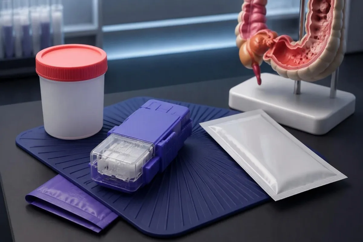
A positive result is not a severity score. Someone having 3 loose stools daily while drinking normally needs a different assessment from someone with persistent vomiting, low urine output, and worsening abdominal tenderness—even if both laboratory reports contain exactly the same wording.
The test method changes the interpretation. A report saying PCR detected identifies a genetic target, whereas a positive culture demonstrates growth of viable organisms; neither method alone tells a clinician whether the bacterium has entered the bloodstream or whether a separate abdominal emergency is present.
An unexpected result deserves clarification rather than automatic treatment. If you have had no diarrhea, have already recovered, or have 2 organisms detected on a multiplex panel, ask why the sample was collected and whether the finding matches the timing and pattern of your illness.
Kantesti iku sawijining Analisa tes getih AI that can help explain accompanying laboratory abnormalities, but stool microbiology requires separate clinical interpretation. Our latar mburi perusahaan describes our role; the immediate decision here depends on symptoms, hydration, examination, and individual risk—not an AI-generated infection label.
How reliable is a Yersinia stool PCR positive result?
A Yersinia stool PCR positive result is usually meaningful when symptoms fit, but PCR detects genetic material rather than proving that live bacteria remain. There is no universal cycle-threshold cutoff that separates active Yersinia illness from residual detection, and 2 different laboratories may use different panels.
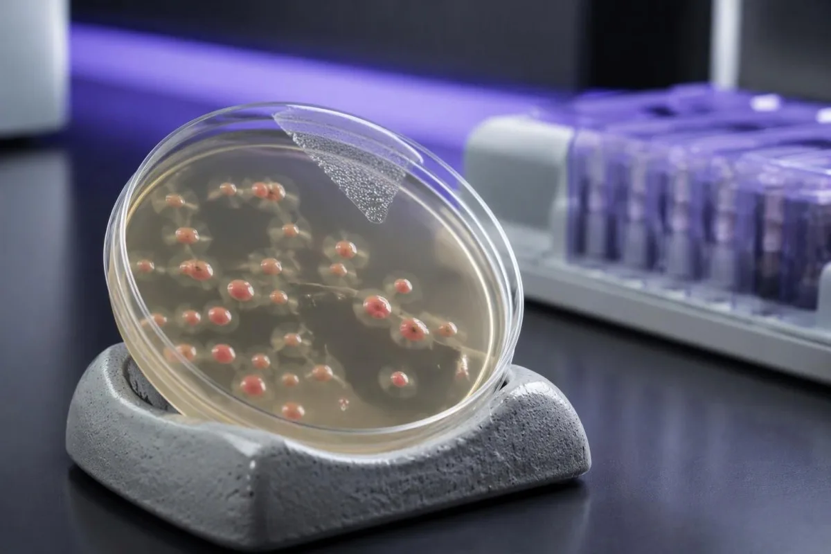
Multiplex stool panels can detect more than 1 organism, and a second positive target can change treatment safety. The 2017 IDSA infectious-diarrhea guideline advises interpreting molecular results clinically because nucleic-acid detection does not necessarily establish viable organisms or identify the principal cause of symptoms (Shane et al., 2017).
Culture has a different purpose from repeating PCR. When susceptibility testing or public-health investigation is needed, ask whether the laboratory can perform kultur refleks; Yersinia may require selective methods, and a standard stool culture does not necessarily include every organism listed on a molecular panel.
A negative culture after positive PCR does not automatically prove that the PCR was wrong. Recent antibiotics, transport conditions, low organism numbers, and culture technique can reduce recovery; the useful distinction is between 2 tests answering different questions, not a simple contest over which result wins.
Reports that say detected or not detected are kualitatif, not measures of bacterial burden; our pandhuan kualitatif vs kuantitatif explains that distinction. Persistent molecular detection also occurs with other organisms, but the timeline described in our panjelasan babagan panyebaran adenovirus should not be transferred directly to Yersinia.
Which Yersinia infection symptoms fit the result?
Yersinia infection symptoms commonly include diarrhea, fever, and abdominal pain; younger children may have bloody diarrhea, while older children and adults can develop prominent right-lower abdominal pain. Symptoms typically start 4–7 days after exposure, with a reported range of 1–14 days.
Timing helps identify plausible exposures without proving their source. If symptoms began 6 days after a shared meal, that meal falls within the usual incubation window; symptoms beginning within a few hours are less typical for newly acquired Yersinia and should prompt consideration of other causes.
Undercooked pork is a recognized source, but contaminated water, unpasteurized milk, and cross-contamination can also transmit Yersinia. Refrigeration is not a complete safeguard: Yersinia can grow at refrigerator temperatures, so storing food at approximately 4°C does not replace adequate cooking and careful separation of raw foods.
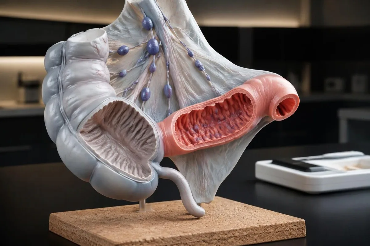
Abdominal pain can outweigh diarrhea. The terminal ileum contains substantial lymphoid tissue, and involvement of this region can produce localized tenderness rather than a diffuse stomach bug; Bottone's review in Clinical Microbiology Reviews describes the intestinal and extraintestinal spectrum of yersiniosis (Bottone, 1997).
Record stool frequency, temperature, fluid intake, and pain location for 24 hours rather than relying only on stool appearance. Our konsistensi tinja can help describe changes, but a stool shape or color cannot identify Yersinia or determine whether antibiotics are needed.
Why can Yersinia mimic appendicitis?
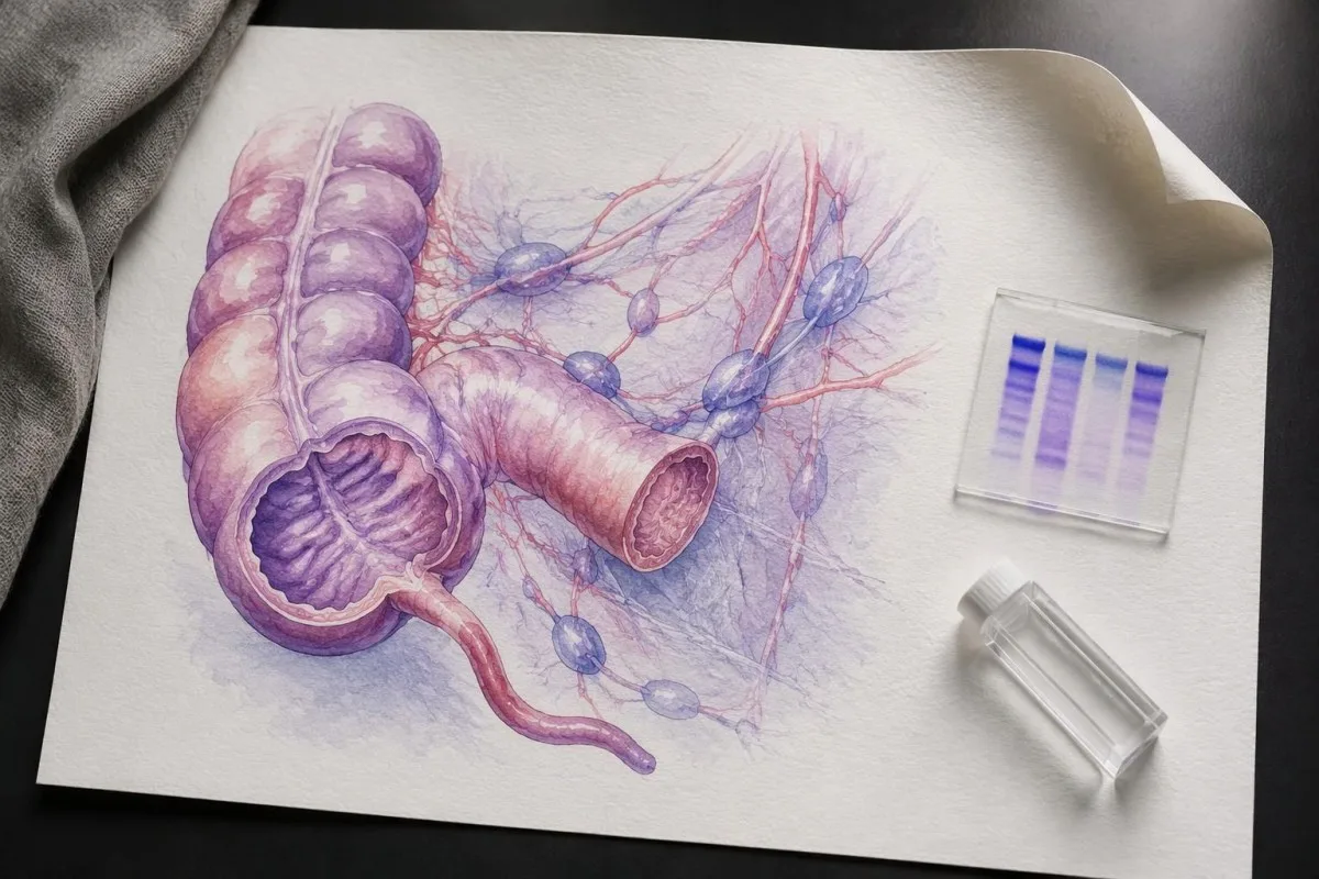
Yersinia can mimic appendicitis by causing terminal ileitis lan mesenteric lymph-node enlargement near the appendix. These 2 processes can produce fever and right-lower abdominal pain; a positive stool result does not exclude simultaneous appendicitis or remove the need for examination and, sometimes, imaging.
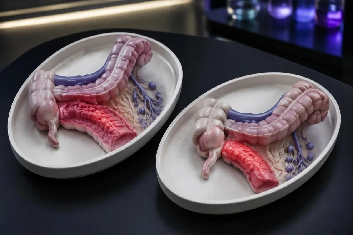
The anatomical explanation matters more than the stool label. The terminal ileum joins the cecum close to the appendix, so irritation of nearby bowel and lymph nodes can create a similar pain pattern; the term pseudoappendicitis describes the mimic, not a reliable diagnosis that can be made at home.
Worsening focal pain needs reassessment even if diarrhea is improving. For example, a hypothetical 16-year-old with fewer loose stools but increasing right-sided tenderness should not wait for repeat stool testing, because improvement in one symptom does not establish that the appendix or surrounding abdomen is safe.
Normal inflammatory markers do not settle the question. A white-cell count of 7.5 × 10⁹/L or a CRP of 3 mg/L can occur early in abdominal disease; our kita explain why normal laboratory results cannot reliably rule out appendicitis.
Ultrasound is often considered first in children and pregnancy, while CT or MRI may be selected according to the clinical situation and local pathways. Kantesti's educational explanations cannot assess guarding, pain with movement, or imaging findings; severe or progressive localized pain warrants urgent in-person assessment rather than a 24-hour home experiment.
Which symptoms require urgent or emergency assessment?
Severe or worsening abdominal pain, confusion, fainting, inability to retain fluids, or substantial intestinal bleeding require urgent assessment. Fever of 39°C or higher merits prompt advice when accompanied by significant illness; a baby younger than 3 months with a temperature of 38°C or higher needs urgent medical assessment.
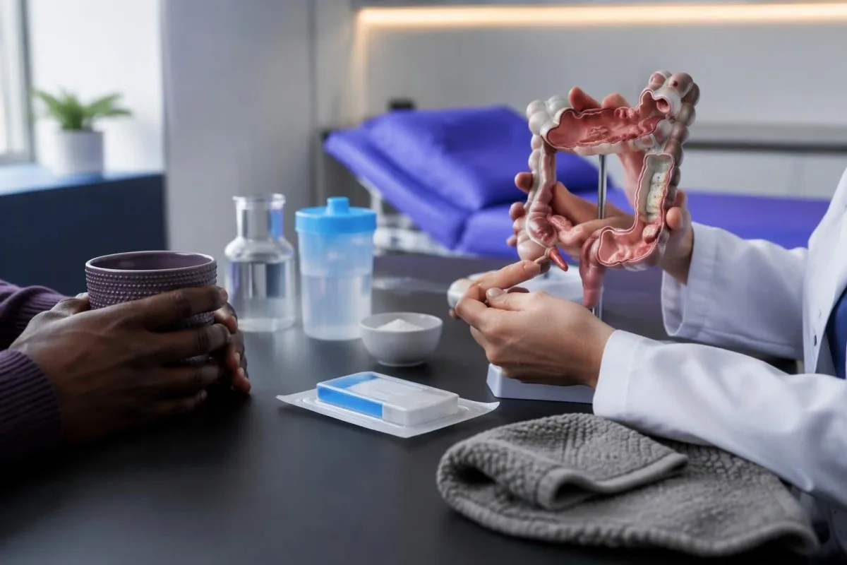
Emergency symptoms are not defined by fever alone. New confusion, very rapid breathing, collapse, cold or mottled extremities, or severe weakness can indicate systemic illness even without a temperature above 39°C; use your local emergency service rather than waiting for a laboratory callback.
Dehydration can become serious before a person looks dramatically unwell. Markedly reduced urine, dizziness on standing, a dry mouth, or inability to keep small sips down should prompt assessment; no urine for roughly 8–12 hours in an adult is concerning, but infants and frail people may need help much earlier.
Visible blood mixed with diarrhea generally deserves same-day advice, while large-volume bleeding, faintness, or new black tarry stool warrants emergency assessment. Our pandhuan getih ing tinja explains why color and quantity matter, but a known Yersinia result should not be used to dismiss ongoing blood loss.
A multiplex panel reporting both Yersinia and Shiga toxin creates a treatment-safety issue. Some antibiotic decisions appropriate for Yersinia may be inappropriate for Shiga toxin-producing E. coli; our Shiga toxin result guidance explains the distinction, and the 2 findings need clinician review before treatment.
When is supportive care enough for Yersinia?
Supportive care is usually enough for uncomplicated Yersinia diarrhea when a person can drink, has no concerning abdominal findings, and is improving. Oral rehydration solution replaces both water and salts; an adult without fluid restrictions can often start with approximately 200–250 mL after each loose stool.
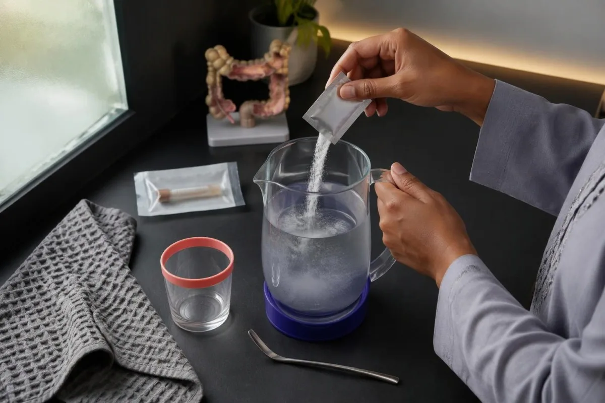
Small, frequent amounts can work better than a large drink during nausea. Try approximately 5–10 mL every 1–2 minutes, increasing gradually if tolerated; prepare commercial oral rehydration sachets using exactly the specified water volume, because making them stronger or weaker changes their salt and glucose balance.
Children need weight-based assessment rather than an adult drinking target. NICE guideline CG84 recommends 50 mL/kg of oral rehydration solution over 4 hours, plus maintenance fluid, for children under 5 with clinical dehydration; a hypothetical 20 kg child would receive 1,000 mL for the deficit under an appropriate care plan (NICE, 2009).
Continue breastfeeding and resume usual age-appropriate food as tolerated rather than fasting for 24 hours. Our childhood gastroenteritis care guide discusses practical hydration observations, although the cause here is bacterial; infants, children with persistent vomiting, and anyone becoming unusually sleepy need reassessment.
Avoid loperamide when diarrhea is bloody, fever is present, or invasive intestinal disease is suspected, and do not give it to children for infectious diarrhea without specific advice. Kidney function may temporarily worsen during fluid loss; our dehydration and eGFR explanation shows why a new abnormal result needs comparison with baseline, not an immediate chronic-disease label.
When are antibiotics appropriate for Yersinia treatment?
Yersinia enterocolitica treatment may include antibiotics for severe illness, bloodstream infection, extraintestinal disease, or selected high-risk patients. The 2017 IDSA guideline lists trimethoprim-sulfamethoxazole as a treatment option, with cefotaxime or ciprofloxacin as alternatives; drug choice depends on susceptibility, patient factors, and infection site.
A positive PCR alone does not establish an antibiotic benefit. The evidence for shortening ordinary, self-limited enteritis is limited, whereas systemic disease creates a much clearer treatment indication; this distinction is reflected in the IDSA guideline's organism-specific recommendations and overall emphasis on targeted treatment (Shane et al., 2017).
Yersinia commonly resists ampicillin and many early-generation cephalosporins because of beta-lactamase activity. Do not assume leftover amoxicillin or a familiar urinary-infection prescription will work; our Salmonella treatment comparison illustrates why identifying a bacterial pathogen is only 1 step in deciding whether and how to treat.
Age, pregnancy, kidney function, allergies, and interacting medicines can change the safest option. Trimethoprim-sulfamethoxazole is generally avoided in infants younger than 2 months, while fluoroquinolones carry important adverse-effect restrictions; a clinician may choose a different agent or hospital-based therapy rather than applying an adult oral regimen to everyone.
There is no single course length that safely covers uncomplicated diarrhea, bacteremia, and a deep-seated focus. Blood cultures, susceptibility results, and response over the first 48–72 hours may guide treatment changes; our Campylobacter antibiotic guide provides another example of why organism-specific recommendations should not be exchanged between infections.
Who is at higher risk of severe Yersinia infection?
Iron overload and deferoxamine treatment are particularly relevant risk factors for severe Yersinia disease. Significant immunosuppression and serious underlying illness also lower the threshold for clinical assessment; a patient with 1 of these risk factors should contact their treating team promptly rather than assuming that ordinary home care is sufficient.
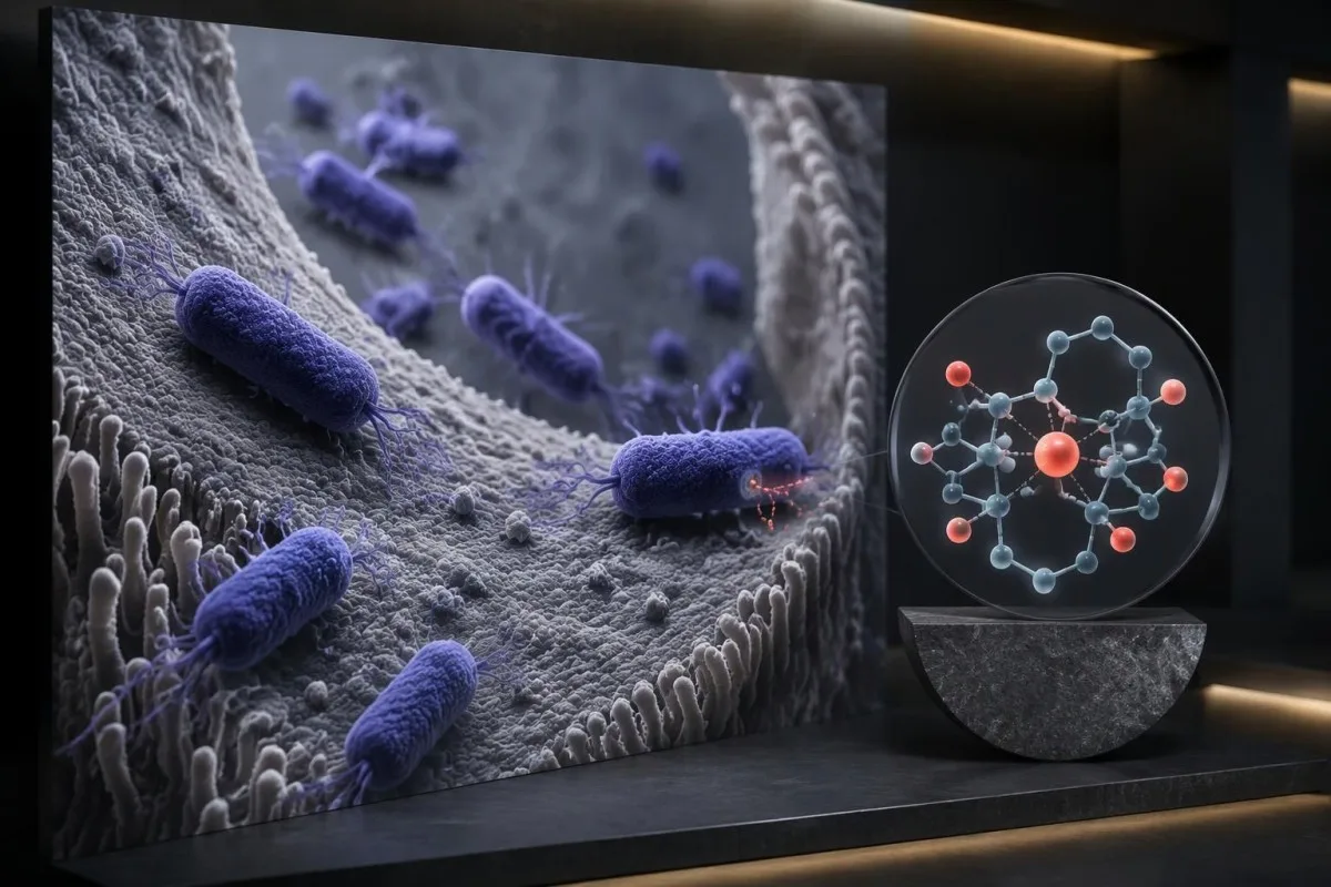
Deferoxamine can bind iron in a form that Yersinia can exploit, helping explain its distinctive association with invasive disease. Bottone's microbiological review discusses iron-dependent virulence and the clinical relevance of iron-loading disorders (Bottone, 1997); this is a biological risk mechanism, not a claim that every patient receiving chelation will become severely ill.
People with transfusion-dependent anemia, known hemochromatosis, or prescribed deferoxamine should tell the assessing clinician about those conditions immediately. If fever or abdominal symptoms develop during treatment, contact the prescribing team the same day; medication interruption or modification is a clinical decision, not a reason to independently abandon long-term chelation.
An isolated ferritin of 800 ng/mL during acute illness does not diagnose iron overload. Ferritin rises as an acute-phase reactant, so previous results, transferrin saturation, transfusion history, and the clinical setting matter; our pandhuan interpretasi panel zat besi explains why a raised storage marker cannot be interpreted alone.
Kantesti AI can help explain why 2 ferritin results taken during different illness states are not directly comparable, but it cannot determine bloodstream spread from ferritin. People receiving chemotherapy, transplant medicines, or substantial immunosuppression need individualized advice; fever, deterioration, or localized pain should trigger assessment even when diarrhea appears relatively mild.
Which blood tests help assess severity and dehydration?
CBC, CRP, electrolytes, and kidney-function tests can help assess the consequences of Yersinia illness, but they do not confirm the stool diagnosis. In an adult, a creatinine increase of at least 0.3 mg/dL, approximately 26.5 µmol/L, within 48 hours meets a standard acute-kidney-injury criterion and needs clinical interpretation.
A high inflammatory marker does not tell you whether the appendix is safe or whether antibiotics will help. For example, a hypothetical CRP of 65 mg/L may accompany several inflammatory conditions; our perbandingan WBC lan CRP explains why disagreement between markers can reflect timing rather than a diagnostic contradiction.
Trend interpretation is more useful than reading a single flagged value. A creatinine rise from 70 to 100 µmol/L over 48 hours can be clinically meaningful even when the second result is close to a laboratory's reference interval; the change and the person's urine output matter more than whether the report prints an asterisk.
Electrolyte losses can cause weakness, cramps, or palpitations, especially when vomiting accompanies diarrhea. A potassium of 2.9 mmol/L is below usual adult reference intervals and warrants prompt clinical review; replacement decisions depend on symptoms, ECG findings, kidney function, and the ability to drink—not simply choosing a high-potassium food.
Kantesti iku sawijining platform interpretasi hasil tes getih AI that can organize accompanying results and explain changes between 2 time points, without diagnosing invasive yersiniosis. Our kanggo BUN lan kreatinin adds context for fluid-loss patterns; blood cultures are a separate clinician-directed test when systemic infection is suspected.
How does Yersinia spread, and when can you return to work?
Yersinia spreads through contaminated food or water and through fecal contamination of hands and surfaces. Many gastroenteritis policies require at least 48 hours without diarrhea or vomiting before returning to school or work, but food handlers, healthcare workers, and childcare staff may face additional local requirements.

Wash hands with soap and water for at least 20 seconds after using the toilet and before preparing food. Alcohol hand rub is not a substitute when hands are visibly soiled; clean frequently touched bathroom surfaces using an appropriate household product and follow its dilution and contact-time instructions.
Avoid preparing food for other people while symptomatic and during the exclusion period recommended locally. Keep raw meat and its utensils separate from ready-to-eat foods, because 1 contaminated cutting board can transfer organisms to food that will not receive further cooking; washing raw meat can also spread droplets around the kitchen.
The usual 48-hour rule is a practical public-health measure, not proof that every organism has disappeared from stool. Our post-recovery contagiousness explanation illustrates the difference between symptom resolution and detection, although Yersinia and norovirus have different biology and should not be assigned identical shedding timelines.
Household contacts without symptoms do not routinely need preventive antibiotics or stool testing just because 1 family member is positive. If several people develop diarrhea after a shared exposure, report that pattern to the clinician or local public-health service; a cluster may justify investigation even when each person's illness seems manageable.
How long does recovery take, and do you need another stool test?
Yersinia diarrhea can last 1–3 weeks and sometimes longer; routine repeat PCR is usually unnecessary after uncomplicated clinical recovery. Diarrhea continuing for 14 days or more deserves review, especially when there is weight loss, blood, persistent fever, or focal abdominal pain rather than a steadily improving pattern.

Clinical improvement means more than a negative laboratory result. Over 2–3 days, falling stool frequency, better drinking, improving appetite, and normalizing urine output are useful recovery signals; worsening focal pain or renewed fever moves the decision back toward assessment even if the diarrhea itself has eased.
A repeat positive PCR cannot reliably distinguish persistent viable organisms from residual genetic material. If symptoms remain significant, the next useful step may be culture, susceptibility testing, a broader stool investigation, or abdominal assessment—not repeatedly ordering the same multiplex test until it becomes negative.
Temporary food intolerance and postinfectious bowel symptoms can follow enteritis, but persistent inflammation needs a different evaluation. Fecal calprotectin can rise during bacterial intestinal illness, so 1 elevated result does not establish Crohn's disease; our calprotectin follow-up guide explains why timing and symptom persistence affect repeat testing.
A hypothetical adult still having diarrhea on day 18 should be assessed for hydration, treatment history, alternative pathogens, and noninfectious causes rather than assuming antibiotic resistance. New diarrhea after antibiotics also deserves review for antibiotic-associated complications; the presence of an earlier Yersinia result does not explain every later gastrointestinal symptom.
Can Yersinia cause joint or skin symptoms after diarrhea?
Yersinia can trigger reactive arthritis and erythema nodosum after the intestinal illness. Reactive joint symptoms often begin approximately 1–4 weeks after enteritis; a painful swollen joint, tender shin nodules, or eye pain should be assessed rather than automatically treated as evidence that intestinal bacteria are still active.
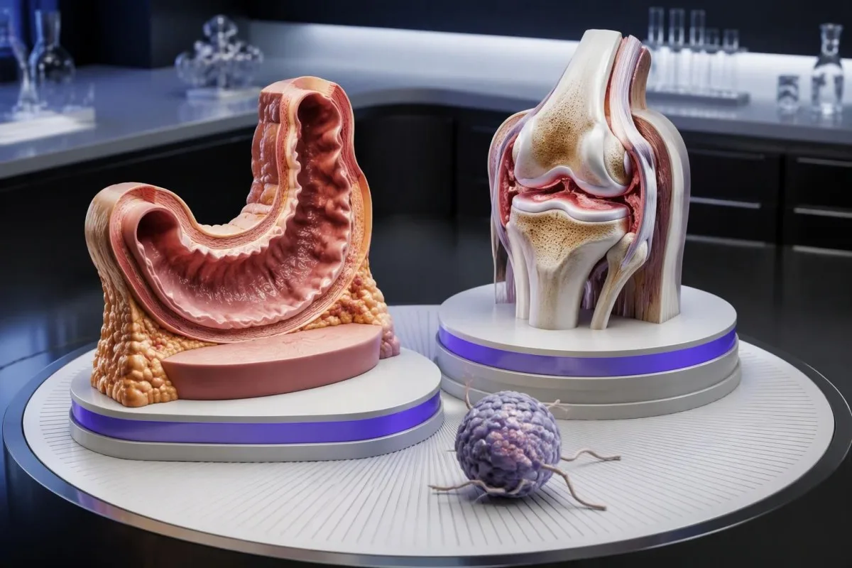
Reactive arthritis is an immune-mediated response, not simply bacteria spreading from stool into every painful joint. Knees and ankles may be affected asymmetrically; a new hot swollen joint with fever still needs urgent assessment because septic arthritis and other causes cannot be excluded from the timing alone.
Erythema nodosum usually produces tender nodules over the shins and has several possible triggers. Appearance 2 weeks after diarrhea can fit a postinfectious pattern, but medicines, inflammatory bowel disease, and other conditions remain relevant; examination is more informative than trying to diagnose the nodules from a positive stool report.
HLA-B27 can be associated with susceptibility or persistence of reactive arthritis, but it is not a routine screening test after uncomplicated yersiniosis. Our HLA-B27 interpretation guide explains why 1 genetic result cannot establish the cause of joint pain or predict an individual person's course.
Antibiotics for the original enteritis have not reliably been shown to prevent postinfectious complications. Joint treatment depends on the assessment and may differ from intestinal treatment; new eye pain, light sensitivity, or reduced vision needs urgent same-day care because an inflammatory eye complication can threaten vision even when diarrhea resolved weeks earlier.
How should you prepare your results for clinical review?
Bring the complete stool report, symptom timeline, medication list, and any accompanying blood results to clinical review. A concise 24–48-hour record of stool frequency, temperature, drinking, urine output, and pain location helps the clinician distinguish uncomplicated recovery from dehydration, appendicitis-like illness, or systemic disease.
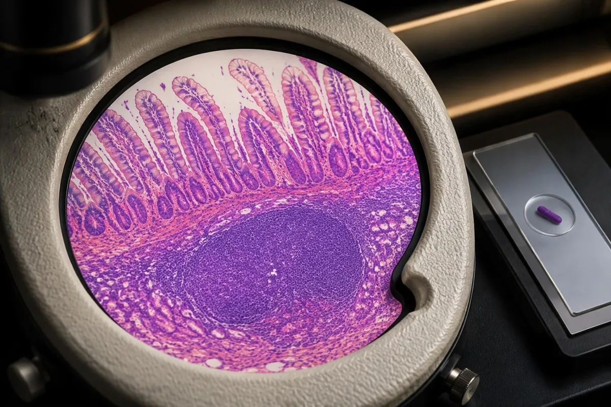
Record when symptoms started and when the sample was collected, because those 2 dates answer different questions. Add whether antibiotics were taken before sampling and whether another organism was detected; these details can explain a culture-PCR mismatch and can materially change the safest treatment discussion.
Kantesti iku sawijining Piranti analisis tes getih berbasis AI that accepts laboratory PDFs or photos and provides explanations in approximately 60 seconds, but that output is not a diagnosis or emergency assessment. Our informasi validasi medis describes evaluation and oversight; performance on blood-report interpretation should not be assumed to establish accuracy for stool-PCR diagnosis.
Check units and collection dates before comparing 2 results. Creatinine reported in mg/dL and µmol/L, or CRP measured before and after hydration, can look contradictory without proper context; an AI explanation should remain anchored to the original report and should never silently replace missing microbiology or examination findings with assumptions.
Laboratory reports can include names, dates of birth, and other identifiers, so share only what is needed for the task and consider consent when handling a family member's results. Our informasi privasi lan data explains our approach; regardless of the interpretation method, do not delay urgent care to complete a digital report review.
Publikasi panaliten lan watesan interpretasi tes getih
The Yersinia treatment discussion is supported by infectious-diarrhea guidance and microbiological literature, not by general blood-test publications. The 2 Zenodo guides listed below provide supplementary laboratory context; they are not Yersinia treatment trials and do not establish that an AI tool can diagnose intestinal infection.
The aPTT and clotting guide addresses coagulation interpretation, which is separate from stool pathogen detection. An uncomplicated positive Yersinia result does not routinely justify ordering aPTT, D-dimer, or Protein C; testing becomes relevant only when a clinician identifies an additional question, such as a severe systemic complication or a separate clotting concern.
The serum-protein guide explains albumin and globulin patterns that may become relevant during prolonged illness. An albumin of 30 g/L can reflect several processes, including inflammation, reduced intake, or protein loss; our pandhuan interpretasi protein serum explains why that value cannot identify Yersinia or determine antibiotic necessity.
Kantesti's Zenodo guides should therefore be read as supporting educational material, while the 2017 IDSA guideline and organism-specific literature support the infection discussion. A repository DOI provides a persistent identifier; it does not, by itself, demonstrate peer review, clinical effectiveness, or validation of a pathogen-specific diagnostic claim.
kita standar konten medis describe how we distinguish source-backed guidance from illustrative examples. Before ending your review, identify 3 practical decisions: whether urgent assessment is needed, whether supportive care is adequate, and whether your risk factors or ongoing symptoms require clinician-directed testing or treatment.
Pitakon sing kerep ditakoni
Apa asil positif Yersinia enterocolitica tegese aku butuh antibiotik?
Asil pemeriksaan feses positif Yersinia enterocolitica ora kanthi otomatis ateges antibiotik dibutuhake. Akeh infeksi sing ora rumit membaik kanthi rehidrasi oral lan perawatan panyengkuyung sajrone 1–3 minggu, sanajan diare bisa tahan luwih suwe. Antibiotik dipikirake kanggo penyakit sing abot, infeksi aliran getih, keterlibatan ing njaba usus, utawa pasien berisiko dhuwur tartamtu. Kelebihan zat besi, perawatan deferoxamine, lan imunosupresi sing signifikan kudu enggal dirembug karo dokter sing nambani.
Apa asil PCR Yersinia saka feses sing positif bisa dadi asil positif palsu?
Hasil PCR tinja Yersinia sing positif kadhangkala bisa ora dinyana-nyana utawa nyasarké, utamané nalika gejalane ora cocog, nanging asil positif ora kena mung disingkiraké. PCR ndeteksi materi genetik lan ora bisa mbuktèkaké manawa organisme sing isih urip isih ana utawa Yersinia njalari saben gejala. Yen 2 patogen dideteksi utawa penyakité wis mari, interpretasi klinis lan kadhangkala kultur luwih migunani tinimbang langsung mbalèni PCR. Kinerja tes gumantung marang cara laboratorium, lan ora ana ambang cycle-threshold Yersinia sing ditrapaké sacara universal kanggo penyakit aktif.
Apa Yersinia bisa nyebabake nyeri kaya apendisitis?
Yersinia bisa nyebabake rasa nyeri kaya usus buntu amarga mengaruhi ileum terminal lan kelenjar getah bening mesenterika ing sakupenge. Rong lokasi anatomi iki cedhak karo usus buntu lan bisa nyebabake nyeri weteng sisih tengen ngisor lan demam. Asil tes tinja positif ora nyingkirake kemungkinan usus buntu sing saktenane, lan asil sel getih putih utawa CRP sing normal ora bisa mesthekake manawa ora ana usus buntu. Nyeri sing abot, terlokalisasi, utawa saya parah mbutuhake pemeriksaan enggal lan kadhangkala ultrasonografi, CT, utawa MRI.
Suwene infeksi Yersinia enterocolitica berlangsung?
Penyakit usus amarga Yersinia kerep berlangsung 1–3 minggu, nanging gejalane bisa terus luwih suwe. Diare sing suwene 14 dina utawa luwih becik dipriksa kanthi klinis, luwih-luwih yen dibarengi bobot awak mudhun, getih, demam, utawa nyeri ing panggonan tartamtu. PCR ulang rutin biasane ora perlu sawisé pulih tanpa komplikasi amarga materi genetik bisa tetep dideteksi tanpa mbuktekake anané infeksi aktif sing isih urip. Gejala sendi bisa diwiwiti kira-kira 1–4 minggu sawisé enteritis lan mbutuhake evaluasi kapisah.
Antibiotik apa sing kanggo ngobati Yersinia enterocolitica?
Pedoman diare infèksius IDSA taun 2017 nyebut trimetoprim-sulfametoksazol kanggo Yersinia enterocolitica, kanthi sefotaksim utawa siprofloksasin minangka alternatif. Yersinia kerep tahan marang ampisilin lan akèh sefalosporin generasi awal, mula antibiotik sisa ora aman digunakaké minangka pengganti resep. Pilihan pengobatan gumantung marang tingkat aboté penyakit, asil tes kepekaan, umur, kandhutan, fungsi ginjel, lan interaksi obat. Ora ana dosis utawa suwéné pengobatan tunggal sing cocog kanggo enteritis tanpa komplikasi, bakteremia, lan kabèh komplikasi ekstrintestinal.
Kapan aku kudu golek perawatan darurat sawisé asil tes Yersinia positif?
Enggal-enggal golek pemeriksaan medis yen lara weteng abot utawa saya parah, ora bisa njaga cairan supaya ora dimuntahaké, jumlah urin mudhun banget, semaput, bingung, utawa ana getihen usus sing cukup akèh. Bayi umur kurang saka 3 sasi kanthi suhu 38°C utawa luwih kudu enggal dipriksa kanthi medis. Wong sing ngalami keluwihan zat besi, lagi diobati nganggo deferoksamin, utawa daya tahan tubuhé mudhun banget kudu enggal njaluk saran sing disesuaikaké karo kahanané, lan ora mung ngandelaké perawatan rutin ing omah. Aja ngenteni asil pemeriksaan tinja sing dibalèni yen gejala nuduhaké dehidrasi, apendisitis, utawa penyakit sistemik.
Apa Yersinia isih nular sawisé diare mandheg?
Yersinia bisa ditularaké liwat kontaminasi feses, lan ilangé gejala ora mbuktekaké yèn pengeluaran bakteri liwat tinja wis mandheg. Akèh kabijakan sekolah lan papan kerja ngenani gastroenteritis mbutuhaké paling ora 48 jam tanpa diare utawa muntah, nanging aturan lokal bisa luwih ketat kanggo wong sing nangani panganan, tenaga kesehatan, utawa staf penitipan anak. Terusna ngumbah tangan kanthi tliti lan aja nyiyapaké panganan kanggo wong liya sajrone periode ora mlebu sing disaranaké. Tes kanggo mesthèkaké wis bebas infèksi biasané ora dibutuhaké sawisé pulih tanpa komplikasi, kajaba dijaluk déning klinisi utawa panguwasa kesehatan umum.
Entuk penjelasan babagan asil tes getih sampeyan
Unggah laporan laboratorium sampeyan lan AI Kantesti bakal nerangake saben biomarker nganggo basa sing prasaja sajrone sawetara detik. Digunakake luwih saka 2 yuta wong ing saindenging donya.
Referensi
- Klein, T., Mitchell, S., & Weber, H. (2026). Rentang Normal aPTT: Pandhuan D-Dimer, Protein C lan Pembekuan Getih. Riset Medis AI Kantesti. DOI
- Klein, T., Mitchell, S., & Weber, H. (2026). Pandhuan Protein Serum: Globulin, Albumin & Rasio A/G ing Tes Getih. Riset Medis AI Kantesti. DOI
- Shane AL dkk. (2017). Pandhuan Praktik Klinis Infectious Diseases Society of America taun 2017 kanggo Diagnosis lan Tata Laksana Diare Infèksius. Penyakit Infèksius Klinis. DOI
- Bottone EJ (1997). Yersinia enterocolitica: the charisma continues. Clinical Microbiology Reviews.
- National Institute for Health and Care Excellence (2009). Diare lan muntah sing disebabake gastroenteritis ing bocah umur kurang saka 5 taun: diagnosis lan penanganan. NICE Clinical Guideline CG84.
- 2M+Tes sing dianalisis
- 127+negara-negara
- 100+Basa
Babagan tim medis


Sinyal Kepercayaan E-E-A-T
Pengalaman
Tinjauan klinis sing dipimpin dokter babagan alur kerja interpretasi lab.
Keahlian
Fokus kedokteran laboratorium babagan carane biomarker tumindak ing konteks klinis.
Kewibawaan
Ditinjau sacara medis dening Dr. Thomas Klein, kanthi kontribusi saka Dr. Sarah Mitchell lan Prof. Dr. Hans Weber.
Kapercayan
Interpretasi adhedhasar bukti kanthi tindak lanjut sing cetha kanggo nyuda rasa kaget.

Sanggahan medis
Artikel iki mung kanggo tujuan edukasi lan ora dadi saran medis. Tansah konsultasi karo panyedhiya layanan kesehatan sing mumpuni kanggo keputusan diagnosis lan perawatan.
Diterbitake · Hubungi tim editorial
Diterbitake dening Kantesti Ltd, kadhaptar ing Inggris & Wales, nomer perusahaan. 17090423, London, Britania Raya.
